


Serial Cryosectioning for the Spatial Transcriptomics of Plant Tissues

发布: 2026年10月20日第16卷第20期 DOI: 10.21769/BioProtoc.5844 浏览次数: 63
评审: Alberto RissoneAnonymous reviewer(s)
 PDF
PDF  Q&A
Q&A





Abstract
Spatial transcriptomics enables genome-wide gene expression profiling while preserving tissue architecture, making it a powerful approach for studying plant developmental transitions. However, preparing small and structurally complex plant tissues for spatial transcriptomics remains technically challenging because samples must be rapidly preserved, precisely oriented, serially sectioned, and accurately positioned within the limited capture area of the Visium slides. Here, we describe an optimized workflow for cryo-embedding, serial cryosectioning, section placement, and data analysis of small plant samples for 10x Genomics Visium spatial transcriptomics. Using maize seedling shoot apices as the target, this protocol includes preparation of custom molds for optimal cutting temperature embedding, rapid fresh sample embedding, serial cryosectioning, section-position marking for Visium HD workflows, and morphological quality assessment of replicate tissue slides before transcript capture. The associated data analysis workflow includes Space Ranger processing, Seurat-based normalization and Harmony integration, anatomical domain annotation, pseudobulk and developmental trend analyses, RNA velocity, pseudotime analysis, transcription factor network analysis, single-cell reference mapping, and 3D transcriptome reconstruction. This computational workflow was developed and tested using maize Visium V1 data, but not Visium HD data. This protocol was used to generate serial spatial transcriptomes of maize shoot apices and developing leaf primordia, enabling reconstruction of gene expression transitions from the shoot apical meristem to sequential leaf developmental stages. The approach is also applicable to other small plant tissues, including Arabidopsis first true leaves and Marchantia thalli.
Key features
• Optimized cryo-embedding and cryosectioning workflow for small plant tissues for 10x Visium spatial transcriptomics.
• A streamlined computational workflow for serial-section processing, spatial-domain annotation, 3D reconstruction, and developmental trajectory analysis.
Keywords: Spatial transcriptomics 3D gene expression
Maize leaf development
Developmental trajectory
Transcription factor networks
Growth-regulating factors
3D gene expression
Maize leaf development
Developmental trajectory
Transcription factor networks
Growth-regulating factors
Graphical overview
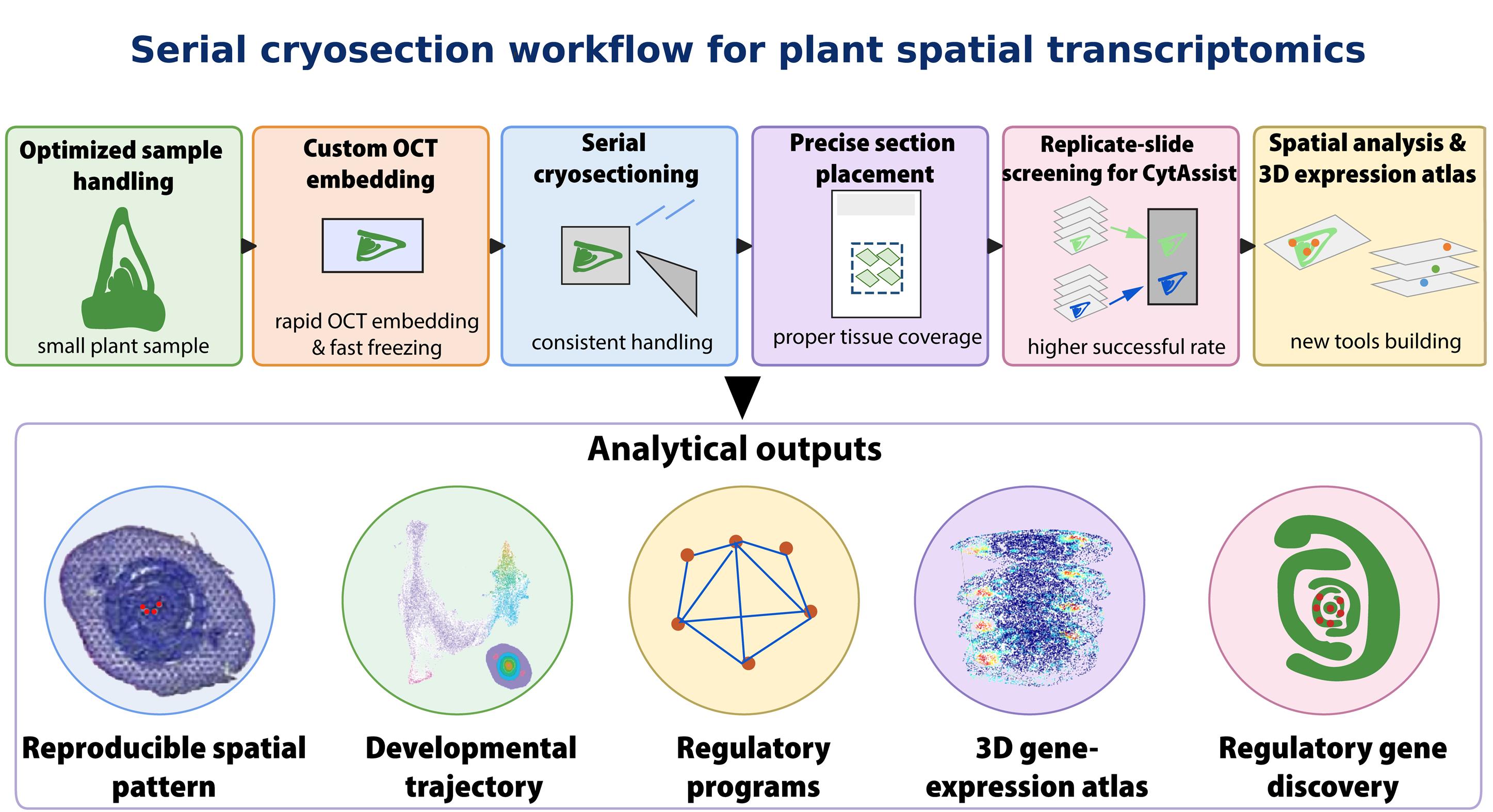
Small plant tissues are processed using an optimized cryo-embedding and serial cryosectioning workflow for plant spatial transcriptomics. Samples are embedded in custom optimal cutting temperature (OCT) embedding molds, rapidly frozen, cryosectioned into serial sections, and positioned within a marked Visium capture area. Replicate tissue slides are screened for tissue integrity, morphology, and placement before 10x Visium CytAssist transcript transfer. The resulting spatial transcriptomes are analyzed to generate reproducible spatial maps, reconstruct developmental trajectories, identify regulatory programs and candidate regulatory genes, and build a 3D gene expression atlas.
Background
Spatial transcriptomics enables transcriptome-wide gene expression profiling while preserving the spatial organization within tissue sections. In plants, this approach is particularly useful for studying developmental transitions that occur across small and structurally complex tissues, such as shoot apices, leaf primordia, vascular tissues, and reproductive organs [1–4]. However, compared with animal and clinical tissues, plant samples often require additional optimization because their small size, extensive intercellular air spaces, tissue fragility, and rigid cell walls make cryosectioning difficult.
Several plant spatial transcriptomics studies have demonstrated the value of spatially resolved gene expression analysis in species such as Arabidopsis, poplar, barley, and maize using a species-agnostic approach [4–7]. These studies showed that spatial transcriptomics can reveal tissue-specific expression patterns and developmental gene expression gradients. However, many published plant applications [4–7] provide limited practical detail on the sample preparation steps that strongly affect data quality, such as fresh-tissue handling, optimal cutting temperature (OCT) embedding medium infiltration, freezing, tissue orientation, section attachment, and accurate placement of small sections within the capture area. These steps are particularly important for small plant samples, where excess OCT, poor orientation, air bubbles, tissue folding, or inaccurate placement can negatively impact tissue morphology, RNA quality, and transcript capture efficiency.
In our previous study, we optimized a 10x Visium-based workflow for serial spatial transcriptomics of maize seedling shoot apices [1]. The method combined rapid fresh-sample handling, OCT immersion before freezing, custom OCT embedding molds, serial cryosectioning, and computational reconstruction of 3D gene expression profiles. Using this workflow, multiple maize shoot cross-sections were placed within one 6.5 × 6.5 mm Visium capture area, enabling spatial transcriptome profiling of 14 biological replicates of shoot apical meristems with developing embryonic leaves, comprising 54 serial sections. Tissue-covered spots were annotated into seven structural domains: SAM, P1_P2, P3, P4, P5, coleoptile, and coleoptile vein.
The protocol described here expands on the experimental details of this workflow and facilitates its broader application to spatial transcriptomics studies of small plant tissues, including Arabidopsis first true leaves and Marchantia thalli. By providing a practical and integrated approach to tissue preparation, this protocol enables the generation of high-quality sections suitable for spatial transcriptomics and downstream 3D gene expression analysis.
Materials and reagents
Biological materials
1. Seeds of the maize cultivar Zea mays cv. White Crystal used in this study were purchased from the local breeder Fong Tien Seed Co. Ltd. (Taiwan). To obtain seedlings with straight, upright stems, seeds were positioned vertically between repeatedly S-folded wet filter paper towels in MagentaTM vessels (Figure 1). Seeds were grown in a growth chamber at 30 °C during the day and 26 °C at night under a 14/10 h light/dark cycle. After 72 h of imbibition, developing leaves containing the coleoptile and shoot apical meristem were carefully dissected and separated from the starchy endosperm (red dashed box in Figure 1C).
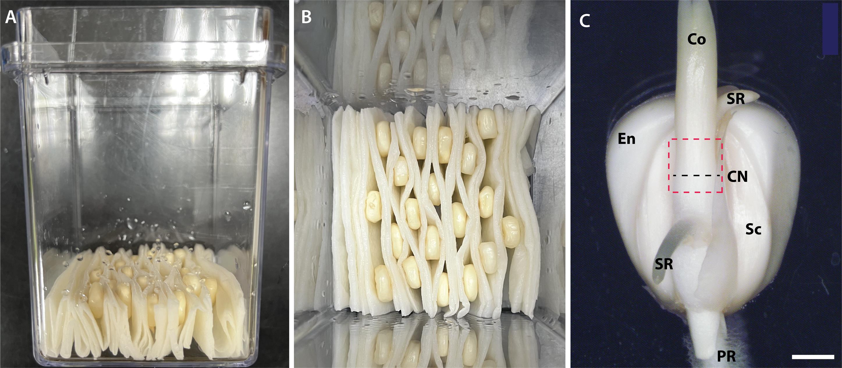
Figure 1. Preparation and sampling of germinating maize seedlings. (A) Maize kernels were positioned upright between folded wet paper towels in MagentaTM vessels to promote straight seedling growth. (B) Top view of seed arrangement. (C) Developing seedling attached to a kernel after 72 h of imbibition, showing roots, endosperm, and coleoptile and embryonic leaf tissues enclosed within the coleoptile. The red dashed box indicates the target region for tissue collection and embedding, and the black dashed line marks the approximate section collection position at the coleoptile node. Co: coleoptile; CN: coleoptile node; En: endosperm; PR: primary root; Sc: scutellum; SR: seminal root. Scale bar, 2 mm.
Reagents
1. Ribonucleoside vanadyl complex 100% (New England Biolabs, catalog number: S1402S)
2. Tissue-Tek OCT 100% (Sakura, catalog number: 4583)
3. RNaseZap RNase decontamination solution (Thermo Fisher, catalog number: AM9780)
4. Isopentane (Alfa Aesar, catalog number: AF-19387-500ML)
5. Phosphate-buffered saline (PBS) (Thermo Fisher, catalog number: AM9624)
6. UltraPure DNase/RNase-free distilled water (Thermo Fisher, catalog number: 10977015)
7. Methanol, molecular biology grade (Fisher Scientific, catalog number: 34860-1L-R)
8. Isopropanol, molecular biology grade (Fisher BioReagents, catalog number: BP2618-4)
Solutions
1. Embedding transition medium 1 (ETM1) (20% OCT) (see Recipes)
2. Embedding transition medium 2 (ETM2) (50% OCT) (see Recipes)
3. Embedding transition medium 3 (ETM3) (100% OCT) (see Recipes)
4. Sample handling buffer (SHB) (see Recipes)
Recipes
1. ETM1
| Reagent | Final concentration | Quantity or volume |
|---|---|---|
| Milli-Q H2O | 4 mL | |
| OCT | 20% | 1 mL |
| Total | 5 mL |
2. ETM2
| Reagent | Final concentration | Quantity or volume |
|---|---|---|
| Milli-Q H2O | 2.5 mL | |
| OCT | 50% | 2.5 mL |
| Total | 5 mL |
3. ETM3
| Reagent | Final concentration | Quantity or volume |
|---|---|---|
| Milli-Q H2O | 0 mL | |
| OCT | 100% | 5.0 mL |
| Total | 5.0 mL |
4. SHB
| Reagent | Final concentration | Quantity or volume |
|---|---|---|
| 1× PBS | 90% | 4.5 mL |
| Ribonucleoside vanadyl complex | 10% | 0.5 mL |
| Total | 5.0 mL |
Laboratory supplies
1. MagentaTM vessel GA-7 (Sigma-Aldrich, catalog number: V8505)
2. Laboratory folded paper towels (Scott, catalog number: 60055)
3. Fisherbrand Superfrost Plus microscope slides (Fisher Scientific, catalog number: FIS12-550-15)
4. Dumont Tweezers Positive Action 3C (Electron Microscopy Sciences, catalog number: 50-949-284)
5. Dissecting scissors (Fisher Scientific, catalog number: 08-940)
6. Hypodermic needles (19G × 1 1/2") (Terumo, catalog number: B130022)
7. Aluminum foil (Diamond Crystal, catalog number: 75 SQFT)
8. Falcon conical 50 mL high clarity PP centrifuge tube (Corning, catalog number: 352098)
9. 60 × 15 mm plastic Petri dishes (Corning, catalog number: BP53-03)
10. 35 × 15 mm plastic Petri dishes (Fisher Scientific, catalog number: 50-147-469)
11. Lint-free laboratory wipes (Texwipe Vectra Alpha 10, catalog number: TX1010)
12. Reagent reservoirs (Corning Costar, sterile disposable reagent reservoirs, catalog number: 07-200-130)
13. Liquid nitrogen or dry ice (local provider)
14. Slide mailer (Fisher Scientific, catalog number: HS15986)
15. Razor blades (Merkur)
16. Single-edge safety razor (Educlub, catalog number: MV-ED0068A)
17. Metal containers for frozen sample transfer (accessories of Leica EMPACT2)
Equipment
1. Growth chamber (Firstek, model: S2703-GC)
2. Freezer (-20 °C) (Nihon Freezer, model: SCF-FG-4002)
3. Freezer (-80 °C) (Nihon Freezer, model: CLN-52UWT)
4. Refrigerator (4 °C) (Nihon Freezer, model: SCF-FG-4002)
5. Dissecting microscope (Nikon, model: SMZ)
6. Rotary pump (Edwards, model: E2S45)
7. Histology vacuum chamber (Tarsons, model: T403030)
8. Brightfield microscope (Evident, model: CX33)
9. Cryostat (Leica, model: CM 1950)
10. Thermal cyclers (Thermo Fisher Scientific, model: VeritiPro Thermal Cycler A48141)
Software and datasets
1. Space Ranger (v2.1.0 for Visium or v4.1.0 for Visium HD 3’, 10x Genomics, https://www.10xgenomics.com/support/software/space-ranger/latest/release-notes/release-notes-for-SR)
2. Loupe Browser (v9.1, 10x Genomics, https://www.10xgenomics.com/support/software/loupe-browser/latest)
3. R environment (v4.6.1, https://www.r-project.org/about.html)
4. Python (v3.11.16, https://www.python.org/)
5. Seurat (v5.5.1, Satija Lab and Collaborators, https://satijalab.org/seurat/)
6. STUtility (v1.1.1, GRG, KTH, https://ludvigla.github.io/STUtility_web_site/)
7. SeuratWrappers (v0.4.0, a collection of Harmony v2, Monocle 3, scVelo v0.2.5, https://github.com/satijalab/seurat-wrappers)
8. SCTransform (v0.4.3, https://github.com/satijalab/sctransform)
9. Velocyto (v0.17.17, https://velocyto.org/velocyto.py/tutorial/index.html#running-the-cli)
10. scDblFinder (v1.26.7, https://github.com/plger/scDblFinder)
11. SCINA (v1.2.0, https://github.com/jcao89757/SCINA)
12. SPOTlight (v1.16.0, https://github.com/MarcElosua/SPOTlight)
13. TO-GCN (https://github.com/petitmingchang/TO-GCN)
14. glmGamPoi (v1.24.0, https://github.com/const-ae/glmGamPoi)
15. Harmony (v2.0.5, https://github.com/immunogenomics/harmony)
16. scVelo (v0.3.4, https://scvelo.readthedocs.io/en/stable/)
17. SeuratObject (v5.4.0, https://github.com/satijalab/seurat-object)
18. Monocle 3 (v1.4.27, https://cole-trapnell-lab.github.io/monocle3/)
19. Napari (v0.7.1, https://napari.org/stable/)
20. Maize shoot data-processing pipeline (https://github.com/bomacchih/maize_shoot_data_process_v2)
21. Maize reference genome (Zm-B73-REFERENCE-NAM-5.0.fa)
22. Maize gene annotation (Zm-B73-REFERENCE-NAM-5.0.51.gtf)
23. Maize shoot scRNA-seq datasets (SRA: SRR11943512 and SRR11943513)
24. Maize shoot Visium datasets (PRJNA805024 and PRJNA804974)
25. Processed datasets for Napari interactive viewer on Zenodo (https://zenodo.org/records/16933147)
26. Processed datasets for bioinformatic analyses on Zenodo (https://zenodo.org/records/22058284)
27. agriGo v2.0 (https://systemsbiology.cau.edu.cn/agriGOv2/)
Procedure
登录/注册后免费查看全文
文章信息
稿件历史记录
提交日期: Jul 19, 2026
接收日期: Sep 10, 2026
在线发布日期: Sep 22, 2026
出版日期: Oct 20, 2026
版权信息
© 2026 The Author(s); This is an open access article under the CC BY license (https://creativecommons.org/licenses/by/4.0/).
如何引用
Wu, C. C., Hsieh, K. T., Yu, C. P., Chen, Y. H., Chou, S. J., Wu, T. Y., Ho, C. K., Wu, S. H., Lu, M. J. and Li, W. H. (2026). Serial Cryosectioning for the Spatial Transcriptomics of Plant Tissues. Bio-protocol 16(20): e5844. DOI: 10.21769/BioProtoc.5844.

 Download Citation in RIS Format
您对这篇实验方案有问题吗?
在此处发布您的问题,我们将邀请本文作者来回答。同时,我们会将您的问题发布到Bio-protocol Exchange,以便寻求社区成员的帮助。
 发布问题
发布问题


