


PIC-RNA-seq for Region-Specific Transcriptomic Analysis of Chicken Limb Bud Progenitors

(*contributed equally to this work) 发布: 2026年10月20日第16卷第20期 DOI: 10.21769/BioProtoc.5843 浏览次数: 63
评审: Alberto RissoneGuohao HanAnonymous reviewer(s)
 PDF
PDF  Q&A
Q&A






Abstract
Region-specific RNA sequencing is a powerful approach for investigating tissue differentiation and dynamic changes in gene expression during embryonic development. The chicken embryo has long served as an important model system in developmental biology. However, the limited availability of tissue-specific reporter lines makes region-specific RNA-seq approaches particularly valuable in this organism. Here, we applied photo-isolation chemistry-based RNA sequencing (PIC-RNA-seq) to the somatic lateral plate mesoderm (sLPM) before the emergence of limb bud progenitor cells (LPCs) and to early LPCs in chicken embryos. These analyses revealed the upregulation of multiple genes, including Hox genes, in LPCs, suggesting the initiation of their regional patterning program. This workflow enables visualization of dynamic changes in the gene expression profile of LPCs and should also be applicable to other tissues in avian embryos.
Key features
• An optimized PIC-RNA-seq protocol for the somatic lateral plate mesoderm (sLPM) and limb bud progenitor cells (LPCs) in chicken embryos.
• Applicable to chicken embryos without the need for tissue-specific transgenic reporter lines.
• Robust generation of high-quality RNA-seq datasets with sufficient sequencing depth, unique molecular identifiers (UMIs), and detected genes for downstream analyses.
• Enables direct comparison between pre-specified sLPM and specified LPCs, allowing the detection of transcriptional changes accompanying early LPC specification.
Keywords: PIC-RNA-seq Chicken embryo
Limb development
Limb bud progenitor cell
Somatic lateral plate mesoderm
Chicken embryo
Limb development
Limb bud progenitor cell
Somatic lateral plate mesoderm
Graphical overview
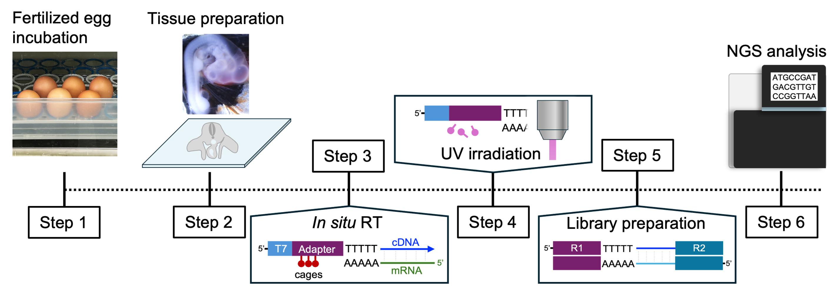
Background
Embryonic development is orchestrated by spatiotemporally regulated gene expression programs that drive cell fate specification, tissue patterning, and organogenesis. Identifying transcriptomic changes within anatomically defined regions is therefore essential for understanding the molecular basis of developmental processes. In particular, the transition from multipotent progenitor populations to lineage-committed cells often occurs within a narrow developmental window, requiring methods that provide both high spatial resolution and robust transcriptomic profiling.
The chicken embryo has long served as a classical model for developmental biology because of its accessibility for in ovo manipulation and live imaging. Despite these advantages, transcriptomic analysis of spatially restricted embryonic tissues remains technically challenging. Fluorescence-based cell isolation generally requires tissue-specific transgenic reporter lines, which are still limited in chickens. Manual microdissection can enrich target tissues but often includes surrounding cells, whereas single-cell RNA sequencing requires tissue dissociation, resulting in the loss of spatial information and introducing additional technical complexity. Integration of spatial transcriptomics with single-cell RNA-seq can provide spatial information on transcriptionally defined cell populations but typically requires additional single-cell datasets and computational mapping or deconvolution, rather than direct transcriptomic profiling of a microscopically defined region of interest (ROI). Laser capture microdissection (LCM) coupled with RNA-seq also enables transcriptomic analysis of microscopically defined regions in tissue sections [1]. However, the spatial resolution and shape of the ROI are constrained by laser-cutting precision and the requirement for physical dissection and recovery of the target tissue. Although LCM facilitates the separate collection and analysis of multiple ROIs from the same tissue section, physical recovery of very small or complexly shaped regions can be technically challenging.
PIC-RNA-seq overcomes many of these limitations by defining ROIs through patterned light irradiation rather than physical tissue isolation [2,3]. The method uses photo-caged primers for in situ reverse transcription, followed by patterned light irradiation to uncage cDNAs specifically within selected ROIs. Only cDNAs from the irradiated regions become competent for subsequent amplification, enabling spatially selective transcriptomic profiling. This approach allows flexible selection of small or irregularly shaped regions at optical resolution. Because tissue selection is performed directly on tissue sections, the method can be applied without tissue dissociation or tissue-specific transgenic reporter lines, making it particularly well suited for developmental studies in organisms with limited genetic resources. We previously employed this approach to investigate the molecular basis of avian sternal diversification [4].
In this protocol, we adapted the original PIC-RNA-seq workflow with minor modifications for the analysis of the somatic lateral plate mesoderm (sLPM) before limb bud progenitor cell (LPC) specification and of specified LPCs in chicken embryos. The optimized workflow consistently yields high-quality sequencing data from small embryonic regions and enables direct comparison of closely related developmental stages. Although demonstrated here using the early limb field, this protocol should be broadly applicable to transcriptomic studies of other spatially defined tissues in avian embryos.
Materials and reagents
Biological materials
1. Fertilized white leghorn chicken Gallus gallus eggs (purchased from Yamagishi poultry farm)
2. Fresh-frozen cryosections (10 μm) of chicken (Gallus gallus) embryos at Hamburger–Hamilton (HH) stages 14 and 19, mounted on MAS-coated slides
Reagents
1. Isopentane (Nacalai Tesque, catalog number: 26404-75)
2. OCT compound (Sakura Finetek, catalog number: 4583)
3. Nuclease-free water (Nacalai Tesque, catalog number: 06442-95)
4. Distilled water (Nacalai Tesque, catalog number: 49506-64)
5. Phosphate-buffered saline (PBS) tablets, pH 7.4 (Takara Bio, catalog number: T9181)
6. 16% formaldehyde (w/v), methanol-free (Electron Microscopy Sciences, catalog number: 15710)
7. Tris-EDTA (TE) buffer solution, pH 8.0 (Nacalai Tesque, catalog number: 32739-31)
8. 0.1 M DTT (Thermo Fisher Scientific, catalog number: Y00147)
9. Proteinase K solution, 20 mg/mL (Kanto Chemical, catalog number: 34060-96)
10. 5 M NaCl (Nacalai Tesque, catalog number: 06900-14)
11. Polyethylene glycol 8,000, 50% (w/v) (Nacalai Tesque, catalog number: 26065-54)
12. 0.5 M EDTA, pH 8.0 (Nacalai Tesque, catalog number: 06894-85)
13. Ethanol (Nacalai Tesque, catalog number: 14713-95)
14. NPOM-caged-dT-CE phosphoramidite (Glen Research, catalog number: 10-1534-95)
15. SuperScript II reverse transcriptase, 200 U/μL, with 5× first-strand buffer and 0.1 M DTT (Thermo Fisher Scientific, catalog number: 18064071)
16. RNaseOUT, 40 U/μL (Thermo Fisher Scientific, catalog number: 10777019)
17. dNTP mix, 10 mM each (New England Biolabs, catalog number: N0447L)
18. MinElute PCR Purification kit (QIAGEN, catalog number: 28006)
19. 5× second-strand buffer (Thermo Fisher Scientific, catalog number: 10812014)
20. E. coli DNA polymerase I, 10 U/μL (Thermo Fisher Scientific, catalog number: 18010025)
21. E. coli DNA ligase, 10 U/μL (Thermo Fisher Scientific, catalog number: 18052019)
22. RNase H, 2 U/μL (Thermo Fisher Scientific, catalog number: 18021071)
23. MEGAscript T7 Transcription kit (Thermo Fisher Scientific, catalog number: AMB13345), which includes ATP, GTP, CTP, UTP, 10× T7 reaction buffer, 10× enzyme mix, and TURBO DNase
24. AMPure XP beads (Beckman Coulter, catalog number: A63881)
25. RNAClean XP beads (Beckman Coulter, catalog number: A63987)
26. Phusion High-Fidelity PCR master mix (New England Biolabs, catalog number: M0531L)
27. Ribonuclease A solution, 10 mg/mL (Nacalai Tesque, catalog number: 30100-31)
28. Nuclear Violet LCS1 (AAT Bioquest, catalog number: 17543)
29. SlowFade Diamond (Thermo Fisher Scientific, catalog number: S36963)
30. PhiX Control v3 (Illumina, catalog number: FC-110-3001)
31. High-sensitivity D5000 ScreenTape (Agilent Technologies, catalog number: 5067-5592)
32. High-sensitivity D5000 reagents (sample buffer and ladder) (Agilent Technologies, catalog number: 5067-5593)
33. Tween-20 (Nacalai Tesque, catalog number: 28353-85), used as a 10% (v/v) stock in the ProK lysis solution
34. Silicone base (Shin-Etsu Chemical, catalog number: KE-106)
35. Curing agent (Shin-Etsu Chemical, catalog number: CAT-RG)
36. Activated charcoal powder (FUJIFILM, catalog number: 037-02115)
Oligonucleotides
The primer design follows Honda et al. [2]. All sequences are represented in the 5′–3′ direction.
1. NPOM-caged RT primer:
GCCGGTAATACGACTCACTATAGGGtttGAGttCtACAGTCCGACGATCNNNNNN[BC]ttTTTTTTTTTTTTTTTTTTTTTTV, where T = unmodified deoxythymidine, t = NPOM-caged dT, N = UMI, [BC] = the 6-nt sample barcode, and V = A, C, or G (a 3′ anchor base). The barcodes used here were CATGAG, AGGATC, AGTGCA, and TCGAAG (one per biological replicate). The caged oligonucleotide was custom-synthesized from NPOM-caged dT-CE phosphoramidite (Reagent list, #14) and OPC-purified by Nihon Gene Research Laboratory. Store at -20 °C, protected from light
2. Read2+N6 primer: GCCTTGGCACCCGAGAATTCCANNNNNN (N = random hexamer)
3. Read1 index primer: AATGATACGGCGACCACCGAGATCTACAC[i5]GTTCAGAGTTCTACAGTCCGA (replace [i5] with the appropriate Illumina index)
4. Read2 index primer: CAAGCAGAAGACGGCATACGAGAT[i7]GTGACTGGAGTTCCTTGGCACCCGAGAATTCCA (replace [i7] with the appropriate Illumina index)
Note: Each pooled library was tagged with a unique dual index (UDI): i5 = CTGCACAA and i7 = TCTCACCT for the HH14 library, and i5 = GTGGTCTT and i7 = GTTCGAGA for the HH19 forelimb bud library. The i5 and i7 sequences listed are those entered into the sequencing sample sheet.
Solutions
1. PBS (see Recipes)
2. 4% PFA in PBS (see Recipes)
3. NPOM-caged RT primer mix (see Recipes)
4. First-strand mix (see Recipes)
5. Second-strand mix (see Recipes)
6. Beads binding buffer (see Recipes)
7. IVT mix (see Recipes)
8. aRNA–N6 primer mix (see Recipes)
9. Reverse transcription mix (see Recipes)
10. Read1 and Read2 index primer mix (see Recipes)
11. Library PCR mix (see Recipes)
12. Proteinase K lysis solution (ProK solution) (see Recipes)
Recipes
Note: Prepare all solutions fresh before use unless otherwise specified. For the reaction mixes, “Final concentration” refers to the concentration in the complete reaction after the component mixes are combined (e.g., the first-strand mix combined with the NPOM-caged RT primer mix, or the IVT mix added to the bead suspension).
1. 10× PBS
| Reagent | Final concentration | Quantity or volume |
|---|---|---|
| NaCl | 80 g/L | 80 g |
| KCl | 2 g/L | 2 g |
| Na2HPO4 | 11.5 g/L | 11.5 g |
| KH2PO4 | 2 g/L | 2 g |
| Distilled water | n/a | to 1 L |
| Total | 10× | 1 L |
2. 4% PFA in PBS
| Reagent | Final concentration | Quantity or volume |
|---|---|---|
| 16% formaldehyde | 4% | 2.5 mL |
| PBS | 1× | 7.5 mL |
| Total | n/a | 10 mL |
Work in a fume hood.
3. NPOM-caged RT primer mix (per section)
| Reagent | Final concentration | Quantity or volume |
|---|---|---|
| Nuclease-free water | n/a | 5 μL |
| NPOM-caged RT primer (500 ng/μL) | 25 ng/μL | 0.5 μL |
| dNTP mix (10 mM each) | 0.5 mM | 0.5 μL |
| Total | n/a | 6 μL |
Keep shielded from light.
4. First-strand mix (per section)
| Reagent | Final concentration | Quantity or volume |
|---|---|---|
| 5× first-strand buffer | 1× | 2 μL |
| 0.1 M DTT | 0.01 M | 1 μL |
| RNaseOUT (40 U/μL) | 2 U/μL | 0.5 μL |
| SuperScript II RT (200 U/μL) | 10 U/μL | 0.5 μL |
| Total | n/a | 4 μL |
5. Second-strand mix (per tube)
| Reagent | Final concentration | Quantity or volume |
|---|---|---|
| 5× first-strand buffer | 0.5× | 2 μL |
| 5× second-strand buffer | 0.5775× | 2.31 μL |
| dNTP mix (10 mM each) | 0.115 mM | 0.23 μL |
| E. coli DNA polymerase I (10 U/μL) | 0.15 U/μL | 0.3 μL |
| E. coli DNA ligase (10 U/μL) | 0.04 U/μL | 0.08 μL |
| RNase H (2 U/μL) | 0.008 U/μL | 0.08 μL |
| Total | n/a | 5 μL |
6. Beads binding buffer
| Reagent | Final concentration | Quantity or volume |
|---|---|---|
| 50% (w/v) PEG 8,000 | 20% | 4 mL |
| 5 M NaCl | 2.5 M | 5 mL |
| Nuclease-free water | n/a | 1 mL |
| Total | n/a | 10 mL |
Stable at room temperature (23–25 °C) for at least 1 year.
7. IVT mix (per tube)
| Reagent | Final concentration | Quantity or volume |
|---|---|---|
| ATP | 7.5 mM | 1.6 μL |
| GTP | 7.5 mM | 1.6 μL |
| CTP | 7.5 mM | 1.6 μL |
| UTP | 7.5 mM | 1.6 μL |
| 10× T7 reaction buffer | 1× | 1.6 μL |
| 10× enzyme mix | 1× | 1.6 μL |
| Total | n/a | 9.6 μL |
8. aRNA–N6 primer mix (per tube)
| Reagent | Final concentration | Quantity or volume |
|---|---|---|
| aRNA (from IVT/purification) | n/a | 4.5 μL |
| Read2+N6 primer (250 ng/μL) | 25 ng/μL | 1 μL |
| dNTP mix (10 mM each) | 0.5 mM each | 0.5 μL |
| Total | n/a | 6.0 μL |
9. Reverse transcription mix (per tube)
| Reagent | Final concentration | Quantity or volume |
|---|---|---|
| 5× first-strand buffer | 1× | 2 μL |
| 0.1 M DTT | 0.01 M | 1 μL |
| RNaseOUT (40 U/μL) | 2 U/μL | 0.5 μL |
| SuperScript II RT (200 U/μL) | 10 U/μL | 0.5 μL |
| Total | n/a | 4 μL |
10. Read1 and Read2 index primer mix (10 μM each)
| Reagent | Final concentration | Quantity or volume |
|---|---|---|
| 100 μM Read1 index primer | 10 μM | 10 μL |
| 100 μM Read2 index primer | 10 μM | 10 μL |
| Nuclease-free water | n/a | 80 μL |
| Total | n/a | 100 μL |
Stable at -20 °C for at least 1 year.
11. Library PCR mix (per tube)
| Reagent | Final concentration | Quantity or volume |
|---|---|---|
| Nuclease-free water | n/a | 10.4 μL |
| Read1 and Read2 index primer mix (10 μM each) | 0.8 μM each | 3.6 μL |
| Phusion High-Fidelity PCR master mix (2×) | 1× | 22.5 μL |
| Secondary RT products | n/a | 8.5 μL |
| Total | n/a | 45 μL |
12. ProK solution
| Reagent | Final concentration | Quantity or volume |
|---|---|---|
| 1× PBS | n/a | 970 μL |
| Tween-20 (10% (v/v) stock) | 0.1% | 10 μL |
| Proteinase K (20 mg/mL) | 0.4 mg/mL | 20 μL |
| Total | n/a | 1 mL |
Laboratory supplies
1. MAS-coated glass slides (Matsunami, catalog number: MAS-01)
2. Round coverslips, 15 mm (Matsunami, catalog number: C015001)
3. PAP pen (BMS, catalog number: BC-PAPPEN-S)
4. Humidified chamber (Cosmo Bio, catalog number: 10HTLS)
5. Handmade silicone-bottom dish: Mix 500 g of silicone base with 50 g of curing agent. Gradually add activated charcoal powder while mixing until opaque (approximately 3–4 tablespoons for a 5-mm-thick layer). Pour into a glass Petri dish, remove air bubbles by briefly applying hot air every 10 min, and leave on a level surface until completely cured (approximately 24 h at room temperature)
6. Handmade aluminum foil cryomold
7. Fine forceps (#55) (Dumont, catalog number: 11255-20)
8. 10 mL syringe (Terumo, catalog number: SS-10SZ)
9. 18G × 1.5” syringe needle (Terumo, catalog number: NN-1838R)
10. 100 mL beaker (Sanpla, catalog number: 01021)
11. Dry ice
12. Curved scissors (AZ ONE, catalog number: 2-540-11)
13. Perforated spoon (Urin Seisakujyo)
Equipment
1. Fluorescence microscope (Leica Microsystems, model: DM6B)
2. Digital mirror device (DMD) (Mightex Systems, model: Polygon1000-G)
3. Dichroic mirror for DMD (Semrock, catalog number: Di02-R442-25336)
4. LED light source for DMD, ~365 nm, 3 W (Prizmatix, model: UHP-F-365LED)
5. Hybridization oven (TAITEC, model: HB-80)
6. Heat shaker (IKA, model: MATRIX Orbital)
7. Aspirator (AXEL, catalog number: 1-6198-01)
8. Magnetic stand (FastGene, catalog number: FG-SSMAG3.2)
9. TapeStation (Agilent Technologies, model: 4200)
10. Illumina NovaSeq 6000 sequencer (Illumina)
11. Cryostat (Thermo Scientific, model: Cryostar NX70)
12. Thermal cycler (Applied Biosystems, model: MiniAmp)
13. Vortex mixer (Scientific Industries, model: Vortex-Genie 2)
14. Benchtop centrifuge (Genereach, model: cubee-O)
Software and datasets
1. UMI-tools (version 1.0.0) [5]; https://github.com/CGATOxford/UMI-tools (free)
2. Trim Galore! (The Babraham Bioinformatics group, version 0.6.6); https://www.bioinformatics.babraham.ac.uk/projects/trim_galore/ (free)
3. HISAT2 (version 2.1.0) [6]; http://daehwankimlab.github.io/hisat2/ (free)
4. featureCounts / Subread (version 2.0.3) [7]; http://subread.sourceforge.net (free)
5. samtools (version 1.23.1) [8]; http://www.htslib.org/ (free)
6. R (CRAN R project, version 4.4.3); https://www.r-project.org (free)
7. DESeq2 (version 1.38.3) [9]; https://bioconductor.org/packages/release/bioc/html/DESeq2.html (free)
8. Fiji (ImageJ; National Institutes of Health, USA; free)
9. Chicken reference genome and annotation: GRCg6a (Ensembl, GCA_000002315.5)
10. Dataset: PIC-RNA-seq of HH14 and HH19 chicken limb bud progenitor regions (deposited in GEO; accession numbers GSE342209)
11. The code used in this protocol has been deposited to GitHub: https://github.com/seungjunekwon/PIC-RNA-Seq_Chick_Limb (accessed July 21, 2026).
Procedure
登录/注册后免费查看全文
文章信息
稿件历史记录
提交日期: Jul 21, 2026
接收日期: Sep 10, 2026
在线发布日期: Sep 22, 2026
出版日期: Oct 20, 2026
版权信息
© 2026 The Author(s); This is an open access article under the CC BY license (https://creativecommons.org/licenses/by/4.0/).
如何引用
Kwon, S. J., Zou, Z., Watanabe, S., Honda, M., Oki, S. and Atsuta, Y. (2026). PIC-RNA-seq for Region-Specific Transcriptomic Analysis of Chicken Limb Bud Progenitors. Bio-protocol 16(20): e5843. DOI: 10.21769/BioProtoc.5843.

 Download Citation in RIS Format
您对这篇实验方案有问题吗?
在此处发布您的问题,我们将邀请本文作者来回答。同时,我们会将您的问题发布到Bio-protocol Exchange,以便寻求社区成员的帮助。
 发布问题
发布问题


