


Identifying Differentially Expressed Proteins via Plasma Exosomal Proteomics
基于血浆外泌体蛋白质组学鉴定差异表达蛋白

发布: 2026年10月05日第16卷第19期 DOI: 10.21769/BioProtoc.5840 浏览次数: 29
评审: Hemant Kumar PrajapatiAnonymous reviewer(s)
 PDF
PDF  Q&A
Q&A
参见作者原研究论文
The authors used this protocol in:
Jan 2026





Abstract
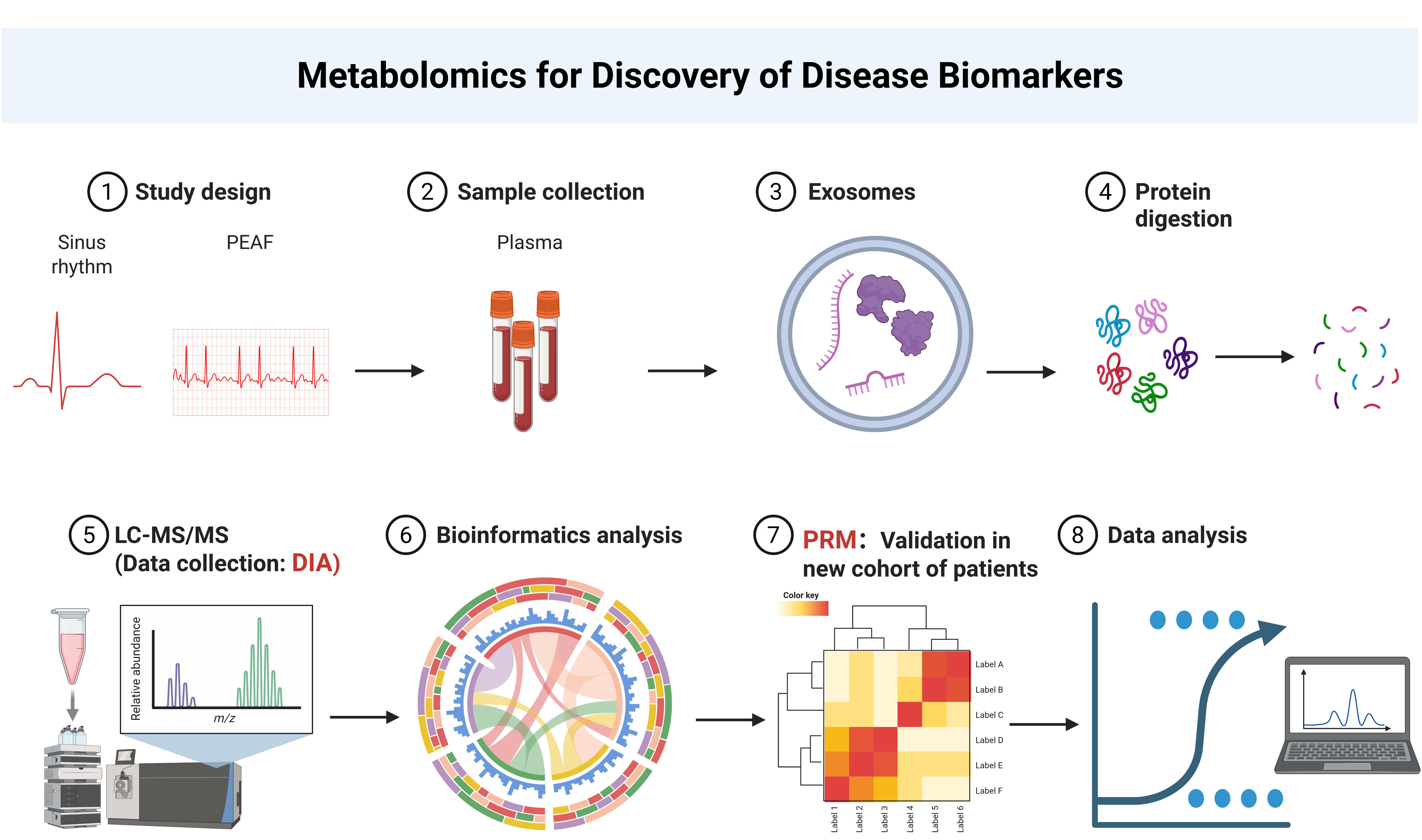
Extracellular vesicle (EV) proteomics can be used to study intercellular communication and find biomarkers of non-invasive diseases. Traditional separation methods (ultracentrifugation, size exclusion chromatography) and data-dependent acquisition (DDA) mass spectrometry usually have the drawbacks of copurification of pollutants, poor reproducibility, and insufficient sampling of low-abundance peptides. This protocol describes a workflow for label-free extracellular vesicle proteomics, which combines size exclusion chromatography for vesicle separation, data-independent acquisition (DIA) for deep discovery, and parallel reaction monitoring (PRM) for targeted verification. Plasma-derived extracellular vesicles are treated by standardized lysis, digestion, and LC-MS/MS procedures, so that the protein group of extracellular vesicles can be identified and quantified robustly. The main advantages of this scheme are that it can achieve high quantitative reproducibility, unbiased detection of low-intensity peptides, and seamless transition from discovery to targeted verification, while also being suitable for liquid biopsy samples and various cardiovascular diseases.
Key features
• Combines size exclusion chromatography (SEC)-based EV isolation with label-free data-independent acquisition (DIA) for deep, unbiased discovery-phase extracellular vesicle proteomics.
• Employs parallel reaction monitoring (PRM) for orthogonal, highly sensitive targeted validation of candidate biomarkers discovered by DIA.
• PRM records the entire, high-resolution fragment ion spectrum for the targeted peptide.
• Developed for atrial fibrillation; adaptable to other cardiovascular diseases, therapy monitoring, and liquid biopsies.
Keywords: Extracellular vesicles (细胞外囊泡) Proteomics (蛋白质组学)
Biomarkers (生物标志物)
Parallel reaction monitoring (平行反应监测)
Mass spectrometry (质谱分析)
Proteomics (蛋白质组学)
Biomarkers (生物标志物)
Parallel reaction monitoring (平行反应监测)
Mass spectrometry (质谱分析)
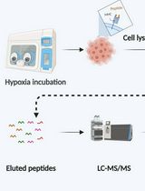
Graphical overview
Background
Extracellular vesicle (EV) proteomics has become a powerful discipline for deciphering intercellular communication networks, identifying biomarkers of non-invasive diseases, and advancing precision medicine [1]. The protein cargo of nanoscale EVs (50–150 nm), derived from the endosome pathway, directly reflects the pathophysiological state of parent cells, making EV proteins an attractive target for liquid biopsy development. [1]. At the sample preparation stage, traditional EV separation methods, such as ultracentrifugation, size exclusion chromatography (SEC), and ultrafiltration, are often affected by the copurification of non-EV contaminants (e.g., abundant plasma proteins, lipoproteins, and protein aggregates), low recovery rates, and low batch-to-batch reproducibility [1,2].
Even after separation, low-abundance EVs are often obscured by the extreme dynamic range of biological fluids, especially in plasma, where high-abundance proteins such as albumin and immunoglobulin dominate the samples, masking disease-related low-abundance proteins [3,4]. In addition, the lack of a standardized end-to-end workflow—from vesicle separation to protein digestion, mass spectrometry collection, and data analysis—leads to poor repeatability and difficulties in cross-study verification [5,6]. At the detection level, the large number of proteins detected in proteomics measurements covers up the inherent heterogeneity among individual EVs, potentially resulting in the loss of rare but functionally important subgroups [7].
In order to address these challenges, proteomics based on mass spectrometry has been widely used to characterize the protein composition of EVs. Data-dependent acquisition (DDA) is the most commonly used mass spectrometry acquisition mode, and precursor ions are selected according to the cracking intensity [8]. Although DDA is suitable for protein identification and spectral library generation, it has some drawbacks, such as insufficient sampling of low-intensity precursors and poor cross-batch quantitative reproducibility [8,9].
In order to overcome these limitations, data-independent acquisition (DIA) has become a promising alternative. DIA systematically fragments all precursor ions within a predefined isolation window in a predefined m/z range, thus enabling unbiased and comprehensive analysis of the precursor ions and providing high quantitative reproducibility for large-scale cohort studies [10]. Unlike DDA, which selects precursor ions based on intensity, DIA enhances the identification and quantification of low-intensity peptides and significantly improves protein-group coverage [10]. For example, by combining SEC with DIA-MS, 2,896 EV-related proteins can be identified and quantified from as little as 200 μL of plasma, representing 3.5 times greater coverage than that reported in previously published studies on melanoma [11]. Although DIA provides in-depth coverage for protein omics in the discovery stage, targeted verification of candidate biomarkers requires an orthogonal strategy with higher quantitative accuracy [12].
Parallel reaction monitoring (PRM) is a targeted MS method implemented on high-resolution instruments, such as Orbitrap, which offers excellent sensitivity and quantitative accuracy [13,14]. Unlike multiple reaction monitoring (MRM), which is based on triple-quadrupole instruments, PRM scans all product ions from the target peptide (FDR:1%) to produce a complete MS/MS spectrum, minimizing ion interference and improving specificity [15]. However, PRM has its inherent limitations: because of the wider MS/MS scanning range and longer cycle time, peptide coverage is affected. In addition, high-resolution mass spectrometers require higher instrument costs and more complex operation and maintenance [16]. The throughput of PRM is also limited to 100–300 targets at a time, which makes it not suitable for target discovery [17,18]. Beyond its application in atrial fibrillation (AF) biomarker discovery, this label-free PRM EV proteomics protocol can be extended to multiple other translational contexts. In the development of liquid biopsy, this pipeline is well-suited to analyzing circulating foreign bodies from minimally invasive samples (such as plasma, urine, or saliva) to identify disease-specific protein signatures [19].
The protocol can also characterize EVs from specific cardiac cell types by targeting cell type–specific surface markers, thereby helping to dissect the contribution of cell specificity to disease pathogenesis.
In treatment monitoring, the same workflow can quantify extracellular proteins longitudinally to track the emergence of treatment response or drug resistance, especially in the context of antiarrhythmic drug therapy or catheter ablation for AF. In addition, for other cardiovascular diseases characterized by progressive structural remodeling, such as heart failure with preserved ejection fraction, hypertrophic cardiomyopathy, and cardiac amyloidosis, this approach provides a minimally invasive method that can monitor disease progression and evaluate treatment effects through a series of extracellular protein profiles. Finally, this standardized label-free PRM EV proteomics scheme provides a multifunctional platform for translational cardiovascular research and has great potential for accelerating the discovery and clinical implementation of EV-based biomarkers for AF and other diseases.
Materials and reagents
Biological materials
1. Human plasma (collected from healthy donors and AF donors at TEDA International Cardiovascular Hospital under ethics approval ([2021]-0325-2))
Reagents
1. Trypsin (Promega, catalog number: V5117)
2. DL-dithiothreitol (Sigma-Aldrich, catalog number: D9163-25G)
3. Iodoacetamide (Sigma-Aldrich, catalog number: V900335-5G)
4. Trifluoroacetic acid (Sigma-Aldrich, catalog number: 302031-1L)
5. Urea (Sigma-Aldrich, catalog number: V900119-500G)
6. Acetonitrile (ThermoFisher Scientific, catalog number: 204433)
7. Methanol (ThermoFisher Scientific, catalog number: A452-4)
8. TEAB (Sigma-Aldrich, catalog number: 140023)
9. Protease Inhibitor Cocktail III (Merck Millipore, catalog number: 539134-10ML)
10. Formic acid (ThermoFisher Scientific, catalog number: A117-50)
11. PBS (Gibco, catalog number: 6124451)
12. PFA (Sigma, catalog number: P6148)
13. Glutaraldehyde (Ted Pella, catalog number: 16051)
14. Uranyl oxalate (Endoscope instrument, catalog number: GZ02625)
Solutions
1. K2EDTA anticoagulant (K2EDTA) (see Recipes)
Recipes
1. K2EDTA
| Reagent | Final concentration | Quantity or volume (example for a 3-mL tube) |
|---|---|---|
| K2EDTA (dipotassium salt) | 1.5–2.2 mg per mL of whole blood (most commonly ~1.5–1.8 mg/mL) | 4.5–6.6 mg (adjust according to tube draw volume) |
| Total | n/a | Dry film; no liquid volume |
Laboratory supplies
1. Solid phase extraction column (SPE strata-X 10 mg/1 mL) (desalination column) (Phenomenex, catalog number: 8B-S100-AAK)
2. Extracellular vesicle extraction column (IZON, catalog number: qEVoriginal Columns/SP1)
3. PAGE Silver Staining kit (Solarbio, catalog number: G7210)
4. 0.22-μM microporous filter membrane (Merk Millipore Ltd., catalog number: 0000212310)
5. Ultrafiltration centrifuge tube 0.5 mL/10 KD (Millipore, catalog number: UFC501024)
6. K2EDTA-coated vacuum blood collection tubes (BD, catalog number: 367856)
7. PierceTM C18 tips, 100 μL bed (96 tips) (Thermo Scientific, catalog number: 87784)
8. BCA Protein Assay kit (Beyotime, catalog number: P0011)
9. Exosomoe Purification Filter (EPF column) (Umibio, catalog number: UR52136)
10. Formvar (SS13.1) carbon-loaded copper mesh (Pelco, catalog number: 01753-F)
Equipment
1. Low-temperature centrifuge (Eppendorf, model: centrifuge 5427R)
2. Vacuum concentrator (Eppendorf, model: Concentrator plus)
3. Constant temperature water bath (Shanghai Boxun Medical Biological Instrument Co., Ltd., catalog number/model: DK-8D)
4. Constant temperature blast drying oven (Shanghai Yiheng Scientific Instrument Co., Ltd., catalog number/model: DHG-9240A)
5. Micro quantitative analyzer (IMPLEN, model: NP80Touch)
6. Automated solid phase extraction instrument (TECAN, model: Resolvex A100)
7. Room-temperature centrifuge (SCILOGEX, catalog number/model: SCI-24)
8. Scanner (Epson, model: V600)
9. Electrophoresis apparatus (Bio-Rad, model: 1645050)
10. TimsTOF Pro 2 mass spectrometry (Bruker, model: Tims TOF PRO 2)
11. NanoElute UHPLC system (Bruker, model: NanoElute)
12. Electron microscope (Jeol, model: JEM-1230)
Software and datasets
1. MaxQuant (https://www.maxquant.org/maxquant/) (Max Planck Institute of Biochemistry, Martinsried, Germany, v.1.6.15.0)
2. GraphPad Prism 8.0.2
Procedure
登录/注册后免费查看全文
文章信息
稿件历史记录
提交日期: Jun 10, 2026
接收日期: Aug 24, 2026
在线发布日期: Sep 23, 2026
出版日期: Oct 5, 2026
版权信息
© 2026 The Author(s); This is an open access article under the CC BY-NC license (https://creativecommons.org/licenses/by-nc/4.0/).
如何引用
Zhang, L. and He, G. (2026). Identifying Differentially Expressed Proteins via Plasma Exosomal Proteomics. Bio-protocol 16(19): e5840. DOI: 10.21769/BioProtoc.5840.

 Download Citation in RIS Format
分类
医学 > 心血管疾病
生物化学 > 蛋白质 > 定量
您对这篇实验方案有问题吗?
在此处发布您的问题,我们将邀请本文作者来回答。同时,我们会将您的问题发布到Bio-protocol Exchange,以便寻求社区成员的帮助。
 发布问题
发布问题


