


In-Gel Tryptic Digestion and HCD/EThcD LC-MS/MS for Mapping Autophosphorylation Sites
结合胶内胰蛋白酶酶解与 HCD/EThcD LC-MS/MS 鉴定蛋白质自磷酸化位点

发布: 2026年11月05日第16卷第21期 DOI: 10.21769/BioProtoc.5836 浏览次数: 325
评审: Laxmi Narayan MishraPrajita PandeyJoyce Chiu
 PDF
 Q&A

参见作者原研究论文
The authors used this protocol in:

Oct 2026




Abstract
This protocol describes a targeted workflow for identifying and quantitatively comparing kinase autophosphorylation sites, using in-gel proteolytic digestion, LC-MS/MS analysis, phosphosite localisation, and extracted ion chromatogram (XIC) analysis. The detailed protocol begins with an excised SDS-PAGE gel band containing the immunoprecipitated kinase of interest. Standard cell culture, expression, immunoprecipitation, and SDS-PAGE procedures can be used to generate this starting material according to the experimental system under investigation; the specific conditions used for NEK1 are described in our associated research article. Here, the protocol is applied to characterise phosphorylation sites on NEK1—a serine/threonine kinase implicated in amyotrophic lateral sclerosis (ALS)—using a homozygous NEK1 knockout cell background in which GFP-tagged wild-type or kinase-dead NEK1 constructs are re-expressed. This experimental system enables accurate assessment of kinase-dependent phosphorylation events in the absence of endogenous NEK1. A key methodological feature of the workflow is the use of two parallel MS2 fragmentation strategies: higher energy collisional dissociation (HCD) and electron-transfer/HCD (EThcD). EThcD generates complementary b/y and c/z ion series, providing superior localisation confidence for labile phosphorylation events that are difficult to resolve using HCD alone. Relative phosphopeptide abundance across conditions can be assessed using extracted ion chromatograms generated in FreeStyle, or using Skyline software with normalisation to the total ion current (TIC), as performed in the associated NEK1 study. Although demonstrated here using NEK1, the protocol is readily adaptable to other kinases amenable to immunoprecipitation and will be of interest to researchers studying kinase signalling, post-translational modification biology, and disease-associated phosphorylation events.
Key features
• Uses a homozygous NEK1 knockout cell background with re-expressed GFP-tagged constructs for discrimination of kinase-activity-associated candidate autophosphorylation events from background.
• Parallel HCD and EThcD fragmentation runs per sample for complementary phosphosite localisation; EThcD is particularly advantageous for labile or multiply phosphorylated peptides.
• Quantitative wild-type vs. kinase-dead comparison using extracted ion chromatograms normalised to reference peptides or total ion current, with worked m/z calculations and FreeStyle screenshots.
• Readily adaptable to any kinase amenable to immunoprecipitation and for which a kinase-dead control construct can be generated.
Keywords: Phosphoproteomics (磷酸化蛋白质组学) Autophosphorylation (自磷酸化)
In-gel digestion (胶内酶解)
LC-MS/MS (液相色谱-串联质谱)
EThcD (电子转移/高能碰撞解离)
HCD (高能碰撞解离)
Extracted ion chromatogram (提取离子色谱图)
Autophosphosite mapping (自磷酸化位点鉴定)
NEK1 (NEK1)
Graphical overview
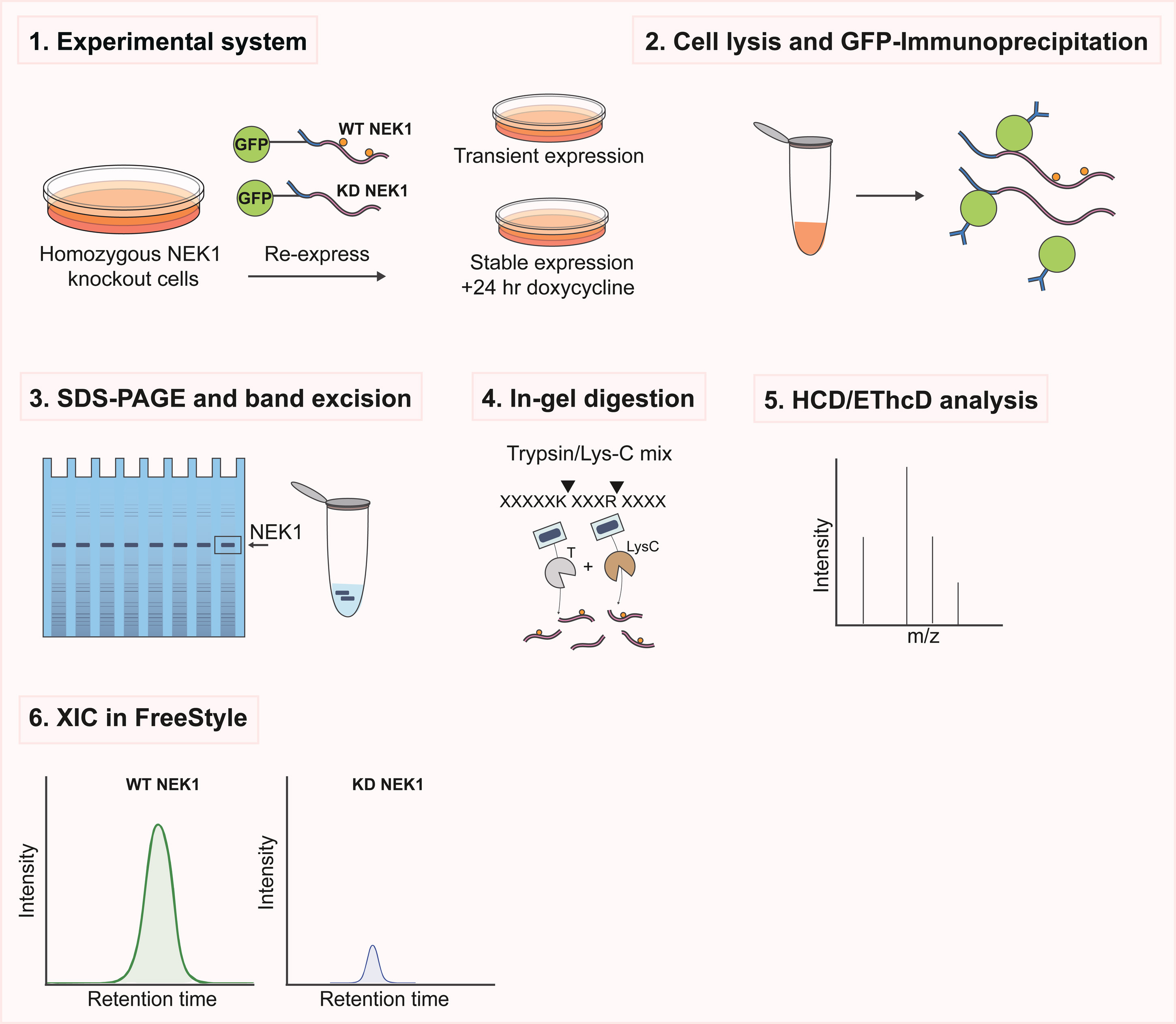
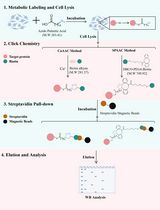
Schematic overview of the experimental workflow for kinase autophosphorylation site mapping using complementary higher energy collisional dissociation (HCD) and electron-transfer/HCD (EThcD) mass spectrometry. Steps 1–3 comprise standard cell culture, immunoprecipitation, and SDS-PAGE procedures that can be performed using established protocols appropriate to the experimental system; the specific conditions used for NEK1 are described in the associated research article [1]. Homozygous NEK1 knockout cells expressing GFP-tagged wild-type (WT) or kinase-dead (KD) NEK1 were used to enable comparison of kinase activity–dependent phosphorylation events in the absence of endogenous NEK1. Subsequent steps (4–6), described in detail in this protocol, comprise excision of the target protein band, in-gel trypsin/Lys-C digestion, LC-MS/MS analysis using complementary HCD and EThcD fragmentation for phosphosite identification and localisation, and quantitative extracted ion chromatogram (XIC) analysis.
Background
Identifying autophosphorylation sites on a specific kinase in cells presents distinct methodological challenges. Whole-proteome phosphoproteomic approaches are poorly suited to this task: endogenous kinases are often expressed at low abundance, and kinase-derived phosphopeptides are vastly outnumbered by background, making bona fide autophosphorylation events difficult to identify with confidence.
The approach described here addresses these limitations through (i) the use of a homozygous knockout cellular background to eliminate endogenous kinase signal; (ii) immunoprecipitation and SDS-PAGE-based enrichment of the target kinase prior to MS analysis; (iii) identifying kinase-activity-dependent phosphorylation events and prioritising candidate autophosphorylation sites; and (iv) complementary higher energy collisional dissociation (HCD) and electron-transfer/HCD (EThcD) fragmentation to increase phosphosite localisation confidence, particularly where HCD alone provides ambiguous site assignment.
This protocol was deployed to identify autophosphorylation sites on NEK1, a serine/threonine kinase whose variants are among the most common genetic causes of amyotrophic lateral sclerosis (ALS). Using this approach, we identified three rigorously validated NEK1 autophosphorylation sites (pS14, pT156, and pS418), further confirmed by phosphospecific antibodies; activation-loop pT156 was then used as an activity readout and shown to be altered by several ALS-associated missense variants [1]. The protocol is, however, applicable to any kinase for which specific immunoprecipitation can be achieved, and a kinase-dead control can be generated [2].
Materials and reagents
Biological materials
1. NEK1 knockout U-2 OS Flp-In T-REx cells (generated by CRISPR-Cas9 nickase, biallelic frameshift confirmed by MiSeq; available upon request from A.R.M.)
2. GFP-NEK1 wild-type and GFP-NEK1 kinase-dead (D146A) expression constructs (available upon request from A.R.M.)
Note: The NEK1 knockout cell line and GFP-NEK1 expression constructs are specific to the experimental system used to validate this protocol. The downstream workflow can be adapted to other kinases using an appropriate cellular background, expression system, and kinase-dead control.
Reagents
1. Colloidal Blue Staining kit (protein gel stain) (Thermo Fisher Scientific, catalog number: LC6025)
2. 1 M triethylammonium bicarbonate (TEAB) buffer (Fisher Scientific, catalog number: 15215753)
3. Bond-BreakerTM TCEP solution, neutral pH (0.5 M) (reducing agent) (Thermo Fisher Scientific, catalog number: 77720/Fisher UK 10657344)
4. Iodoacetamide (IAA) (alkylating agent) (Sigma-Aldrich, catalog number: I1149)
5. LC-MS-grade water (solvent) (Fisher Scientific, catalog number: 10434902)
6. 100% acetonitrile, LC-MS grade (solvent) (Fisher Scientific, catalog number: 10055454)
7. Ammonium bicarbonate (compound) (Sigma-Aldrich, catalog number: A6141)
8. Trypsin/Lys-C protease mix (enzyme) (Thermo Fisher Scientific, catalog number: A41007)
9. CaCl2 (enzyme stabilizer) (Sigma-Aldrich, catalog number: C1016)
10. Formic acid (LC-MS solvent modifier) (Merck, catalog number: 1.00263)
11. n-Dodecyl-β-D-maltoside (DDM) (LC-MS additive/detergent) (Thermo Fisher Scientific, catalog number: 89902)
12. Trifluoroacetic acid (TFA) (LC-MS grade) (LC-MS solvent modifier) (Thermo Fisher Scientific, catalog number: 85183)
13. Pierce HeLa protein digest standard (Thermo Fisher Scientific, catalog number: 88329)
Solutions
1. 50 mM TEAB (see Recipes)
2. 120 mM TCEP (see Recipes)
3. 0.5 M iodoacetamide (see Recipes)
4. Trypsin/Lys-C digestion buffer (see Recipes)
5. 80% ACN/0.5% TFA (see Recipes)
6. Peptide resuspension buffer (see Recipes)
7. 50 mM ammonium bicarbonate (see Recipes)
8. 50 mM ammonium bicarbonate in 50% acetonitrile (see Recipes)
9. 0.1% TFA (see Recipes)
10. LC solvent A (see Recipes)
11. LC solvent B (see Recipes)
Recipes
1. 50 mM TEAB
a. To prepare 10 mL, combine 0.5 mL of 1 M TEAB stock with 9.5 mL of LC-MS-grade water.
b. Prepare fresh on the day of use.
2. 120 mM TCEP (in 50 mM TEAB)
a. Use 0.5 M Bond-BreakerTM TCEP solution, neutral pH, as the stock solution.
b. To prepare 1 mL, combine 240 μL of 0.5 M TCEP stock with 760 μL of 50 mM TEAB.
3. 0.5 M iodoacetamide (in 50 mM TEAB)
a. To prepare 1 mL, dissolve 92.5 mg of IAA in 50 mM TEAB and adjust to a final volume of 1 mL.
b. Prepare fresh immediately before use and protect from light throughout preparation and use.
4. Trypsin/Lys-C digestion buffer
a. Prepare 50 mM TEAB containing 2.5 mM CaCl2.
b. Resuspend trypsin/Lys-C protease mix according to the manufacturer's instructions and prepare sufficient digestion solution to provide 2 μg of enzyme per sample.
c. For each sample, prepare 100 μL of digestion solution containing 2 μg of trypsin/Lys-C.
d. Prepare fresh immediately before use.
5. 80% ACN/0.5% TFA
a. To prepare 10 mL, combine 8 mL of LC-MS-grade acetonitrile, 50 μL of TFA, and LC-MS-grade water to a final volume of 10 mL.
b. Prepare fresh on the day of use.
6. Peptide resuspension buffer
a. Prepare fresh using LC-MS-grade water and 0.1% formic acid.
b. Add n-dodecyl-β-D-maltoside (DDM) to a final concentration of 0.015% (w/v).
c. Reconstitute dried peptide pellets with 10 min of gentle shaking before transferring to a suitable vial and loading the LC autosampler.
7. 50 mM ammonium bicarbonate
a. To prepare 10 mL, dissolve 39.5 mg of ammonium bicarbonate in LC-MS-grade water and adjust to a final volume of 10 mL.
b. Prepare fresh on the day of use.
8. 50 mM ammonium bicarbonate in 50% acetonitrile
a. To prepare 10 mL, combine 5 mL of 100 mM ammonium bicarbonate (79.1 mg in 10 mL of LC-MS-grade water) with 5 mL of LC-MS-grade acetonitrile.
b. Prepare fresh on the day of use.
9. 0.1% (v/v) TFA
To prepare 10 mL, add 10 μL of TFA to LC-MS-grade water to a final volume of 10 mL. Used only for the optional C18 clean-up in Stage 6.
10. LC solvent A: 0.1% (v/v) formic acid in LC-MS-grade water
To prepare 1 L, add 1 mL of formic acid to 999 mL of LC-MS-grade water.
11. LC solvent B: 80% (v/v) acetonitrile, 0.08% (v/v) formic acid in LC-MS-grade water
To prepare 1 L, combine 800 mL of LC-MS-grade acetonitrile, 0.8 mL of formic acid, and LC-MS-grade water to 1 L.
Laboratory supplies
1. 1.5 mL protein low-binding microcentrifuge tubes (Sarstedt, catalog number: 72.706.600)
2. 1–200 μL round gel loading tips (Starlab, catalog number: I1022-0600), for removing wash solutions from around gel pieces without aspirating them
3. Disposable sterile scalpels (Swann-Morton, catalog number: DS11)
4. Clean glass plate or disposable Petri dish as a cutting surface, rinsed with LC-MS-grade water immediately before use
5. LC-MS vials and caps (FisherbrandTM 11 mm Snap Ring Micro-Vial, Plastic, catalog number: 11717597; VWR Snap Caps for Snap Ring Vials ND11, catalog number: 548-0016)
6. Powder-free nitrile gloves (worn at all times)
7. Aluminium foil for protecting iodoacetamide-containing samples from light
8. Microcentrifuge tube racks
9. C18 desalting tips (e.g., Waters, catalog number: WAT0200805), required only if the optional peptide clean-up step is performed
Equipment
1. ThermoMixer (Eppendorf)
2. SpeedVac vacuum concentrator (Thermo, model: Savant SPD140DDA)
3. Scalpel and clean cutting surface
4. Dry ice
5. Orbitrap Lumos Tribrid mass spectrometer (Thermo Scientific) with EASY-nESI source
6. Dionex Ultimate 3000 nano-LC system
7. EASY-SprayTM HPLC Column (Thermo ScientificTM, catalog number: ES903, 2 µm, 100 Å, 75 µm × 50 cm)
8. Acclaim PepMap 100 C18 trapping column (Thermo ScientificTM, catalog number: 164564, 150 mm × 0.1 mm × 5 µm)
9. Nano-LC fittings and consumables: nanoViper fingertight fittings and PEEK/fused-silica capillary tubing (20–50 µm i.d.) for all post-column connections; use the minimum practicable tubing length to limit extra-column dispersion
Note: Recommended maintenance: replace the trapping column every ~500 injections or sooner if carryover increases; clean the ion transfer tube monthly; run a standard peptide mixture (e.g., Pierce HeLa protein digest standard) before each sample batch to confirm chromatographic and spectral performance.
Software and datasets
1. FreeStyle software v1.5 (Thermo Fisher Scientific; available via https://thermo.flexnetoperations.com/control/thmo/login), used here for XIC extraction and integration
2. Proteome Discoverer 2.4 (Thermo Scientific), used for database searching and phosphosite localisation
3. Mascot v2.8.3 (Matrix Science; requires institutional license), used as the search engine within the associated workflow
4. AlphaMap (Voytik et al. [3]; open-source Python package)
5. Skyline v25.1 (MacCoss Lab, University of Washington), used for TIC-normalised XIC analysis in the associated NEK1 study
Note: Proteome Discoverer and Mascot are commercial products and require a paid institutional licence; Mascot additionally requires access to a local or hosted Mascot server. FreeStyle is distributed free of charge by Thermo Fisher Scientific but requires registration of a user account and reads only Thermo .RAW files. Skyline is free and open source and reads Thermo, Bruker, SCIEX, Agilent, and Waters formats. Open-source alternatives: Laboratories without Proteome Discoverer/Mascot licences can perform equivalent database searching and phosphosite localisation using FragPipe/MSFragger with PTM-Prophet, MaxQuant with the Andromeda search engine and its PTM score, or the Trans-Proteomic Pipeline (Comet with PTMProphet). For XIC generation and integration, Skyline is a fully open-source substitute for FreeStyle and is the software used for the quantification reported in the associated research article [1]. Localisation-probability thresholds should be applied on the equivalent scale reported by the chosen tool.
Procedure
登录 / 注册后阅读全文
文章信息
稿件历史记录
提交日期: Jun 21, 2026
接收日期: Aug 30, 2026
在线发布日期: Sep 16, 2026
出版日期: Nov 5, 2026
版权信息
© 2026 The Author(s); This is an open access article under the CC BY-NC license (https://creativecommons.org/licenses/by-nc/4.0/).
如何引用
Agarwal, S., Gourlay, R., Soares, R. F. and Mehta, A. R. (2026). In-Gel Tryptic Digestion and HCD/EThcD LC-MS/MS for Mapping Autophosphorylation Sites. Bio-protocol 16(21): e5836. DOI: 10.21769/BioProtoc.5836.

 Download Citation in RIS Format
分类
生物化学 > 蛋白质 > 翻译后修饰
生物科学 > 生物技术 > 质谱
系统生物学 > 蛋白质组学 > 磷酸化蛋白质组学
您对这篇实验方案有问题吗?
在此处发布您的问题,我们将邀请本文作者来回答。同时,我们会将您的问题发布到Bio-protocol Exchange,以便寻求社区成员的帮助。
 发布问题


