


An In Vitro Model to Study Drugs That Affect Macrophage Adhesion to Murine Brain Endothelial Cells After Proinflammatory Insults of LPS and Pilocarpine
用于研究促炎刺激后药物对巨噬细胞黏附小鼠脑内皮细胞影响的体外模型

发布: 2026年09月20日第16卷第18期 DOI: 10.21769/BioProtoc.5825 浏览次数: 606
评审: Sébastien GillotinManasa VL ChanduriAnonymous reviewer(s)
 PDF
PDF  Q&A
Q&A





Abstract
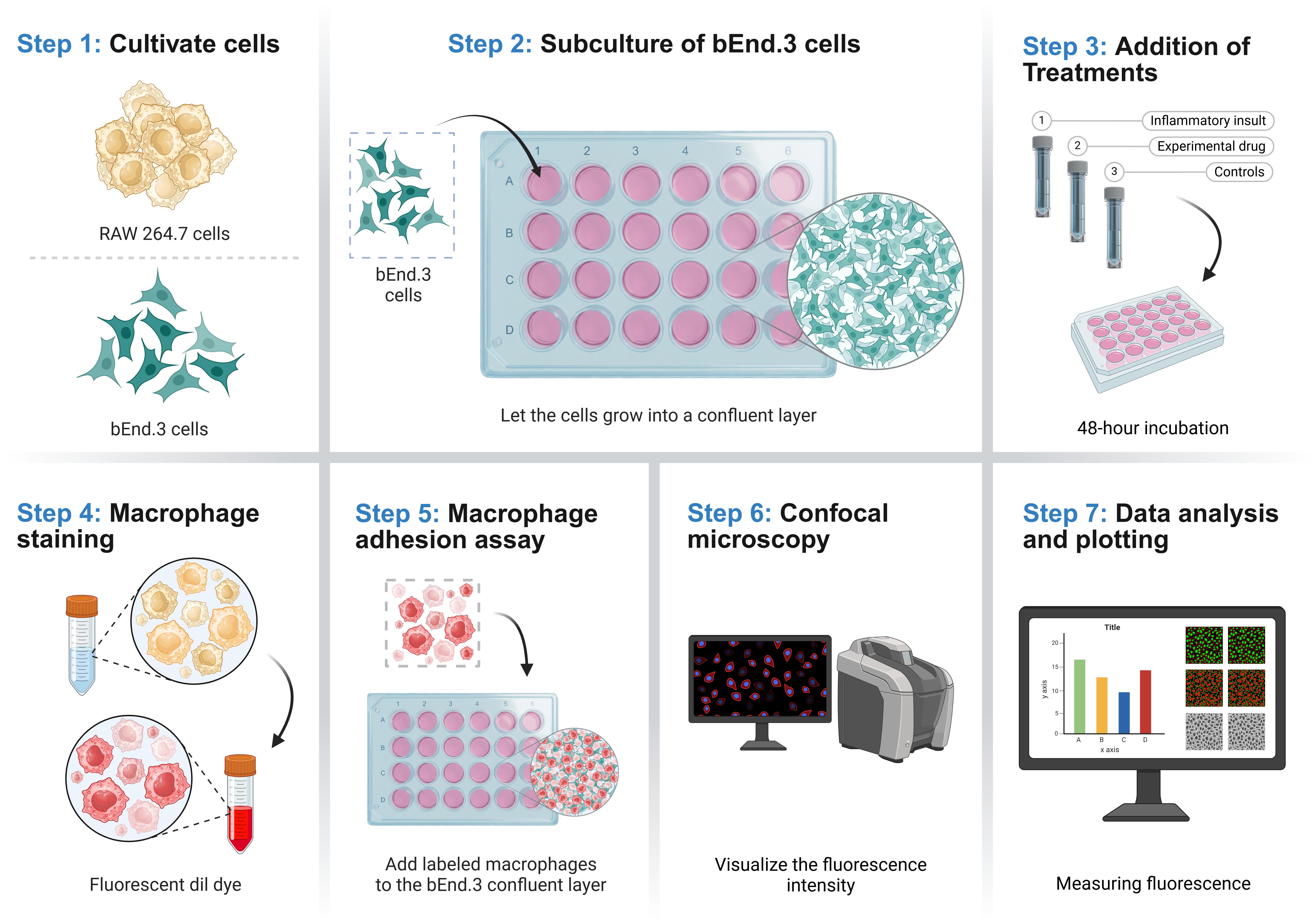
Neuroinflammation disrupts blood–brain barrier (BBB) integrity, promoting leukocyte recruitment into the central nervous system and contributing to the progression of neurological disorders. This protocol describes a reproducible macrophage adhesion assay to evaluate interactions between immune cells and brain endothelial cells and to screen compounds with potential anti-inflammatory activity. Murine brain endothelial cells (bEnd.3) were cultured to confluency and exposed to inflammatory stimuli, such as lipopolysaccharide (LPS) or pilocarpine, a cholinergic muscarinic receptor agonist reported to induce inflammatory responses through seizure-associated neuroinflammatory mechanisms, in the presence or absence of candidate therapeutic compounds. In these studies, the natural flavonoid quercetin and the synthetic alkyl-lysophospholipid edelfosine were tested for their effects on macrophage adhesion. After 48 h of treatment, fluorescently labeled murine macrophages (RAW 264.7) were added to the endothelial monolayer, and adherent cells were quantified by fluorescence microscopy. The assay was validated using dexamethasone as an anti-inflammatory control and inflammatory stimulation with LPS or pilocarpine. As expected, dexamethasone reduced macrophage adhesion, whereas both LPS and pilocarpine significantly increased adhesion, demonstrating the assay's sensitivity to changes in endothelial inflammatory status. Overall, this protocol provides a reliable and accessible platform for investigating endothelial–immune cell interactions under neuroinflammatory conditions and for evaluating therapeutic compounds that may preserve BBB function and reduce inflammatory cell recruitment in neurological disease models.
Key features
• This protocol creates an in vitro model that simulates the luminal interface of the blood–brain barrier (BBB).
• The protocol accounts for the 48-h interval required for maximal physiological expression of adhesion molecules at the cell surface.
• The protocol quantifies macrophage adhesion to brain endothelial cells following inflammatory stimulation with lipopolysaccharide (LPS) or pilocarpine.
• It enables evaluation of therapeutic candidates for their ability to modulate macrophage–brain endothelial cell adhesion under neuroinflammatory conditions.
Keywords: Endothelial cells (内皮细胞) Inflammation (炎症)
Quercetin (槲皮素)
Edelfosine (依地福新)
Lipopolysaccharides (脂多糖)
Pilocarpine (毛果芸香碱)
Macrophages (巨噬细胞)
Blood–brain barrier (血脑屏障)
Inflammation (炎症)
Quercetin (槲皮素)
Edelfosine (依地福新)
Lipopolysaccharides (脂多糖)
Pilocarpine (毛果芸香碱)
Macrophages (巨噬细胞)
Blood–brain barrier (血脑屏障)
Graphical overview
Background
Neuroinflammation is a central pathological process in the progression of neurodegenerative diseases (NDs), including epilepsy, Alzheimer’s disease (AD), Parkinson’s disease (PD), and others [1–3]. These disorders are characterized by chronic activation of the innate immune system within the central nervous system (CNS), involving microglia, astrocytes, and the recruitment of peripheral immune cells [3–5]. While acute inflammatory responses may initially serve protective functions by promoting debris clearance and tissue repair, persistent and dysregulated neuroinflammation contributes to progressive neuronal damage and synaptic dysfunction [6,7]. Blood–brain barrier endothelial cells (BECs) are also disrupted during neuroinflammation through signaling from activated microglia, astrocytes, and infiltrating immune cells [8–10]. Proinflammatory cytokines such as TNF-α, IL-1β, and IL-6, together with reactive oxygen species (ROS), impair endothelial function by disrupting tight junctions, increasing transcellular transport, and promoting enzymatic degradation of the barrier [11–14]. These changes compromise BBB integrity and facilitate additional immune cell infiltration into the central nervous system [13]. These inflammatory signals, together with matrix metalloproteinases (MMPs), compromise the integrity of the blood–brain barrier (BBB) by disrupting tight junction proteins such as occludins and claudins, leading to increased vascular permeability [15,16]. BBB dysfunction further exacerbates neuroinflammation by allowing the infiltration of circulating monocytes and lymphocytes into the brain parenchyma, creating a feed-forward inflammatory cycle [3]. Such immune cell infiltration has been reported across multiple neurodegenerative conditions. In AD, BBB disruption often precedes significant cognitive decline and facilitates the entry of peripheral immune cells and circulating Aβ into the brain [2,6,17]. In temporal lobe epilepsy (TLE), the most prevalent form of epilepsy, BBB disruption is a hallmark pathological feature that contributes to the maintenance of chronic seizures and promotes neuroinflammatory processes [18,19]. BBB dysfunction is often persistent and has been associated with increased seizure frequency, disease severity, and resistance to antiseizure medications [20]. Although infiltrating macrophages can facilitate the clearance of cellular debris and support tissue repair, they also release proinflammatory mediators that may exacerbate neuronal damage and perpetuate neuroinflammation [21,22]. Consequently, therapeutic strategies aimed at preserving BBB integrity and modulating neuroinflammatory responses—including inhibition of matrix metalloproteinase (MMP) activity, suppression of proinflammatory cytokine production, and promotion of reparative immune phenotypes—have emerged as promising approaches to limit disease progression and improve neurological outcomes [23].
Anti-inflammatory compounds that modulate endothelial activation have emerged as promising strategies to protect BBB function [24]. Both natural and synthetic molecules may regulate inflammatory signaling and immune cell interactions at the BBB. For example, a synthetic compound, edelfosine, which targets phospholipase Cβ signaling [25,26], has shown anti-inflammatory effects in BBB endothelial cells exposed to seizure-inducing stimuli such as pilocarpine [27]. Quercetin is a natural anti-inflammatory molecule that has recently been shown to promote neuroprotection by preserving BBB integrity, reducing inflammation and oxidative stress, preventing neuronal death, and enhancing synaptic function [28]. In this context, the present study investigates whether the natural flavonoid quercetin and the synthetic lysophospholipid edelfosine can attenuate macrophage adhesion under proinflammatory conditions.
Macrophage–endothelial adhesion assays are widely used to investigate leukocyte recruitment during inflammation and to evaluate the anti-inflammatory potential of therapeutic compounds. Common approaches include static adhesion assays, flow-based systems, live-cell imaging, and transwell migration assays, each providing complementary information on leukocyte–endothelial interactions [29–31]. Static assays remain the most widely used because they are simple, reproducible, and suitable for quantitative drug screening, although they do not reproduce physiological shear stress or leukocyte rolling. Despite these limitations, static assays remain the preferred approach for initial drug screening because they provide highly reproducible, quantitative measurements under controlled experimental conditions, enabling rapid evaluation of multiple therapeutic candidates before validation in more physiologically complex models. In contrast, flow-based assays better mimic the vascular microenvironment but require specialized equipment and have lower throughput. Confocal imaging provides high-resolution visualization of adhesion dynamics, whereas transwell assays combine adhesion with transmigration but are less suited for quantitative screening [29,32].
Materials and reagents
Biological materials
1. bEnd.3 (ATCC, CRL-2299); organism: Mus musculus (mouse); tissue: brain, cerebral cortex; disease endothelioma; cell type: endothelial cell
2. Raw cell 264.7 (ATCC, TB1-71); organism: Mus musculus (mouse); tissue: ascites; disease: Abelson murine leukemia virus-induced tumor; cell type: macrophage
Reagents
1. Lipopolysaccharide (LPS) (Sigma-Aldrich, catalog number: L2630)
2. Dexamethasone (MedChemExpress, catalog number: HY-14648)
3. Quercetin (MedChemExpress, catalog number: HY-18085)
4. Dimethyl sulfoxide (DMSO) (Santa Cruz Biotechnology, catalog number: sc-358801)
5. Pilocarpine (Sigma-Aldrich, catalog number: P6503)
6. Edelfosine (Sigma-Aldrich, catalog number: SML0332)
7. Dil (1,1′-Dioctadecyl-3,3,3′,3′-tetramethylindocarbocyanine perchlorate) (Thermo Fisher, catalog number: D3911)
8. Sodium bicarbonate (Sigma-Aldrich, catalog number: S5761)
9. Dulbecco’s modified Eagle medium (DMEM) (Thermo Fisher, catalog number: 12800017)
10. Phosphate-buffered saline (PBS) (Sigma-Aldrich, catalog number: P5368-10PAK)
11. Penicillin and streptomycin (Thermo Fisher, catalog number: 15140122)
12. Fetal bovine serum (Gibco, catalog number: A52567-01)
13. GibcoTM trypsin-EDTA (0.25%), phenol red (Fisher Scientific, catalog number: 25-200-072)
14. Formalin solution, neutral buffered, 10% (Sigma-Aldrich, catalog number: HT501128)
15. Trypan Blue solution 0.4% (Millipore-Sigma, catalog number: T8154)
Solutions
1. Edelfosine stock solution, 1 mM (see Recipes)
2. PBS solution (see Recipes)
3. Pilocarpine stock solution, 10 mM (see Recipes)
4. Dexamethasone stock solution, 50 mM (see Recipes)
5. LPS stock solution, 1 mg/mL (see Recipes)
6. Quercetin stock solution, 5 mM (see Recipes)
7. Complete DMEM (see Recipes)
Recipes
1. Edelfosine stock solution, 1 mM
| Reagent | Initial concentration | Quantity or volume | Final working concentration |
|---|---|---|---|
| Edelfosine (523.73 g/mol) | 1 mM | 5.24 mg | 1 μM |
| DMSO | 10 mL | 0.1% |
According to the molecular weight of edelfosine (523.73 g/mol), prepare a stock solution at 1 mM diluted in DMSO. Accurately weigh the edelfosine, dissolve it in DMSO to the desired volume to prepare the standard solution, and mix thoroughly until completely dissolved.
2. PBS solution
| Reagent | Initial concentration | Quantity or volume |
|---|---|---|
| PBS | 1× | 1 packet |
| Deionized water | 1 L |
Following the manufacturer's instructions, add dry powder from the packet to 1 L of deionized water and stir; this will yield PBS 0.01 M, NaCl 0.138 M, KCl 0.0027 M, pH 7.4 at 25 °C. Sterilize by filtration and store at room temperature.
3. Pilocarpine stock solution, 10 mM
| Reagent | Initial concentration | Quantity or volume | Final working concentration |
|---|---|---|---|
| Pilocarpine (208.26 g/mol) | 10 mM | 20.83 mg | 10 μM |
| PBS solution | 1× | 10 mL |
According to the molecular weight of pilocarpine (208.26 g/mol), prepare a stock solution at 10 mM in PBS solution. Accurately weigh the pilocarpine, dissolve it in PBS solution to the desired volume to prepare the standard solution, and mix thoroughly until completely dissolved.
4. Dexamethasone stock solution, 50 mM
| Reagent | Initial concentration | Quantity or volume | Final working concentration |
|---|---|---|---|
| Dexamethasone (392.46 g/mol) | 50 mM | 196.23 mg | 50 μM |
| DMSO | 10 mL | 0.1% |
According to the molecular weight of dexamethasone (392.46 g/mol), prepare a 50 mM stock solution in DMSO. Accurately weigh the dexamethasone, dissolve it in DMSO to the desired volume to prepare the stock solution, and mix thoroughly.
5. LPS stock solution
| Reagent | Initial concentration | Quantity or volume | Final working concentration |
|---|---|---|---|
| LPS | 1 mg/mL | 10 mg | 100 ng/mL |
| PBS solution | 1× | 10 mL |
According to the molecular weight of LPS, prepare a stock solution at 1 mg/mL in PBS. Accurately weigh the LPS and dissolve it in PBS solution to the desired volume to prepare the standard solution.
6. Quercetin stock solution, 5 mM
| Reagent | Initial concentration | Quantity or volume | Final working concentration |
|---|---|---|---|
| Quercetin (302.24 g/mol) | 5 mM | 15.1 mg | 5 μM |
| DMSO | 10 mL | 0.5% |
According to the molecular weight of quercetin (302.23 g/mol), prepare a stock solution at 5 mM diluted in DMSO. Accurately weigh the quercetin, dissolve it in DMSO to the desired volume to prepare the standard solution.
7. Complete DMEM
| Reagent | Initial concentration | Quantity or volume | Final working concentration |
|---|---|---|---|
| DMEM | 445 mL | ||
| FBS | 50 mL | 10% | |
| Penicillin-streptomycin | 100× | 5 mL | 1% |
| Sodium bicarbonate | 1.85 g |
Laboratory supplies
1. 25 cm2 cell culture flasks (Corning, catalog number: 430639)
2. 75 cm2 cell culture flasks (Corning, catalog number: 430641U)
3. VWR tissue culture plate (24-well plate) (VWR, catalog number: 10861-558)
4. 2 mL serological pipettes (Fisherbrand, catalog number: 15604009)
5. 5 mL serological pipettes (Costar Stripette, catalog number: 13625005)
6. 10 mL serological pipettes (Costar Stripette, catalog number: 12825057)
7. 1.5 mL microtubes (Thermo Scientific, catalog number: 02-682-002)
8. 15 mL NuncTM conical sterile polypropylene centrifuge tubes (Thermo Scientific, catalog number: 339650)
9. 50 mL NuncTM conical sterile polypropylene centrifuge tubes (Thermo Scientific, catalog number: 339652)
10. 2 mL sterile cryovials (Greiner Bio-One, catalog number: 5612-6263)
11. P-10 micropipette (Eppendorf, catalog number: 3123000020)
Equipment
1. Class 2 Biosafety Cabinet (purifier biological safety cabinet) (Labconco, model: Logic/3440809)
2. SureTherm 180 CO2 incubator (Benchmark Scientific, catalog number: H3565-180)
3. Brightfield microscope (Nikon, model: Eclipse Ts2)
4. Water bath (Four E’S Scientific, model: WB201)
5. Haier Ultra Low Temperature (ULT) Freezer (Qingdao Haier Biomedical, model: DW-86L578J)
6. HERMLE benchtop centrifuge (Benchmark Scientific, model: Z327-K)
7. Keyence fluorescence microscope (Keyence, model: BZ-X700)
Software and datasets
1. ImageJ [developed by Wayne Rasband at the U.S. National Institutes of Health (NIH), version: Fiji]
2. GraphPad (GraphPad Software, Inc., version: 10)
Procedure
登录/注册后免费查看全文
文章信息
稿件历史记录
提交日期: Jun 25, 2026
接收日期: Aug 9, 2026
在线发布日期: Sep 3, 2026
出版日期: Sep 20, 2026
版权信息
© 2026 The Author(s); This is an open access article under the CC BY-NC license (https://creativecommons.org/licenses/by-nc/4.0/).
如何引用
Rivera-Rivera, A. J., Gracia-Ayala, P. N., Velázquez Pulliza, A. L., Martins, A. H. and Ferrer-Acosta, Y. (2026). An In Vitro Model to Study Drugs That Affect Macrophage Adhesion to Murine Brain Endothelial Cells After Proinflammatory Insults of LPS and Pilocarpine. Bio-protocol 16(18): e5825. DOI: 10.21769/BioProtoc.5825.

 Download Citation in RIS Format
分类
神经科学 > 神经系统疾病 > 血脑屏障
细胞生物学 > 基于细胞的分析方法 > 细胞粘附
免疫学 > 免疫细胞功能 > 巨噬细胞
您对这篇实验方案有问题吗?
在此处发布您的问题,我们将邀请本文作者来回答。同时,我们会将您的问题发布到Bio-protocol Exchange,以便寻求社区成员的帮助。
 发布问题
发布问题


