


Visualizing the Osteocyte Lacuno-Canalicular System via a Rapid 10-Minute Silver Nitrate Staining Method
采用快速10分钟硝酸银染色法观察骨细胞陷窝-小管系统

发布: 2026年08月05日第16卷第15期 DOI: 10.21769/BioProtoc.5784 浏览次数: 228
评审: Raniki KumariPankaj MoghaAnonymous reviewer(s)
 PDF
PDF  Q&A
Q&A
参见作者原研究论文
The authors used this protocol in:

Apr 2026





Abstract
The conventional Ploton silver method employs a high-concentration 50% (w/v; 2.943 mol/L) silver nitrate solution for histological staining and characterization of the osteocyte lacuno-canalicular system (LCS). However, it is limited by prolonged staining times (55 min) and by risks of LCS ultrastructural damage and/or incomplete impregnation. To address these limitations, we developed the Wu–Wang silver nitrate staining method, which uses a 1 mol/L silver nitrate solution under elevated temperature (50–70 °C) to achieve rapid, effective, and high-contrast visualization of the osteocyte LCS within 10 min. We further demonstrate that this novel method enables robust LCS visualization across multiple vertebrate species. Compared with the Ploton method, the Wu–Wang method substantially reduces staining time and overcomes staining limitations inherent to prolonged exposure to concentrated silver nitrate solutions. This rapid and efficient staining method supports more accurate quantitative analysis of LCS morphology and facilitates systematic investigation of osteocyte and LCS morphogenesis, as well as the pathological mechanisms underlying bone and joint disease.
Key features
• A novel rapid method uses 1 mol/L silver nitrate at 50–70 °C, enabling high-contrast LCS staining within 10 min.
• A rapid silver nitrate staining method provides superior, consistent LCS visualization across multiple vertebrate species.
• The novel method serves as a fast, efficient histological tool to investigate osteocytes, LCS, and bone–joint pathology.
Keywords: Wu–Wang silver nitrate staining method (Wu–Wang硝酸银染色法) Ploton silver nitrate staining method (Ploton硝酸银染色法)
Osteocyte (骨细胞)
Lacuno-canalicular system (LCS) (陷窝-小管系统(LCS))
Bone (骨)
Ploton silver nitrate staining method (Ploton硝酸银染色法)
Osteocyte (骨细胞)
Lacuno-canalicular system (LCS) (陷窝-小管系统(LCS))
Bone (骨)
Graphical overview
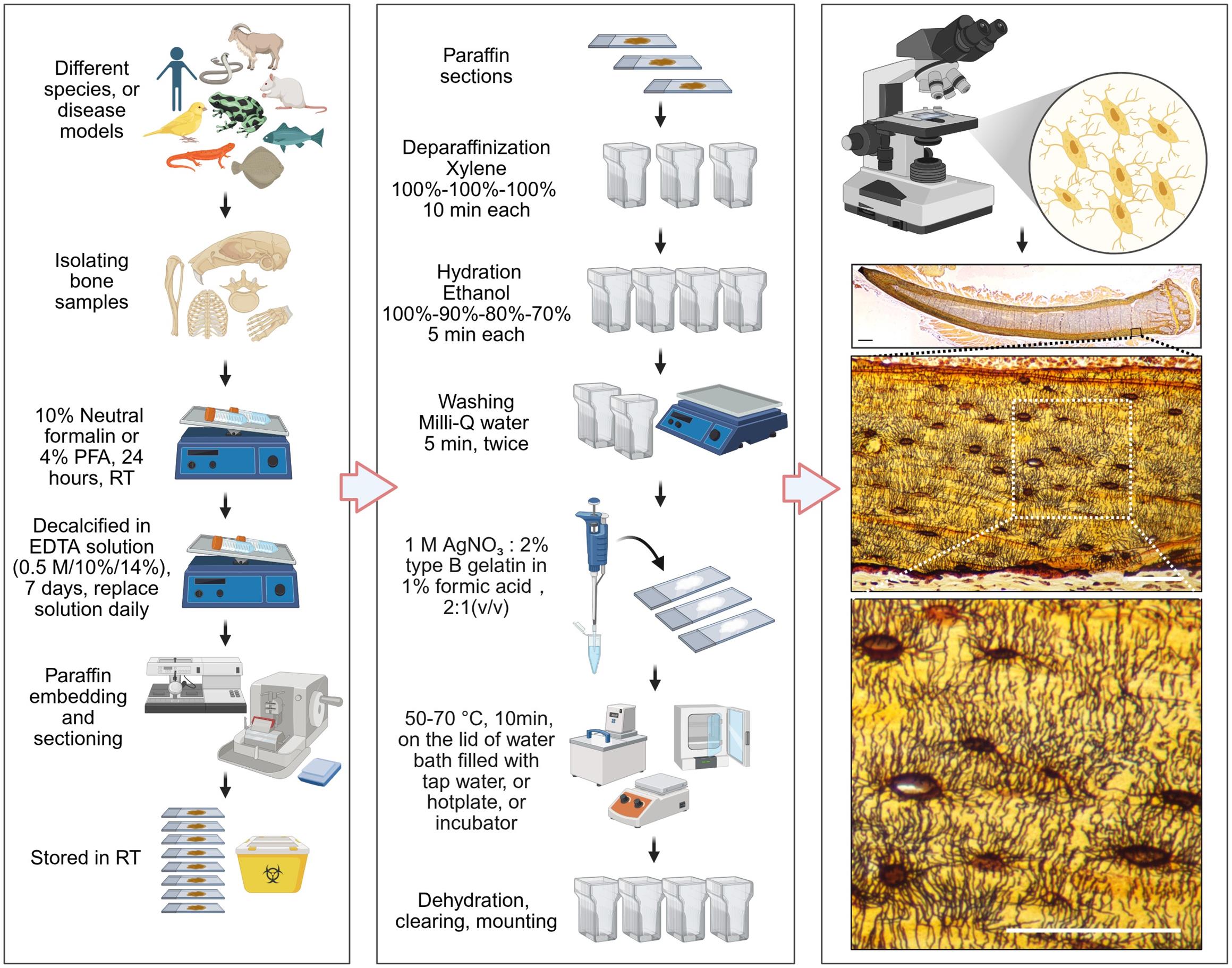
Workflow of the Wu–Wang silver nitrate staining method for osteocyte lacuno-canalicular system (LCS) visualization. Created with BioRender (https://BioRender.com).
Background
Osteocytes, derived from osteoblasts and embedded within the mineralized bone matrix, play an essential role in both bone development and the progression of bone and joint diseases [1–2]. Osteocytes reside in lacunae and extend long cellular processes into an interconnected network of canaliculi, which together constitute the lacuno–canalicular system (LCS) [3–4]. Through LCS architecture, osteocytes function as mechanosensors and signal transducers, thereby regulating bone remodeling and maintaining mineral homeostasis [5–6]. Abnormalities in LCS structure during aging or disease can alter remodeling dynamics and disrupt mineral homeostasis, ultimately contributing to a range of musculoskeletal and degenerative disorders [7–10].
Despite the importance of the LCS, detailed morphological analyses and a comprehensive understanding of LCS development remain challenging, largely due to the lack of rapid, robust, and reproducible histological staining approaches. The currently widely used Ploton silver staining method often yields variable results, with inconsistencies arising from differences in reagent concentration and composition [11–14]. In previous studies [15–16], we showed that using lower silver nitrate concentrations (0.5–1 mol/L) enables effective and clear visualization of the LCS while reducing tissue damage and improving staining adequacy compared with the Ploton formulation (50% w/v; 2.943 mol/L). Nevertheless, the staining time of our optimized method (60 min) remained comparable to that of the Ploton method (55 min), which may limit its practicality and broader adoption. Building on our earlier observations that temperature can facilitate silver staining [16], and informed by literature describing how temperature affects silver-based staining reactions [17–19], we systematically evaluated the effect of incubation temperature on LCS silver nitrate staining [20]. Surprisingly, we discovered and established a rapid and efficient staining condition: incubation with 1 mol/L silver nitrate at 50–70 °C for 10 min [20].
Herein, we present a detailed protocol for this novel histological staining method, including step-by-step procedures and practical notes (see Graphical overview). We further show that this novel method enables rapid, high-contrast, and clear visualization of osteocyte LCS in mouse skeletal tissues as well as in bone specimens from diverse vertebrate species. Widespread adoption and application of this rapid and efficient histological method will advance our understanding of osteocyte and LCS morphogenesis among osteocyte biologists and evolutionary developmental biologists, while simultaneously providing a practical histological tool for investigating the pathological mechanisms of bone and joint diseases in clinical research.
Materials and reagents
Biological materials
1. 8-month-old male C57BL/6J mice (Jiangsu GemPharmatech Co., Ltd.)
2. Domestic pigs (Sus scrofa domesticus), domestic black-spotted frogs (Pelophylax nigromaculatus), Chinese three-keeled pond turtles (Mauremys reevesii), domestic pigeons (Columba livia domestica), grass carp (Ctenopharyngodon idella), and Asian swamp eels (Monopterus albus), obtained from local food markets
3. Zebrafish (Danio rerio) and rare gudgeons (Gobiocypris rarus), obtained from local aquarium suppliers
Reagents
1. Silver nitrate solution, 1 mol/L (Bolinda Technology, catalog number: P1929026)
Note: Alternatively, commercially available silver nitrate powder can be purchased and used to prepare a 1 mol/L silver nitrate solution with 18.2 MΩ/cm Milli-Q water.
2. Type-B gelatin (Sangon Biotech, catalog number: A600908–0500)
3. Formic acid (FA) 88% (Sinopharm, catalog number: 10010118)
4. EDTA decalcification solution 0.5 mol/L (Servicebio, catalog number: G1105-500ML)
Note: Alternatively, commercially available 10% or 14% EDTA decalcification solutions can also be used. Rapid decalcification solutions, such as formic acid–based decalcification solutions, should be avoided because they result in substantially poorer LCS staining quality than EDTA decalcification solutions.
5. Neutral formalin fixative 10% (Wexis, catalog number: 311010014)
Note: Alternatively, commercially available 4% paraformaldehyde (PFA) (Servicebio, catalog number: G1101-500ML) can be used.
6. PBS (Servicebio, catalog number: G2156-1L)
7. Paraffin (Leica, catalog number: 39601006)
8. Ethanol (SinoPharm, catalog number: 10009218)
9. Xylene (SinoPharm, catalog number: 10023418)
10. Neutral balsam mounting medium (Solarbio, catalog number: G8590)
Solutions
1. 1% (v/v) FA solution (see Recipes)
2. 2% (w/v) gelatin in 1% FA solution (see Recipes)
3. Silver nitrate staining solution (see Recipes)
Recipes
1. 1% (v/v) FA solution
| Reagent | Final concentration | Quantity or volume |
|---|---|---|
| 88% FA | 1% (v/v) | 1.136 mL |
| 18.2 MΩ/cm Milli-Q water | n/a | 98.864 mL |
| Total | n/a | 100 mL |
The prepared solution can be stored at room temperature and does not require light protection.
2. 2% (w/v) gelatin in 1% FA solution
| Reagent | Final concentration | Quantity or volume |
|---|---|---|
| Type-B gelatin | 2% (v/w) | 2 g |
| 1% FA solution | n/a | 100 mL |
| Total | n/a | 100 mL |
The prepared solution can be stored at room temperature and does not require light protection. See General note 1.
3. Silver nitrate staining solution
| Reagent | Final concentration | Quantity or volume |
|---|---|---|
| 1 mol/L silver nitrate solution | 33.3% (v/v) | 0.4 mL |
| 2% (w/v) gelatin in 1% FA solution | 66.7% (v/v) | 0.2 mL |
| Total | n/a | 0.6 mL |
The staining solution should be prepared immediately before use and protected from light throughout the preparation process. See General note 2.
Laboratory supplies
1. Slides (Citotest, catalog number: 80312–3161)
2. Cover slides (Citotest, catalog number: 80340–0130)
3. Pipettes (Gilson, models: P1000 and P200)
4. 50 mL tubes (Greiner, catalog number: 227651)
5. Embedding cassettes (Citotest, catalog number: 31050102W)
6. Forceps and scissors
Equipment
1. High-precision water bath (Jinghong, model: DKB-501S)
Note: Alternatively, a temperature-controlled incubator with precise temperature regulation or a temperature-controlled hotplate can also be used.
2. Automatic benchtop tissue processor (Leica, model: TP1020)
3. Tissue embedding station (Kedi, model: KD-BM)
4. Rotary microtomes (Leica, model: RM2135)
5. Histology water bath (Leica, model: Histobath HI1210)
6. Ultrapure water purification system (Millipore, model: Milli-Q Elix® Essential)
7. Nikon microscope (Nikon, model: ECLIPSE 200)
Procedure
登录/注册后免费查看全文
文章信息
稿件历史记录
提交日期: May 21, 2026
接收日期: Jul 10, 2026
在线发布日期: Jul 19, 2026
出版日期: Aug 5, 2026
版权信息
© 2026 The Author(s); This is an open access article under the CC BY license (https://creativecommons.org/licenses/by/4.0/).
如何引用
Wu, J., Dai, W. and Wang, L. (2026). Visualizing the Osteocyte Lacuno-Canalicular System via a Rapid 10-Minute Silver Nitrate Staining Method. Bio-protocol 16(15): e5784. DOI: 10.21769/BioProtoc.5784.

 Download Citation in RIS Format
分类
细胞生物学 > 细胞成像 > 固定细胞成像
细胞生物学 > 组织分析 > 组织形态学
细胞生物学 > 组织分析 > 组织染色
您对这篇实验方案有问题吗?
在此处发布您的问题,我们将邀请本文作者来回答。同时,我们会将您的问题发布到Bio-protocol Exchange,以便寻求社区成员的帮助。
 发布问题
发布问题


