


Actin Quantification Using the Filamentous Actin Segmentation Tool (FAST)
利用丝状肌动蛋白分割工具(FAST)定量分析肌动蛋白

发布: 2026年07月20日第16卷第14期 DOI: 10.21769/BioProtoc.5765 浏览次数: 527
评审: Elena A. OstrakhovitchAnonymous reviewer(s)
 PDF
PDF  Q&A
Q&A
参见作者原研究论文
The authors used this protocol in:

Feb 2026





Abstract
Studying actin-filament assembly into distinct subcellular structures can provide insights into both physiological cellular processes and the mechanisms of disease. However, there are a limited number of tools that can quantify the organization and abundance of different actin structures from confocal microscopy images of cells expressing Lifeact or fixed and stained with phalloidin. Filamentous actin segmentation tool (FAST) is a deep learning model trained with a unique approach of antibody-assisted annotation, resulting in accurate and efficient quantification of distinct classes of actin structures. Here, we detail the protocol for using antibody-assisted annotation to generate datasets that could be applied to train machine learning models. Additionally, we provide step-by-step instructions for applying FAST on phalloidin-stained or live-cell confocal imaging data using our pretrained model. FAST is open source and freely available, with user-friendly notebooks that enable quantification of different classes of actin structure, without the need for structure-specific antibodies. As such, FAST can be a practical tool for researchers investigating the role of cytoskeletal organization in a range of processes.
Key features
• This antibody-assisted labeling approach can be used for identifying different classes of actin structure and generating labeled datasets for training machine learning algorithms.
• The trained FAST model then enables the detection of distinct classes of actin structure without the need for multiple structure-specific antibodies.
• FAST generates segmentation masks that can be used to quantify the abundance and organization of detected classes.
• This protocol provides a graphical user interface for fine-tuning custom phalloidin-stained images and provides instructions on using trained model on Ilastik interface.
Keywords: Actin (肌动蛋白) Cytoskeleton (细胞骨架)
Fluorescence microscopy (荧光显微镜)
Confocal microscopy (共聚焦显微镜)
Cell segmentation (细胞分割)
Deep learning (深度学习)
Cytoskeleton (细胞骨架)
Fluorescence microscopy (荧光显微镜)
Confocal microscopy (共聚焦显微镜)
Cell segmentation (细胞分割)
Deep learning (深度学习)
Graphical overview
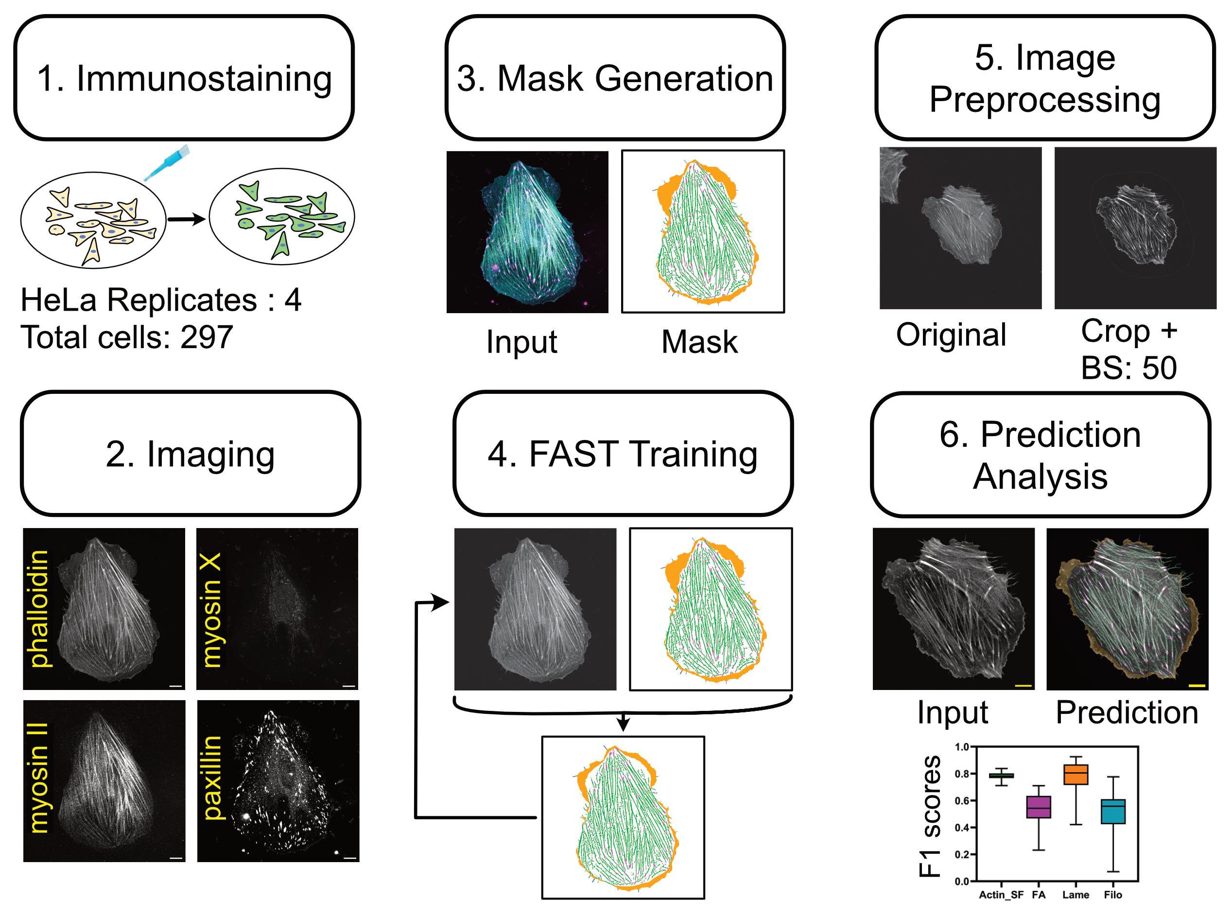
Overview of the pipeline. (1) This tool was trained and validated with confocal microscopy images of HeLa cells that had been fixed and stained with standard immunohistochemistry protocols. (2) Multichannel images with phalloidin, myosin II, myosin X, and paxillin were collected. (3) Masks were created semi-automatically, where phalloidin was used to detect actin and stress fibers (green). The absence of myosin II was used to identify lamellipodia and lamellar regions (orange), the presence of myosin X puncta at the end of thin protrusions was used to detect filopodia (cyan), and paxillin was used to detect focal adhesions (magenta). (4) The phalloidin image and the multilabel mask were used to train FAST. (5) For an unseen dataset, images were preprocessed to remove background (with rolling ball radius 50) and crop single cell prior to inference. (6) Predicted masks based on the phalloidin image alone (Input) can be used as a basis for higher level actin substructure quantification (Prediction) and analysis including F1 scores to measure accuracy (bottom) of actin and stress fibers (Actin_SF, cyan), focal adhesions (FA, magenta), lamellipodia and lamellar regions (Lame, orange), and filopodia (Filo, blue).
Background
Actin is one of the most ubiquitous proteins in eukaryotic cells and is involved in diverse cellular processes, ranging from separating daughter cells during cytokinesis, determining cell shape, mechanotransduction, and driving cell motility [1–3]. This functional versatility is enabled through the dynamic assembly of filamentous actin (F-actin) into diverse higher-order structures, chief of which are lamellipodial and lamellar networks, filopodia, stress fibers, and focal adhesions [4,5]. These structures are mainly defined by their morphology (e.g., lamellipodia and lamellar networks are thin sheet-like extensions, whereas filopodia are finger-like protrusions), the spatial organization of actin filaments in the structure (e.g., stress fibers are formed from bundling of actin filaments), and their association with specific actin regulatory proteins (e.g., focal adhesions, located at sites of contact with the environment surface, contain an array of actin-associated proteins, such as paxillin) [6–9]. Confocal microscopy is well-suited for characterizing the organization of actin filaments into different subcellular structures, where a combination of fluorescently labeled phalloidin with fluorescently conjugated antibodies targeting actin regulatory proteins has been widely used [10]. While this approach can offer high specificity, its effectiveness strongly depends on the quality of the signal and the binding specificity of the antibodies [11]. Additional limitations include the availability of specific antibodies and cross-reactivity arising from shared host species. To address these challenges, we developed a deep learning model, named filamentous actin segmentation tool (FAST), with a unique approach using antibody-assisted labeling, to create high-quality ground truth annotations for detecting multiple classes of actin structures [12]. This protocol details the pipeline used to create datasets that were used to train and validate FAST. The protocol begins with instructions for the preparation of a multichannel dataset by immunostaining and imaging of HeLa cells. While this antibody-assisted annotation approach enables users to generate high-quality custom datasets, we also describe using a pretrained FAST model to quantify the custom phalloidin-stained or live-cell confocal imaging datasets. As part of that, we provide a user-friendly application for fine-tuning image preprocessing parameters.
Materials and reagents
Biological materials
1. HeLa cells (ATCC CCL-2TM)
Reagents
1. Myosin X antibody with mouse host (Novus Biologicals, catalog number: NBP2-88926), store at -20 °C
2. Human myosin IIA (GeneTex, catalog number: GTX33939), store at -20 °C
3. Anti-paxillin antibody (Y113) with rabbit host (Abcam, catalog number: ab32084), store at -20 °C
4. Phalloidin-iFluor 647 reagent (Abcam, catalog number: ab176759), store at -20 °C
5. Goat anti-mouse IgG (H+L) (Alexa Fluor® 405) (Life Technologies Inc, catalog number: A31553), store at 4 °C
6. Goat anti-human IgG (H+L) (Alexa Fluor® 488) (Life Technologies Inc, catalog number: A11013), store at 4 °C
7. Goat anti-rabbit IgG H&L (Alexa Fluor® 568) (Abcam, catalog number: ab175471), store at -20 °C
8. Dulbecco’s modified Eagle’s medium (DMEM) with 4.5 g/L D-Glucose and phenol red (Gibco, catalog number: 11965118), store at 4 °C
9. Phosphate-buffered saline (PBS), 1× with calcium and magnesium (Corning, catalog number: 21-030-CV), store at 4 °C
10. Trypsin-EDTA solution, 0.25% (Gibco, catalog number: 25200056), store at 4 °C
11. Fetal bovine serum (FBS), heat-inactivated (Thermo ScientificTM, catalog number: 10270106), store at -20 °C
12. Penicillin-streptomycin 100× solution (Cytiva, catalog number: SV30010), store at -20 °C
13. PIPES, sodium salt (1.5) reagent grade (BioShop, catalog number: PIP663), store at room temperature
14. Ethylene glycol-bis(2-aminoethylether)-N,N,N',N'-tetraacetic acid (EGTA), ultra-pure, min. 98% (BioShop, catalog number: EGT101), store at room temperature
15. Magnesium chloride (MgCl2) (Quality Biological, catalog number: 351-033-721EA), store at room temperature
16. TritonTM X-100, pH 9.7, non-ionic, liquid (Sigma-Aldrich, catalog number: T8787-50ML), store at room temperature
17. Fibronectin (Corning, catalog number: 354008), store at 4 °C
18. Sucrose (Ward’s Science, catalog number: 470302–808), store at room temperature
19. PierceTM 16% formaldehyde (PFA) (w/v), methanol-free (Thermo ScientificTM, catalog number: 28908), store at room temperature
20. HyCloneTM HyPure water, cell culture grade (Cytiva, catalog number: SH30529.03), store at room temperature
21. Bovine serum albumin (BSA), BioReagent (Sigma-Aldrich, catalog number: A9418), store at -20 °C
22. Sodium azide solution (Sigma-Aldrich, catalog number: 08591-1ML-F), store at 4 °C
Solutions
1. Cell culture medium (see Recipes)
2. Cytoskeletal buffer (see Recipes)
3. Fixing agent (see Recipes)
4. Permeabilization solution (see Recipes)
5. Blocking solution (see Recipes)
6. Sodium azide solution (see Recipes)
7. Primary antibody solution (see Recipes)
8. Secondary antibody solution (see Recipes)
Recipes
1. Cell culture medium
| Reagent | Final concentration | Quantity or volume |
|---|---|---|
| DMEM (+ 4.5 g/L D-Glucose) | 89% | 450 mL |
| FBS | 10% | 50 mL |
| Penicillin-streptomycin | 1% | 5 mL |
| Total | 100% | 505 mL |
Supplement 450 mL of DMEM with 50 mL of heat-inactivated FBS and 5 mL of penicillin-streptomycin. Mix gently to avoid foaming. Store at 2–8 °C for 2–4 weeks. Use sterile technique.
2. Cytoskeletal buffer
| Reagent | Final concentration | Quantity or volume |
|---|---|---|
| PIPES | 80 mM | 1.21 g |
| EGTA | 5 mM | 95 mg |
| MgCl2 | 2 mM | 20.3 mg |
| Culture-grade water | - | 50 mL |
Mix 30–35 mL of dH2O with 1.21 g of PIPES, 95 mg of EGTA, and 20.3 mg of MgCl2. Raise the pH to 6.8 using KOH. Make the final volume of solution to 50 mL by adding dH2O. Sterilize using a 0.22 μm filter. Store at 2–8 °C.
3. Fixing agent
| Reagent | Final concentration | Quantity or volume |
|---|---|---|
| Sucrose | 0.1 g/mL | 1 g |
| Cytoskeletal buffer (Recipe 2) | 0.1× (buffer stock) | 1 mL |
| 16% PFA | 4% | 2.5 mL |
| Culture-grade water | - | 6.5 mL |
Add 1 g of sucrose to 1 mL of cytoskeletal buffer and mix well with 6.5 mL of culture-grade water. Finally, add 2.5 mL of 16% PFA aliquot and mix gently to avoid foaming. Store at 2–8 °C for 2–4 weeks.
4. Permeabilization solution
| Reagent | Final concentration | Quantity or volume |
|---|---|---|
| TritonTM X-100 | 0.1% | 10 μL |
| PBS | - | 10 mL |
Make permeabilization solution by adding 10 mL of PBS to 10 μL of TritonTM X-100. Mix gently to avoid foaming. Store at 2–8 °C for 2–4 weeks.
5. Blocking solution
| Reagent | Final concentration | Quantity or volume |
|---|---|---|
| BSA | 2 mg/mL | 100 μL |
| PBS | - | 10 mL |
Make blocking solution by adding 100 μL of 200 mg/mL BSA stock to 10 mL of PBS and mix gently to avoid foaming. Store at 2–8 °C for 2–4 weeks.
6. Sodium azide solution
Supplement 1 mL of PBS with 1 μL of sodium azide. Store at 2–8 °C for 2–4 weeks. Use sterile technique.
7. Primary antibody solution
| Reagent | Final concentration | Quantity or volume |
|---|---|---|
| Myosin X | 5 μg/mL | 1 μL |
| Myosin IIA | 5 μg/mL | 1 μL |
| Paxillin | 5 μg/mL | 1 μL |
| Phalloidin | 5 μg/mL | 1 μL |
| Blocking solution (Recipe 5) | - | 200 μL |
Make primary antibody solution by adding 1 μL of each of the antibodies listed above to 200 μL of blocking solution and mix gently to avoid foaming.
8. Secondary antibody solution
| Reagent | Final concentration | Quantity or volume |
|---|---|---|
| Goat anti-mouse (Alexa Fluor® 405) | 5 μg/mL | 1 μL |
| Goat anti-human (Alexa Fluor® 488) | 5 μg/mL | 1 μL |
| Goat anti-rabbit (Alexa Fluor® 568) | 5 μg/mL | 1 μL |
| Blocking solution (Recipe 5) | - | 200 μL |
Make secondary antibody solution by adding 1 μL of each of the antibodies listed above to 200 μL of blocking solution and mix gently to avoid foaming.
Laboratory supplies
1. Cell culture flask with filter cap, 25 cm2 (FroggaBio, catalog number: FB-T25-200)
2. 0.22 um PES syringe filter, 25 mm, sterile (FroggaBio, catalog number: SF0.22PES)
3. 8-well chambered cover glass with #1.5 high performance cover glass, 57 mm × 25 mm base (Cellvis, catalog number: C8-1.5H-N)
4. FroggaBio 15 mL conical tubes (rack) (FroggaBio, catalog number: TR15-500)
Equipment
1. Microscope (Nikon Eclipse Ti2 inverted microscope utilizing a CrestOptics X-Light V3 spinning disk integrated with a Photometrics Kinetix camera with 60× 1.2 Numerical Aperture Plan Apo VC water immersion objective)
2. Class II biological safety cabinet (ESCO)
3. CO2 incubator (Thermo Scientific, model: 3110)
4. Freezer, -20 °C
5. Refrigerator, 2–8 °C
Software and datasets
Dataset, model, and software are provided in Table 1 along with version and license information. Additionally, Google Colab notebooks are provided for training to simulate a GUI on a cloud computing platform. Inference and preprocessing are performed with Ilastik [13] and Fiji/ImageJ [14], respectively.
Note: While FAST can be run freely on Google Colab, the availability of GPU resources on the cloud is subject to usage and the subscription type of the user.
Table 1. Software and datasets for data analysis. FAST can be downloaded from GitHub (https://github.com/Carleton-CTE-Lab/FAST-Protocol/archive/refs/heads/main.zip), and the corresponding model link is available on the Google Colab notebook. Dataset is publicly available on Zenodo (https://zenodo.org/records/18135376).
| Type | Software/dataset/resource | Version | Date | License | Access |
|---|---|---|---|---|---|
| Dataset | Zenodo (https://zenodo.org/records/18135376) | V1 | Jan 2, 2026 | Creative Commons | Free |
| Model | Zenodo (https://zenodo.org/records/18627454) | V2 | Feb 13, 2026 | Creative Commons | Free |
| Software | GitHub (https://github.com/Carleton-CTE-Lab/FAST-Protocol) | fast_protocol (tag) | Apr 9, 2026 | MIT | Free |
| Software tool | Fiji/ImageJ (https://imagej.net/software/fiji/) | 2.17.0 | Aug 12, 2025 | GPLv3+ | Free |
| Software tool | Supervisely (https://app.supervisely.com/projects) | 6.15.40 | Dec 23, 2025 | Apache-2.0 | Paid (has free tier) |
| Workflow manager | Ilastik (https://www.ilastik.org/) | 1.4.1.post1 | May 5, 2025 | GPLv3+ | Free |
Procedure
登录/注册后免费查看全文
文章信息
稿件历史记录
提交日期: Apr 13, 2026
接收日期: Jun 9, 2026
在线发布日期: Jun 29, 2026
出版日期: Jul 20, 2026
版权信息
© 2026 The Author(s); This is an open access article under the CC BY-NC license (https://creativecommons.org/licenses/by-nc/4.0/).
如何引用
Aljapur, V., Gardner, A., Carayanniotis, J. and Harris, A. R. (2026). Actin Quantification Using the Filamentous Actin Segmentation Tool (FAST). Bio-protocol 16(14): e5765. DOI: 10.21769/BioProtoc.5765.

 Download Citation in RIS Format
分类
生物信息学与计算生物学
细胞生物学 > 细胞成像 > 共聚焦显微镜
细胞生物学 > 细胞成像 > 荧光
您对这篇实验方案有问题吗?
在此处发布您的问题,我们将邀请本文作者来回答。同时,我们会将您的问题发布到Bio-protocol Exchange,以便寻求社区成员的帮助。
 发布问题
发布问题


