


Separating Chromera velia Zoospores From Culture and Estimating Their Average Motility Speed and Lifespan
从培养物中分离Chromera velia游动孢子并测定其平均运动速度和寿命



发布: 2026年07月20日第16卷第14期 DOI: 10.21769/BioProtoc.5756 浏览次数: 282
评审: Veronika KselíkováAnonymous reviewer(s)
 PDF
PDF  Q&A
Q&A
参见作者原研究论文
The authors used this protocol in:

Dec 2023
Advertisement






Abstract
Chromera velia is an apicomplexan alga uniquely positioned as the closest photosynthetic relative to apicomplexan parasites (Sporozoa), which include the human pathogens that cause malaria (Plasmodium) and toxoplasmosis (Toxoplasma). Under favorable conditions, C. velia forms motile zoospores that contribute to dispersal and possibly host interaction. However, zoospores coexist with other developmental stages in culture, making their isolation technically challenging. Previous studies characterized the phototactic behavior of zoospores in several taxa, yet this response has not been used to separate motile zoospores from mixed cultures. Other reported methods for zoospore recovery relied instead on physical or chemical principles such as passive filtration, differential centrifugation, or column-based purification, all of which can compromise zoospore motility and viability through mechanical shear or osmotic changes. To address this limitation, we developed a non-invasive, simple, and effective method for rapid zoospore isolation depending entirely on their negative phototaxis response. Using a directional light gradient, the method enables reliable collection of active, motile zoospores without specialized equipment or chemical treatments. Our protocol is straightforward to reproduce, relies on standard laboratory equipment, can be completed in under two hours, and yields a zoospore fraction of sufficient quality for live-imaging, motility assays, and downstream molecular and -omics applications. It may also be adapted to other flagellated protists with light-responsive motile stages.
Key features
• Requires basic experience with algal culturing and access to a light-controlled environment.
• A typical 7-day pre-cultivation period is needed for Chromera velia to initiate and reach peak zoosporogenesis.
• Provides a rapid procedure for isolating C. velia zoospores that can be fully completed within 1–2 h, including material preparation.
• Enables downstream experiments with clean, motile zoospore populations for physiological, behavioral, molecular, or -omics analyses.
Keywords: Chromera velia (Chromera velia) Zoospore isolation (游动孢子分离)
Negative phototaxis (负趋光性)
Motility assay (运动能力检测)
Life cycle (生命周期)
Zoospore isolation (游动孢子分离)
Negative phototaxis (负趋光性)
Motility assay (运动能力检测)
Life cycle (生命周期)
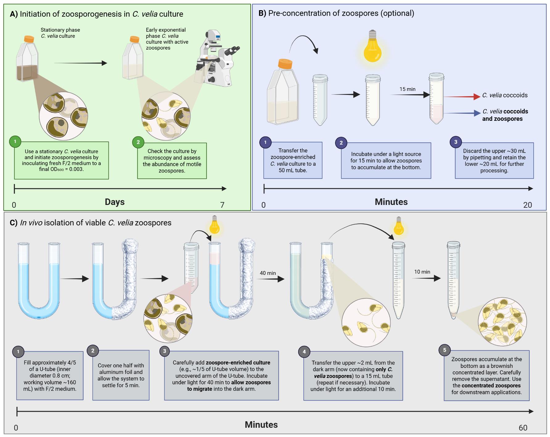
Graphical overview
Background
Sitting at the evolutionary crossroads between photosynthesis and parasitism, Chromera velia (Apicomplexa, Apicomonada [1]) is a photoparasitic alga and the closest known photosynthetic relative of apicomplexan parasites, the group encompassing the causative agents of malaria (Plasmodium) and toxoplasmosis (Toxoplasma). Chromera velia has been isolated from the scleractinian coral Plesiastrea purpurea using a method developed for isolating intracellular coral symbionts. These symbionts, usually dinoflagellates, reside within coral tissue and support the host by supplying photosynthetically derived carbon, receiving carbon dioxide and protection in return. Chromera velia was initially presumed to have a similar life strategy [2]. However, Cumbo et al. [3] demonstrated that, unlike dinoflagellate symbionts that invade the endoderm of coral larvae via the oral pore, C. velia was also found in the ectoderm, indicating active penetration from the outside. Additionally, transcriptomic analyses of C. velia–infected coral larvae revealed expression patterns indicative of a host–pathogen interaction rather than a mutualistic association [4].
Chromera velia has a complex life cycle that is fully maintained under laboratory conditions (Figure 1). It includes immotile stages such as coccoid cells (Figure 2A), autosporangia, zoosporangia (Figure 2B), and cysts, as well as highly motile, biflagellate zoospores (Figure 2C). The process that produces zoospores is called zoosporogenesis [5]. The zoospore is the only life stage believed to penetrate host cells, as it contains a primitive apical complex, in the form of a conoid (pseudoconoid), a structure known from parasitic apicomplexans [6,7]. Thus, C. velia plays a significant role in our understanding of the evolutionary origin of parasitism [8].
Beyond C. velia, zoosporogenesis and the release of motile cells depend heavily on the biology and life cycle of each organism and its adaptation to its environment. The release of zoospores can be triggered by various biotic and abiotic factors, including osmotic stress, salinity, temperature, pH, nutrient availability, wave motion, and light intensity [9]. In C. velia, zoosporogenesis is induced particularly in response to specific light wavelengths and medium composition [5,10]. Appearance of zoospores has been well documented under 12/12 h light/dark (LD) conditions [2,5]. Zoospore numbers peak between 7 and 10 days after culture inoculation and 6 h after the onset of illumination [6,10]. Except under red light, C. velia zoospores display negative phototaxis in response to all tested light spectra, actively swimming away from the light source. Their average swimming speed is estimated at approximately 2 cm/min [10]. A persistent challenge in the study of C. velia has been the reliable isolation of clean zoospore populations, free from coccoid and other non-motile stages, which is crucial for downstream applications such as gene expression analysis, behavioral assays, and physiological studies. The phototactic behavior of zoospores has been characterized in several taxa, including Ulkenia sp. zoospores [11], Allomyces sp. [12], and Ulva sp. [13]; however, these studies focused on identifying the wavelengths eliciting positive or negative phototaxis and did not exploit this response as a separation principle. To our knowledge, the present protocol is the first to use phototaxis directly as a means of purifying motile zoospores from a mixed culture. Previously reported methods for recovering zoospores or other flagellated stages in related systems rely instead on physical or chemical principles. Passive filtration through nylon or polycarbonate meshes has been used to collect zoospores in oomycetes and chytrids [14,15], differential centrifugation has been applied to actinomycete zoospores [16], and column-based purification, such as anion-exchange chromatography of Eimeria sporozoites [17], has been conducted, all of which can compromise zoospore motility and viability by mechanical shear or osmotic change. By contrast, our method is non-invasive, and its selectivity is driven entirely by the behavior of the zoospores—negative phototaxis. Using a directional light source and a standard U-shaped glass tube, only actively swimming zoospores migrate into the collection arm of the U-tube, yielding a population of intact, motile cells suitable for live-cell imaging, motility assays, and downstream molecular analyses. Furthermore, this low-cost approach may be adapted for other flagellated protists exhibiting light-responsive motility.
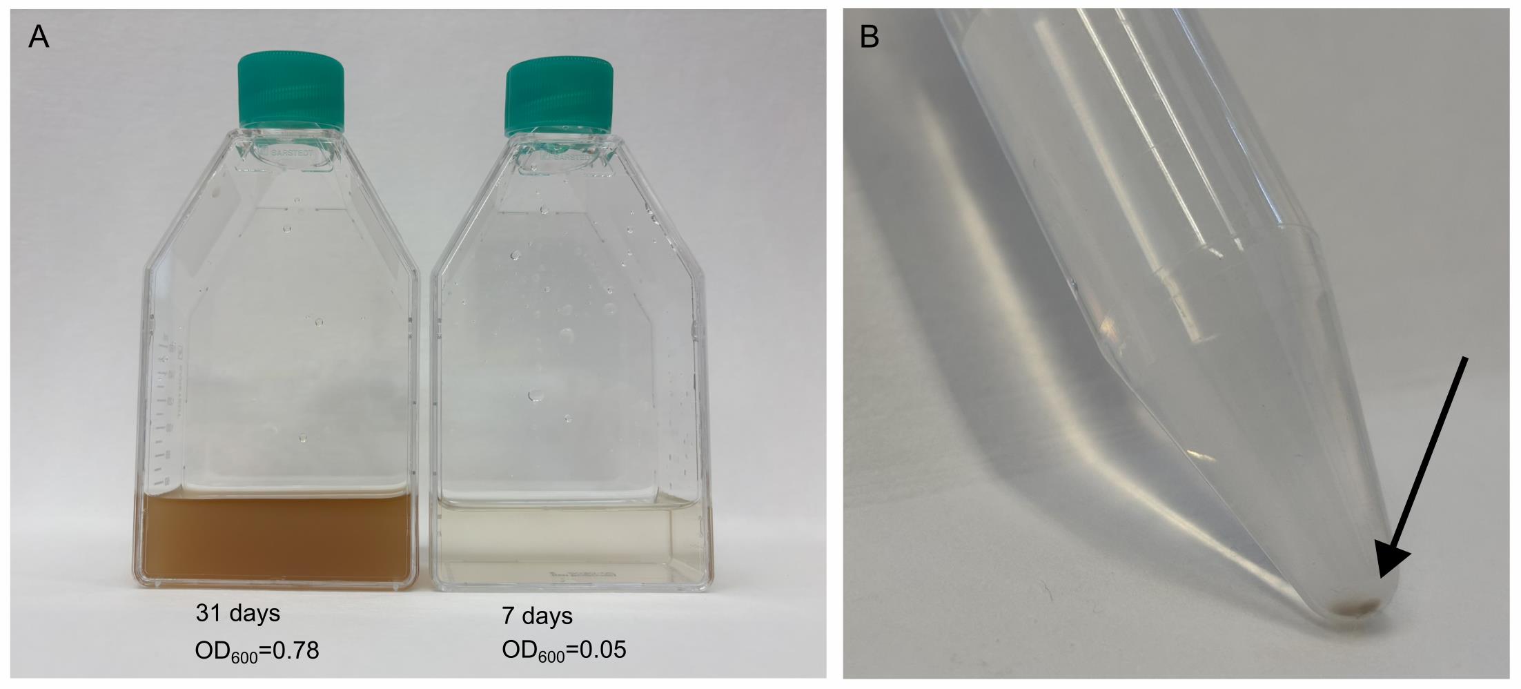
Figure 1. Representative stages of Chromera velia cultures and isolated zoospores. (A) A 31-day-old stationary culture (OD600 = 0.78) was used as inoculum, compared with a 7-day-old early exponential culture (OD600 = 0.05) enriched in zoospores used for isolation. (B) Concentrated zoospore fraction visible as a brownish layer at the tip of the tube after isolation (arrow).
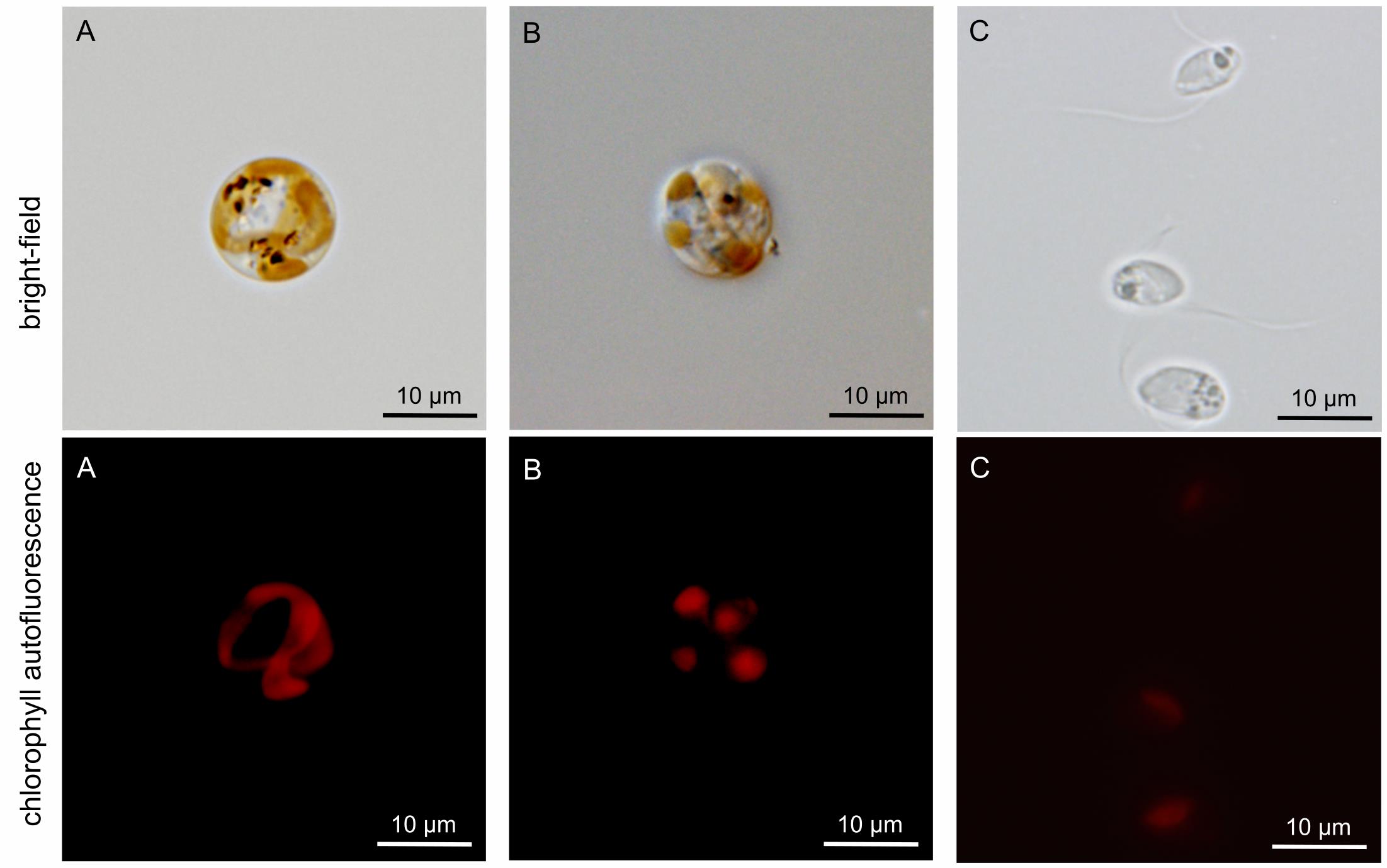
Figure 2. Brightfield light microscopy and chlorophyll autofluorescence of Chromera velia developmental stages. Upper row: Brightfield images. Lower row: Corresponding chlorophyll autofluorescence (red signal). (A) Coccoid cell containing a single, well-developed plastid. (B) C. velia zoosporangium containing six zoospores. (C) Isolated zoospores fixed with 4% paraformaldehyde.
Materials and reagents
Biological materials
1. Chromera velia [2] (National Centre for Marine Algae and Microbiota, Maine, USA; catalog number: CCMP 2878)
Reagents
1. Sea salt: Tropic Marin PRO REEF (Tropic Marin, catalog number: 10581)
2. Guillard F/2 marine enrichment solution (Sigma-Aldrich, catalog number: G0154)
3. Distilled water (Milli-Q or equivalent)
4. Azoxystrobin (Azo) (TraceCERT®) (Sigma-Aldrich, catalog number: 43854)
5. Dimethyl sulfoxide (DMSO) (≥99.5%, suitable for plant cell culture) (Sigma-Aldrich, catalog number: D4540)
6. Ethanol 70% (v/v), prepared from absolute or 96% ethanol with distilled water; for surface sterilization, typical exposure time 1–2 min (optional)
7. Bleach solution 1% (v/v), prepared by diluting commercial bleach stock solution [Savo Original, Unilever; ~3%–5% (w/v) sodium hypochlorite] 1:5 in distilled water (optional)
Solutions
1. Guillard’s medium (F/2 medium) (see Recipes)
2. Azoxystrobin stock solution (2 mM) (see Recipes)
Recipes
1. F/2 medium
| Reagent | Final concentration | Quantity or volume |
|---|---|---|
| Tropic Marin PRO REEF sea salt | 33.3 g/L | 33.3 g |
| Guillard F/2 marine enrichment solution (50×) | 1× | 20 mL |
| Azoxystrobin | 2 mM stock | 1 mL |
| Distilled water (Milli-Q or equivalent) | - | To 1 L |
| KOH or HCl to adjust pH to 7.9–8.1 | - | - |
Note: Prepare the medium using distilled water (Milli-Q or equivalent). Sterility is achieved by filtration (0.22 μm).
2. Azoxystrobin stock solution (2 mM)
Prepare the stock solution by dissolving azoxystrobin powder in DMSO (≥99.5%) to a final concentration of 2 mM.
Laboratory supplies
1. Cell culture flask, T-75, surface: suspension, filter cap (Sarstedt, catalog number: 83.3911.502)
2. Micropipette tips with filter, 5 mL (Dualfilter T.I.P.S.®) (Eppendorf, catalog number: 0030078616)
3. Micropipette tips with filter, 1 mL (Axygen MaxyMum Recovery) (Corning Life Sciences, catalog number: TF-1000-R-S)
4. Micropipette tips filter, 200 μL (Axygen MaxyMum Recovery) (Corning Life Sciences, catalog number: TF-200-R-S)
5. TPP® centrifuge tubes, 50 mL (Sigma-Aldrich, catalog number: Z707708)
6. TPP® centrifuge tubes, 15 mL (Sigma-Aldrich, catalog number: Z707724)
7. U-shaped glass tube, 50 cm long [custom-made; borosilicate glass (SIMAX), outer diameter 22 mm, wall thickness 1.8 mm, height ~350 mm, spacing between arms 50 mm, KAVALIERGLASS, a.s.; see Figure S1 for detailed specifications] Note: Comparable commercially available or laboratory-assembled U-shaped tubes with similar geometry and approximate dimensions can be used as an alternative.
8. 96-well round-bottom plates (Techno Plastic Products, catalog number: 92696)
9. Aluminum foil (standard laboratory-grade)
Equipment
1. White light source (PowerGlo, Hagen Group®, or equivalent; 380–720 nm, 100 μmol/m2/s)
2. Portable lux meter (e.g., Delta OHM, model: HD 2102.1)
3. Spectrometer (Sven Bio Labs, model: Byonoy Absorbance 96)
4. Inverted light microscope (Motic or equivalent, 100× magnification)
5. Temperature-controlled incubator or growth chamber set to 26 °C (WTW, model: TS 606CZ/2)
Procedure
登录/注册后免费查看全文
文章信息
稿件历史记录
提交日期: Mar 6, 2026
接收日期: Jun 4, 2026
在线发布日期: Jun 24, 2026
出版日期: Jul 20, 2026
版权信息
© 2026 The Author(s); This is an open access article under the CC BY-NC license (https://creativecommons.org/licenses/by-nc/4.0/).
如何引用
Richtová, J., Ennaceur, D. and Oborník, M. (2026). Separating Chromera velia Zoospores From Culture and Estimating Their Average Motility Speed and Lifespan. Bio-protocol 16(14): e5756. DOI: 10.21769/BioProtoc.5756.

 Download Citation in RIS Format
分类
微生物学 > 微生物细胞生物学 > 细胞分离和培养
微生物学 > 微生物生理学 > 趋光性
植物科学 > 藻类学 > 细胞分离与培养
您对这篇实验方案有问题吗?
在此处发布您的问题,我们将邀请本文作者来回答。同时,我们会将您的问题发布到Bio-protocol Exchange,以便寻求社区成员的帮助。
 发布问题
发布问题


