


Histological Processing of Organoids for Immunostaining
类器官免疫染色的组织学处理方法

发布: 2026年07月20日第16卷第14期 DOI: 10.21769/BioProtoc.5750 浏览次数: 1124
评审: Samantha HallerShilpa BishtNeha Saxena
 PDF
PDF  Q&A
Q&A
参见作者原研究论文
The authors used this protocol in:
Oct 2026





Abstract
Organoids are three-dimensional cell structures derived from stem cells that recapitulate the architecture and function of native tissues. Histological analysis of organoids is essential for assessing their structure, cellular composition, and responses to experimental conditions. However, their small size and fragility make standard paraffin embedding workflows difficult. Here, we describe a robust and reproducible protocol for the fixation, paraffin embedding, and sectioning of human organoids, enabling high-quality histological and immunostaining analysis. The method involves direct fixation within the culture matrix and inclusion in HistoGel to prevent organoid loss during processing. The protocol is compatible with hematoxylin–eosin (H&E) staining and multiplex immunofluorescence. Critical steps, troubleshooting, and adaptations for intestinal and cardiac organoids are discussed. This cost-effective and accessible method supports long-term preservation and detailed structural analysis of organoid models.
Key features
• Enables reliable fixation, embedding, and sectioning of fragile human organoids for histological and immunofluorescence analysis.
• Uses HistoGel inclusion to prevent organoid loss during paraffin embedding and processing.
• Compatible with multiple organoid types (intestinal, cardiac, etc.) and staining methods (H&E, immunofluorescence).
• Produces high-quality paraffin blocks and sections suitable for long-term storage.
Keywords: Paraffin embedding (石蜡包埋) Organoids (类器官)
HistoGel (HistoGel)
Histology (组织学)
Immunofluorescence (免疫荧光)
FFPE (福尔马林固定石蜡包埋(FFPE))
Tissue processing (组织处理)
Organoids (类器官)
HistoGel (HistoGel)
Histology (组织学)
Immunofluorescence (免疫荧光)
FFPE (福尔马林固定石蜡包埋(FFPE))
Tissue processing (组织处理)
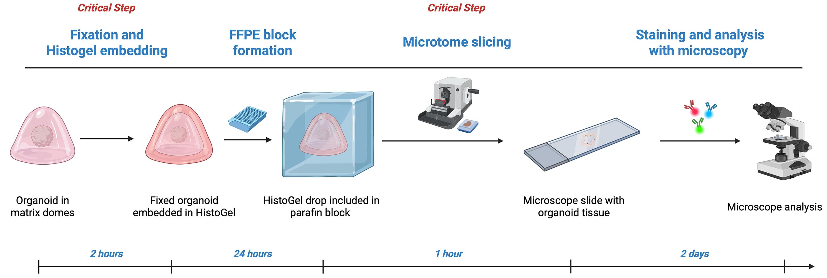
Graphical overview
Background
Organoid histology: Organoids are in-vitro 3D cell structures generated from human pluripotent or adult stem cells. Both methods depend on the expansion and differentiation abilities of stem cells to create organ-specific, self-organized structures [1]. The key advantage of organoids is their cellular diversity, architecture, and functions, which resemble those of in vivo organs. Consequently, organoids have become highly valuable for modeling tissue development, adult physiology, and diseases [2]. Additionally, organoids hold significant potential for clinical uses such as personalized medicine, drug screening, and cell therapy [3]. Laboratory analysis of organoids typically involves histological techniques, which can be performed in various ways. One standard method is paraffin embedding, where tissue is fixed, dehydrated, and sliced into thin sections after being embedded in paraffin [4]. Paraffin embedding enables the long-term storage of organoids with minimal tissue degradation and is compatible with techniques such as immunohistochemistry or immunofluorescence. However, due to their limited size, ranging from a few hundred micrometers to a few millimeters, handling, processing, and embedding individual organoids in paraffin is challenging. In this protocol, we outline a versatile method for processing and embedding organoids in paraffin wax for histological analysis. This method requires only basic histology skills for tissue processing and staining. We validated the efficiency and robustness of this technique by applying it to two types of human organoids: intestinal organoids and cardiac ventricular chamber-specific organoids, called cardioids [5,6]. This approach, which involves direct fixation of organoids within their culture matrix combined with the use of a HistoGel, ensures efficient inclusion of organoids in paraffin without risk of loss during processing. Additionally, we describe various staining protocols applicable to different types of organoids.
Comparison with other methods: Histological analysis of in vitro organoids can be performed using various methods, each with its own advantages and limitations. Whole-mount staining offers a quick and efficient way to analyze organoids as a whole, while maintaining their three-dimensional structure. This technique is particularly useful for obtaining 3D volume rendering images [7,8]. However, whole-mount staining also presents several technical challenges. Direct whole-mount staining of organoids in their culture matrix can cause excessive background fluorescence and artifacts, while removing organoids from the culture matrix may result in loss of organoids during staining because of their small size. Whole-mount staining also often requires clearing methods, some of which use solvents incompatible with certain antibodies [9]. This technique limits the number of stainings that can be performed on the same organoid. Moreover, colorimetric staining, such as hematoxylin–eosin, cannot be used for whole-mount staining, although some alternatives have been developed [10]. Tissue sectioning is another technique used for organoid histological analysis. This method is especially useful for examining tissues at a sub-cellular level and allows for multiple stains on the same organoid across different sections. Two approaches can be employed: frozen tissue sections and paraffin-embedded sections. Frozen tissue sections involve quickly freezing the organoids, embedding them in a medium like OCT, and then sectioning them with a cryostat before staining. Although frozen sections better preserve the natural protein structure of antigens, they may cause tissue morphology to degrade due to freezing artifacts. The paraffin-embedded section consists of fixing and dehydrating tissue samples before embedding them in paraffin wax and sectioning with a microtome. The main benefit of paraffin sections is their excellent preservation of tissue morphology, along with cost-effective storage for indefinite periods. Additionally, paraffin-embedded sections can be used for immunohistochemistry and immunofluorescence and are compatible with multiplexing staining, which uses multiple immunostains on the same tissue section, as well as computational 3D reconstruction techniques using a series of tissue sections [11].
Materials and reagents
Biological materials
1. Organoids (300 μm to 3 mm in size) embedded in matrix domes
Reagents
1. Double-deionized water produced by the Synergy water purification system and ion exchange resin water system (Merck Millipore, catalog number: SYNSVHFFR-KIT and Veolia water system, catalog number: CFCFGU201181)
2. Methanol-stabilized formaldehyde 30% in solution (Carl ROTH, catalog number: 4235)
3. Sodium azide (NaN3) (Sigma-Aldrich, catalog number: S8032)
4. Potassium chloride (KCl) (Sigma-Aldrich, catalog number: P5405)
5. Potassium dihydrogen phosphate monobasic (KH2PO4) (Sigma-Aldrich, catalog number: P0662)
6. Sodium chloride (NaCl) (Sigma-Aldrich, catalog number: S7653)
7. Sodium phosphate dibasic heptahydrate (Na2HPO4·7H2O) (Sigma-Aldrich, catalog number: S9390)
8. Glycine, aminoacetic acid (Sigma-Aldrich, catalog number: G8898)
9. Butanol-2-Methyl-1-butanol (Sigma-Aldrich, catalog number: 133051-100ML)
10. Xylene histological grade [C6H4(CH3)2] (Sigma-Aldrich, catalog number: 534056-500ML)
11. Absolute ethanol (CH3CH2OH) (Sigma-Aldrich, catalog number: 1070172511); when necessary, dilute ethanol with double-deionized water
12. Epredia HistoGel specimen processing gel (Epredia, catalog number: HG4000012)
13. Epredia Paraffin Type 9 (Epredia, catalog number: 8337)
14. Hematoxylin GILL II (Diapath, catalog number: CP813)
15. Trichloroacetic acid (Thermo Fisher Scientific, catalog number: 7402)
16. Lithium carbonate (Microm Microtech, catalog number: 45006 29)
17. Eosin (Diapath, catalog number: C0362)
18. Saffron (VWR, catalog number: 27481.105)
19. Diamount mounting medium (Diapath, catalog number: 030400)
20. Dako Target retrieval solution, citrate pH 6.1, 10× (Agilent, catalog number: S1699)
21. ReadyProbes hydrophobic barrier pap pen (Life Technologies, catalog number: R3777)
22. ProLong Gold anti-attenuation mounting medium (Life Technologies, catalog number: P36930)
23. Dako protein block solution, ready to use (Agilent, catalog number: XO909)
24. Dako antibody diluent, ready to use (Agilent, catalog number: SO809)
25. Antibody (depending on application)
Solutions
1. PBS 10× (see Recipes)
2. PBS/glycine (see Recipes)
3. 2% formaldehyde (see Recipes)
Recipes
1. PBS 10×
| Reagent | Final concentration (g/L) | Final concentration (mM or M) | Quantity or volume |
|---|---|---|---|
| Double-deionized water | 1× | n/a | 1,000 mL |
| Potassium chloride | 2 g/L | 26.8 mM | 2 g |
| Potassium dihydrogen phosphate monobasic | 2 g/L | 14.7 mM | 2 g |
| Sodium chloride | 80 g/L | 1,369 M | 80 g |
| Sodium phosphate dibasic heptahydrate | 21.6 g/L | 80.6 mM | 21.6 g |
Dilute 10× solution with double-deionized water for 1× utilization. Store at 4 °C for up to one year.
2. PBS/glycine
| Reagent | Final concentration (g/L) | Final concentration (μM or M) | Quantity or volume |
|---|---|---|---|
| PBS 10× | 10× | n/a | 1,000 mL |
| Glycine | 75 g/L | 1 M | 75 g |
Dilute 10× solution with double-deionized water for 1× utilization. Store at 4 °C for up to six months.
3. 2% Formaldehyde
| Reagent | Initial concentration | Final concentration | Quantity or volume |
|---|---|---|---|
| PBS | 10× | 1× | 6 mL |
| Double-deionized water | 1× | 1× | 50 mL |
| Formaldehyde (methanol-stabilized) | 30% | 2% | 4 mL |
Prepare fresh before use.
Laboratory supplies
1. Complete staining sets: well, cover, slide rack (Epredia, catalog number: 121)
2. Slides for microscopy Epredia SuperFrost Plus Gold (Epredia, catalog number: K5800AMNZ72)
3. Lid of 48-wells plate (Corning, catalog number: 3548)
4. Immunostaining slide moisture chambers (Evergreen, catalog number: 240-9020-Z10)
5. Dissection Fine-Pointed Forceps (Fisherbrand, catalog number: 08-880)
6. 660 mL glass jar (Dutscher, catalog number: PI82PC660)
7. White Pasteurizable lid, TO 82 ring (Dutscher, catalog number: CPAST82BL)
8. Razor blade compatible with microtome (ThermoFisher Scientific, catalog number: 3050835)
9. 12-piece blue HB pencil set (Lyreco, catalog number: 013LYRK7720/1)
10. Epredia Cryotom Cryostat Accessories, Camel HairBrush (Epredia, catalog number: 1910)
11. Epredia Shandon disposable embedding molds 15 × 15 mm (Epredia, catalog number: 41741)
12. Epredia Shandon disposable embedding molds 24 × 30 mm (Epredia, catalog number: 41743)
13. Fisherbrand graduated transfer pipettes, non-sterile, LDPE, clear (Fisher Scientific, catalog number: 13439108)
14. Epredia Cassette II slotted tissue cassettes (Epredia, catalog number: B851729)
15. Biopsy foam pads (Simport Scientific, catalog number: M476-1)
Equipment
1. 115 V Programmable Dry Bath Incubator (Boekel Scientific, catalog number: 115001)
2. Paraffin dispenser (Bio-Optica Milano, catalog number: 40-200-101)
3. Cooling plate (Micro Microtech, catalog number: EC 350-2)
4. Decloaking ChamberTM NxGen (Biocare Medical, catalog number: DC2012-220V)
5. Block Module, 16 mm tubes (Boekel Scientific, catalog number: 110016)
6. Stuart Mini Gyro-Rocker SSM3 (Fisher Scientific, catalog number: 10034264)
7. Microtome Microm (Epredia, catalog number: HM355S)
8. EprediaTM STP 120 Spin Tissue Processor (Epredia, catalog number: STP 120)
9. Automated slide stainer Gemini AS (Epredia, catalog number: A81500001)
10. Epifluorescence microscopes (Zeiss, Axio Observer 7 with LED Colibri 7 and HAMAMATSU C11440 digital camera; Nikon, Eclipse Ti2 with DS-Fi3 brightfield camera and DS-Qi2 fluorescence camera)
Procedure
登录/注册后免费查看全文
文章信息
稿件历史记录
提交日期: Jan 9, 2026
接收日期: May 19, 2026
在线发布日期: Jun 29, 2026
出版日期: Jul 20, 2026
版权信息
© 2026 The Author(s); This is an open access article under the CC BY license (https://creativecommons.org/licenses/by/4.0/).
如何引用
Brossard, L., Perreaux, V., Vales, S., Bonneau, L., Godin, S., Bibonne, A., Noël, T., Bachir, L., Khan, A., Lamirault, G., Gaignerie, A., Gaborit, N. and Mahe, M. M. (2026). Histological Processing of Organoids for Immunostaining. Bio-protocol 16(14): e5750. DOI: 10.21769/BioProtoc.5750.

 Download Citation in RIS Format
分类
干细胞 > 类器官培养
细胞生物学 > 组织分析 > 组织染色
细胞生物学 > 细胞成像 > 固定组织成像
您对这篇实验方案有问题吗?
在此处发布您的问题,我们将邀请本文作者来回答。同时,我们会将您的问题发布到Bio-protocol Exchange,以便寻求社区成员的帮助。
 发布问题
发布问题


