


Ex Vivo Assessment of Extracellular Acidification Rate in Murine Intestinal Tissue
小鼠肠道组织细胞外酸化率的离体测定方法

(§Technical contact: alexander.lesser@case.edu) 发布: 2026年07月20日第16卷第14期 DOI: 10.21769/BioProtoc.5749 浏览次数: 517
评审: Wendy Leanne HempstockSAPTARSHI MAJIAnonymous reviewer(s)
 PDF
PDF  Q&A
Q&A
参见作者原研究论文
The authors used this protocol in:
Jan 2026
Advertisement







Abstract
Seahorse metabolic assays are now widely utilized across numerous fields for performing functional assessments of glycolysis and mitochondrial function in adherent or suspension cell culture samples. Seahorse assays measure extracellular acidification rate (ECAR) and oxygen consumption rate (OCR) as a means of assessing glycolysis and mitochondrial function, respectively. Currently, the vast majority of Seahorse metabolic assays are performed using in vitro samples due to the current established standardized method. However, a uniform approach to assess real-time functional measurements of glycolysis and mitochondrial function in ex vivo tissue samples remains elusive. In particular, this protocol was designed to assess glycolysis in ex vivo murine intestinal samples through ECAR measurements using the Agilent Seahorse XFe24 platform with corresponding Islet Capture microplates and screens. This protocol was developed to provide functional measurements of glycolytic metabolism in murine intestinal tissue samples. This protocol details a method to assess glycolysis in tissue samples and represents the next stage of ex vivo metabolic methods to complement existing standardized in vitro approaches. While this protocol was developed to assess ECAR in ex vivo murine intestinal samples, the same approach can be applied to assessing mitochondrial respiration through measurements of OCR in other tissue types. Overall, this protocol expands the purview of Seahorse metabolic assays through the inclusion of tissue samples and provides the framework to interrogate organ-level metabolism in the context of systemic nutrient metabolism and physiology.
Key features
• Protocol for applying Seahorse metabolic assays to tissue samples.
• Specifically designed to assess glycolysis through extracellular acidification rate (ECAR) in ex vivo murine intestinal samples.
• Provides functional metabolism data to complement gene and protein expression data.
• Protocol can be adapted to measure mitochondrial function through assessments of oxygen consumption rate (OCR) in other tissue types.
Keywords: Tissue (组织) Seahorse (Seahorse代谢分析)
Glycolysis (糖酵解)
Extracellular acidification rate (细胞外酸化率)
Metabolism (代谢)
Intestine (肠道)
Murine (小鼠)
Oxygen (氧消耗率)
Seahorse (Seahorse代谢分析)
Glycolysis (糖酵解)
Extracellular acidification rate (细胞外酸化率)
Metabolism (代谢)
Intestine (肠道)
Murine (小鼠)
Oxygen (氧消耗率)
Graphical overview
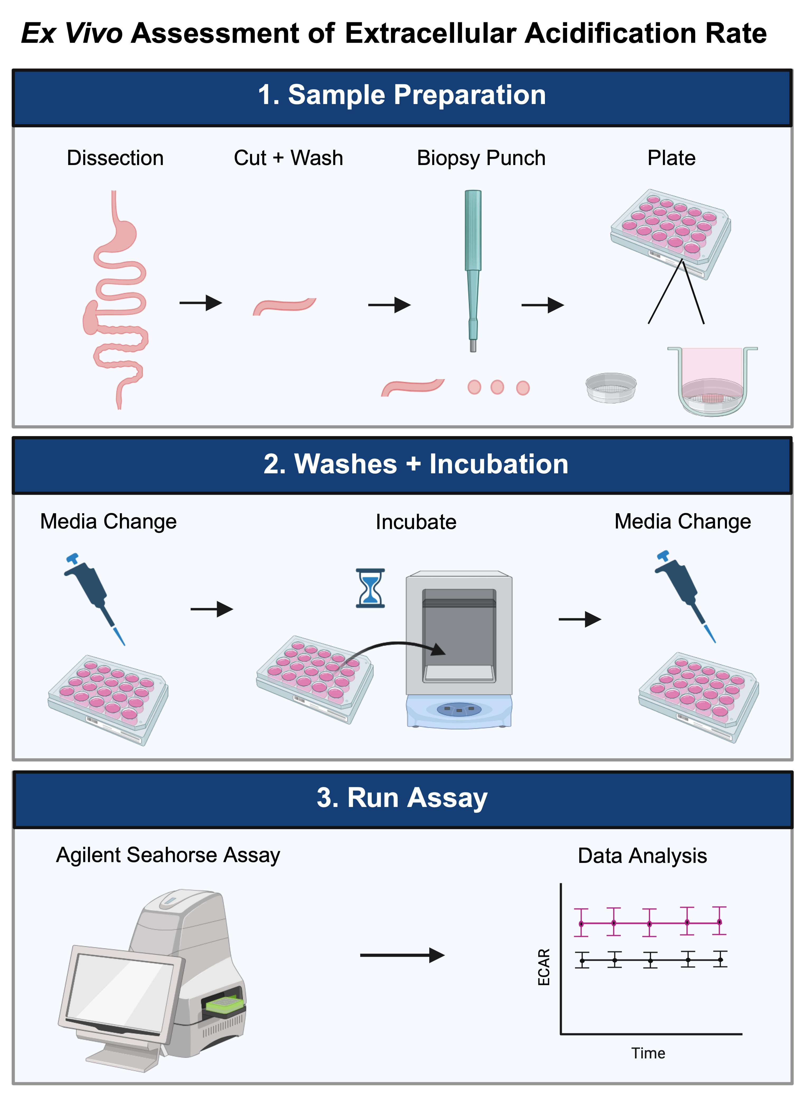
Overview of ex vivo assessment of extracellular acidification rate in murine intestinal tissue
Background
Seahorse metabolic assays have been widely adopted across numerous fields, spanning immunology, cancer, genetics, neuroscience, pharmacology, and others, to provide real-time measurements of cellular metabolism in live cells. Seahorse assays offer a standardized method to interrogate glycolysis and mitochondrial function through assessments of extracellular acidification rate (ECAR) and oxygen consumption rate (OCR), respectively. Extracellular acidification rate is calculated from changes in pH over time that reflect differences in proton efflux to act as a surrogate for glycolysis measurements. Oxygen consumption rate is determined through measurements of dissolved oxygen levels and reflects mitochondrial respiration. pH changes and oxygen levels are measured by sensor probes within the Seahorse Bioanalyzer every few seconds directly above the cell monolayer in standard assays. The vast majority of Seahorse metabolic studies are performed using in vitro samples due to existing standardized approaches for adherent or suspension cell culture samples. However, the ability to assess ECAR and OCR in ex vivo tissue samples broadens the scope of Seahorse assays to better assess organ-level metabolism, as it pertains to systemic energy utilization and physiology in a manner beyond cell culture.
Several groups have performed Seahorse assays in various tissue types, ranging from intestine to brown and white adipose tissue, the retina, and the hippocampus [1–6]. However, a uniform approach does not exist for using ex vivo tissue samples for Seahorse experiments. Various sample preparation methods have been employed, including using 200–300 µm slices and various size biopsy punches to generate uniform sample sizes with and without tissue mincing [1–6]. Current ex vivo methods have focused primarily on assessing basal oxygen consumption rate and adapting the Mito Stress assay for interrogating mitochondrial function for tissue samples [2–6]. The majority of publications utilized the Agilent XF24 well platform and the corresponding Islet Capture screens, as it provides an established existing framework to hold the tissue samples in place [1–4,6].
In particular, this protocol was designed to measure the extracellular acidification rate of ex vivo murine intestinal samples. In short, our work first identified an increased in vivo glucose uptake from the blood supply to the cystic fibrosis (CF) mouse intestine [1]. Transcriptional and protein-level evidence indicated an enrichment of glycolysis gene expression and increased protein expression of glucose transport and glycolytic enzymes in the CF intestine [1]. This protocol was designed as a functional approach to assess glycolysis in ex vivo intestinal samples. In particular, this approach utilizes the Agilent Seahorse XFe24 platform with Islet Capture microplates and corresponding Islet Capture screens to hold tissue pieces in place.
This ex vivo approach expands beyond the current in vitro methods and allows for functional assessments of metabolism in tissue samples. While this method was specifically designed for assessing extracellular acidification rate in ex vivo murine intestinal samples, the same approach can be applied to other tissue types to interrogate other metabolic processes. For instance, the current method can be modified to assess mitochondrial function through measurements of OCR in numerous tissue types. Overall, this protocol broadens the scope of Seahorse metabolic assays to include tissue samples to establish a framework for better assessing organ-level metabolism in the context of systemic nutrient metabolism and physiology.
Materials and reagents
Biological materials
1. Wildtype mice (C57BL/6J background), approximately 8–12 weeks of age
Reagents
1. DMEM low glucose, pyruvate (Gibco, catalog number: 11885-084)
2. HEPES (4-(2-hydroxyethyl)-1-piperazineethanesulfonic acid) (Sigma, catalog number: H4034)
3. D-(+)-glucose (Sigma, catalog number: G8270)
4. Glycolytic rate media (Agilent, catalog number: 103575-100)
5. D-(+)-glucose solution (Sigma, catalog number: G8769)
6. Sodium pyruvate solution (Sigma, catalog number: S8636)
7. L-glutamine solution (Sigma, catalog number: G7513)
8. XF calibrant (Agilent, catalog number: 100840-000)
9. 1× phosphate-buffered saline (PBS) (Gibco, catalog number: 20012-027)
10. 70% ethanol (prepared with Koptec, catalog number: V1101)
Solutions
1. Wash media (see Recipes)
2. Assay media (see Recipes)
Recipes
1. Wash media
| Reagent | Final concentration | Quantity or volume |
|---|---|---|
| DMEM low glucose, pyruvate | 5.56 mM glucose 1.0 mM sodium pyruvate 4.0 mM L-glutamine | 500 mL |
| HEPES | 25 mM | 2.98 g |
| D-(+)-glucose (powder) | 25 mM (total glucose) | 1.75 g |
2. Assay media
| Reagent | Final concentration | Quantity or volume |
|---|---|---|
| Glycolytic rate media (DMEM-based, pH adjusted to 7.4, with 5 mM HEPES, no phenol red) | 45 mL | |
| D-(+)-glucose solution | 17.5 mM | 316 μL |
| Sodium pyruvate solution | 1 mM | 450 μL |
| L-glutamine solution | 4.0 mM | 900 μL |
Laboratory supplies
1. Seahorse XFe24 FluxPak (Agilent, catalog number: 102340-100)
2. Seahorse XF24 Islet Capture microplates (Agilent, catalog number: 101122-100)
3. Seahorse XF Islet Capture screen insert tool (Agilent, catalog number: 101135-100)
4. 2 mm Harris Uni-Core biopsy puncher (Harris, catalog number: 7093508, included in Sigma, WHAWB100029)
5. Well tech cutting mat, small (included in Sigma, WHAWB100029)
6. Flat-headed forceps (Fine Scientific Tools, catalog number: 18025-10)
7. Dissecting scissors (Fine Scientific Tools, catalog number: 14058-09)
8. Curved forceps (Fine Scientific Tools, catalog number: 11272-40)
9. 6 cm Petri dish (Falcon, catalog number: 353002)
10. 12-well plates (Corning, catalog number: 3513)
11. Medi-Vac Guardian Canister (Cardinal Health, catalog number: 65651-395)
12. Glass Pasteur pipettes (Fisher Scientific, catalog number: 13-678-20C)
13. 50 mL conical tubes (Falcon, catalog number: 352098)
14. 1,000 μL tips (Thomas Scientific, catalog number: 1159M42 or P1126)
15. Kimwipes (KimTech, catalog number: 34120)
16. 25 mL serological pipettes (VistaLab, catalog number: 4090-0025)
17. Rectangular ice pan, Maxi 9 L (Corning, catalog number: 07210094)
Equipment
1. Seahorse XFe24 Analyzer and Controller (Agilent, catalog number: S7801A)
2. MyTemp Mini Digital Incubator (non-CO2 incubator) (Benchmark, catalog number: H2200-H)
3. Isotemp Digital Waterbath (Fisher Scientific, catalog number: 2320)
4. Gilson P1000 Pipette (Pipetman, catalog number: HA53667)
5. Pipetaid (Drummond, catalog number: 4-000-100)
6. Vacuum connection
7. Relevant institution approved murine euthanasia equipment/method (i.e., isoflurane, bell jar, gauze pads)
Software and datasets
1. Wave Desktop Software (Agilent, V2.6.4.24)
2. Prism (GraphPad, 10.3.0)
Procedure
登录/注册后免费查看全文
文章信息
稿件历史记录
提交日期: Mar 25, 2026
接收日期: Jun 4, 2026
在线发布日期: Jun 17, 2026
出版日期: Jul 20, 2026
版权信息
© 2026 The Author(s); This is an open access article under the CC BY-NC license (https://creativecommons.org/licenses/by-nc/4.0/).
如何引用
Lesser, A. F. and Drumm, M. L. (2026). Ex Vivo Assessment of Extracellular Acidification Rate in Murine Intestinal Tissue. Bio-protocol 16(14): e5749. DOI: 10.21769/BioProtoc.5749.

 Download Citation in RIS Format
分类
生物化学 > 糖类
细胞生物学 > 细胞新陈代谢 > 糖类
细胞生物学 > 组织分析 > 生理学
您对这篇实验方案有问题吗?
在此处发布您的问题,我们将邀请本文作者来回答。同时,我们会将您的问题发布到Bio-protocol Exchange,以便寻求社区成员的帮助。
 发布问题
发布问题


