


Measuring PINK1 Activity in Single Cells Using a PINK1 Kinase Activity Reporter
利用PINK1激酶活性报告器检测单细胞PINK1活性

发布: 2026年07月20日第16卷第14期 DOI: 10.21769/BioProtoc.5746 浏览次数: 366
评审: Sébastien GillotinAnonymous reviewer(s)
 PDF
PDF  Q&A
Q&A
参见作者原研究论文
The authors used this protocol in:

Feb 2026
Advertisement








Abstract
Phosphatase and tensin homolog-induced kinase 1 (PINK1) is a serine/threonine kinase that plays a key role in mitophagy initiation. Loss-of-function autosomal recessive mutations in PINK1 cause early onset Parkinson’s disease (EOPD). Current approaches for studying PINK1 function depend on bulk techniques that can only provide snapshots of activity and could miss the dynamics and cell-to-cell heterogeneity of PINK1 activity or provide an indirect readout of PINK1 activity. Here, we present a protocol using our newly developed phase separation–based PINK1 biosensor (PINK1-SPARK) to observe real-time activity of endogenous PINK1 in single cells. Following transfection of live cells with PINK1-SPARK, cells are treated with mitochondrial depolarizing agents and visualized using widefield or confocal fluorescence microscopy, either following the same cells over time for time-lapse imaging of PINK1 activity or end-point measurements. Thus, PINK1-SPARK is a new tool that enables the measurement of PINK1 activity in single live cells, allowing for further elucidation of the role of PINK1 in mitophagy and cell function.
Key features
• Detailed protocol for use of PINK1-SPARK, a new PINK1 biosensor introduced in Vineall et al. [1].
• PINK1-SPARK, based on phase separation, has a high signal-to-noise, enabling robust detection of PINK1 activity in multiple cell types under multiple activating conditions.
• Enables measurement of real-time endogenous PINK1 activation at the single-cell level.
Keywords: PINK1 (PINK1) Kinase activity reporter (激酶活性报告器)
Biosensor (生物传感器)
Functional imaging (功能成像)
Mitophagy (线粒体自噬)
Fluorescence microscopy (荧光显微镜)
Kinase activity reporter (激酶活性报告器)
Biosensor (生物传感器)
Functional imaging (功能成像)
Mitophagy (线粒体自噬)
Fluorescence microscopy (荧光显微镜)
Graphical overview
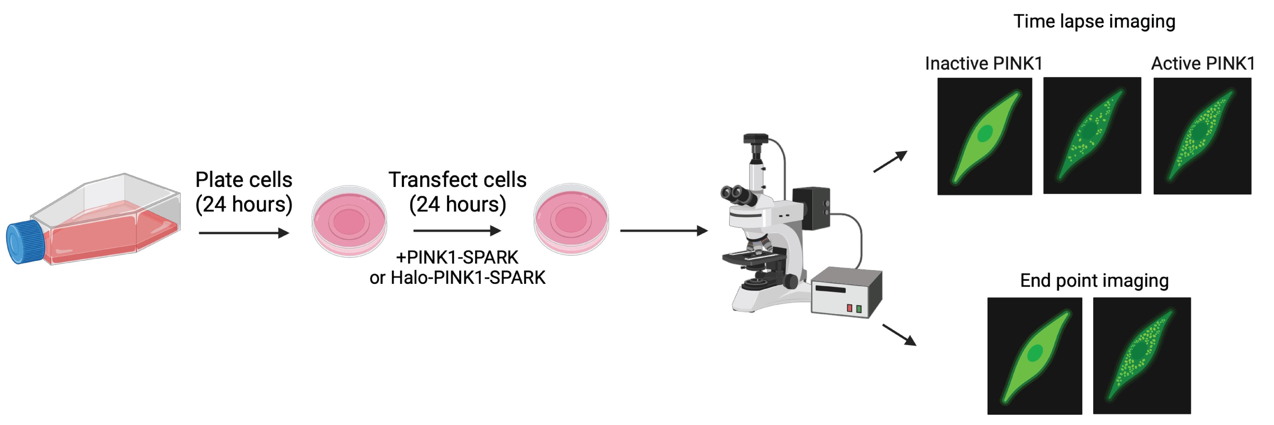
PINK1-SPARK protocol overview. Following plating of cells onto imaging dishes (24 h), cells are transfected with the desired SPARK reporter (24 h) and imaged following treatment with phosphatase and tensin homolog-induced kinase 1 (PINK1) activators or dimethyl sulfoxide (DMSO). Either time-lapse or end-point imaging can be conducted using PINK1-SPARK. PINK1 activity is visualized as the presence of green fluorescent puncta within the cell.
Background
Phosphatase and tensin homolog-induced kinase 1 (PINK1) is a mitochondrial serine/threonine kinase involved in mitophagy, the selective degradation of damaged mitochondria. In healthy mitochondria, PINK1 is constitutively recruited to the outer mitochondrial membrane and imported into the inner membrane space, where it undergoes N-terminal cleavage by proteases and subsequent degradation in the cytosol [2]. Following mitochondrial damage, the inner mitochondrial membrane becomes depolarized, and PINK1 is stabilized in its full-length form. This results in the dimerization, trans-autophosphorylation, and activation of PINK1, allowing it to phosphorylate and activate the E3 ubiquitin ligase Parkin and ubiquitin [3]. Parkin catalyzes the ubiquitination of outer mitochondrial membrane proteins, tagging damaged mitochondria for degradation by autophagic machinery [4,5]. Loss-of-function autosomal recessive mutations in PINK1 are known to cause early onset Parkinson’s disease (EOPD) [6], leading to the onset of symptoms, including tremor, rigidity, and bradykinesia, at a mean age of 31 years [7]. While advances have been made in understanding the role of PINK1 in mitochondrial dynamics and involvement of PINK1 in the development of Parkinson’s disease, current tools available to study PINK1 typically only provide snapshots of PINK1 activity, provide indirect readouts of PINK1 activity, or rely on overexpression of PINK1 [8–10]. We therefore sought to develop a kinase activity reporter capable of characterizing endogenous PINK1 activity dynamics in live cells using both time-lapse microscopy and end-point imaging.
Fluorescent protein-based kinase activity reporters (KARs) offer a unique approach to study kinase activity dynamics in single cells with high spatiotemporal resolution [11]. KARs have been developed for several kinases, including AMP-activated protein kinase (AMPK), protein kinase A (PKA), and protein kinase C (PKC) [12]. One KAR design—separation of phase-based activity reporter of kinase (SPARK)—takes advantage of liquid–liquid phase separation for the detection of kinase activity [13]. In this KAR design, phosphorylation of the SPARK construct increases the local concentration of fluorophores, observed as puncta, resulting in a simple readout of kinase activity that can be multiplexed with other biosensors [14]. SPARK-based biosensors have been made for kinases, including PKA, ataxia-telangiectasia mutated (ATM), and AMPK [13–15]. As the SPARK design enables both time-lapse imaging and snapshots of kinase activity in single cells with high signal-to-noise, we sought to design a SPARK-based KAR for PINK1 (Figure 1).
While several methods exist to measure mitochondrial function, none are capable of directly reporting PINK1 activity in real time. Western blot analysis has so far been the standard for measuring PINK1 activity, but it only provides bulk readouts of activity at specific points in time. Other fluorescence protein–based tools have been created to provide readouts of mitophagy, for example, MT-Keima and Mito-QC [16]. Both rely on pH-dependent changes in fluorescent proteins to visualize mitochondria being taken up by autophagic machinery, but this is an indirect readout of PINK1 activity, as PINK1-independent mitophagy mechanisms have been described [17]. A more recent tool is MitoPain, which uses a mitochondrial PINK1 accumulation index to quantify mitochondrial stress by comparing PINK1-GFP and RFP-Omp25 signals [10]. While MitoPain allows for the quantification of mitochondrial stress, this reporter does not provide a direct readout of PINK1 activity and requires overexpression of PINK1. Our newly developed PINK1-SPARK utilizes the PINK1 phosphomotif from ubiquitin, a canonical PINK1 substrate, to overcome these limitations and measure endogenous PINK1 activity. Furthermore, we developed a HaloTag-based PINK1-SPARK for multiplexing of PINK1 activity with other markers of mitochondrial damage. We validated the use of PINK1-SPARK in a variety of cell lines, including PINK1 knockout HeLa cervical cancer cells, and found minimal response of PINK1-SPARK in PINK1 knockout HeLa cells. While PINK1-SPARK has a large dynamic range, its spatial resolution falls short of that of other KARS, notably Förster resonance energy transfer (FRET)-based KARS, which can measure kinase activity at distinct subcellular locations [11]. Additionally, we did not test the reversibility of PINK1-SPARK, as PINK1 inhibitors are nonspecific [18]; thus, PINK1-SPARK functions best as a “turn on” reporter of PINK1 activity. Nevertheless, PINK1-SPARK represents a significant advancement in the detection and quantification of endogenous PINK1 activity. In this protocol, we provide an overview of how to use PINK1-SPARK to study endogenous PINK1 activity at the single-cell level.
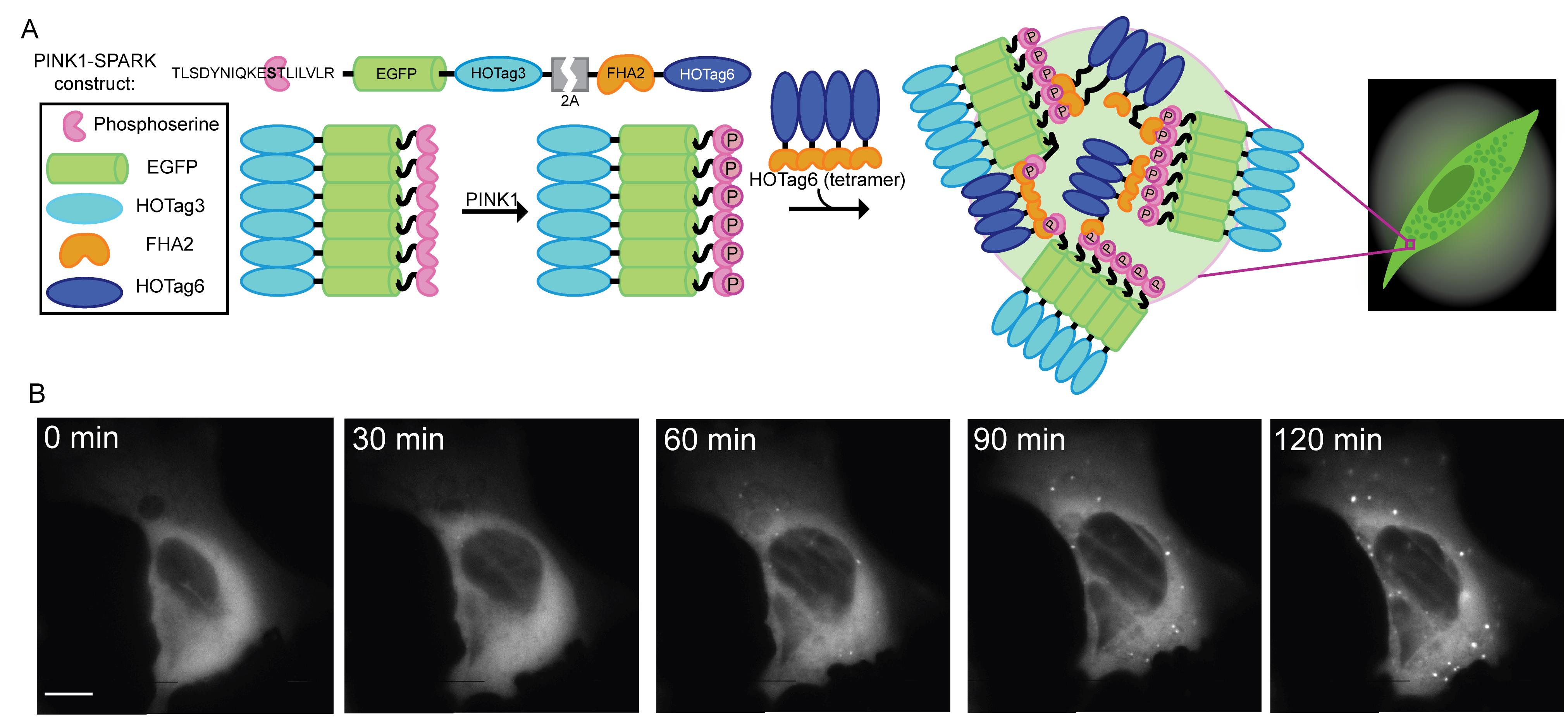
Figure 1. PINK1-SPARK protocol overview. (A) General scheme of reporter function. The first component consists of the tensin homolog-induced kinase 1 (PINK1)-specific ubiquitin consensus sequence, EGFP, and the hexameric homooligomeric tag, HOTag3. The phosphoamino acid binding domain, FHA2, and the tetrameric HOTag6 make up the second component. PINK1 phosphorylates Ser65 within its consensus sequence, allowing recognition and binding by FHA2. The resulting proximity of multivalent interactions promotes phase separation and allows for the visualization of PINK1 activity as green fluorescent droplets. (B) Time-lapse images of U2OS cells transiently expressing PINK1-SPARK following the addition of 10 μM CCCP. Adapted from Vineall et al. [1] under a CC BY-NC-ND 4.0 license.
Materials and reagents
Biological materials
1. HeLa cervical cancer cells (ATCC, CRM-CCL-2)
Note: While we describe the use of PINK1-SPARK in HeLa cells in this protocol, this method can be applied to any cell line that can be easily transfected; we have previously used this protocol in U2OS (Figure 1) and SHSY5Y cells.
2. PINK1-SPARK plasmid (Addgene, catalog number: 248086)
3. PINK1-SPARK S/A plasmid (Addgene, catalog number: 248087)
4. Halo-PINK1-SPARK plasmid (Addgene, catalog number: 248088)
5. Halo-PINK1-SPARK S/A plasmid (Addgene, catalog number: 248089)
Reagents
1. Dulbecco’s modified Eagle medium (DMEM) (Thermo Scientific, catalog number: 10569010)
2. Hank’s buffered saline solution (HBSS) (Thermo Scientific, catalog number: 14185052)
3. Opti-MEM (Thermo Scientific, catalog number: 31985070)
4. FuGene 4K (Promega, catalog number: E5911)
5. Fetal bovine serum (FBS) (Thermo Scientific, catalog number: A4736101)
6. Penicillin/streptomycin (pen/strep) (Thermo Scientific, catalog number: 15140122)
7. Ultrapure water (Fisher Scientific, catalog number: 10-977-023)
8. HEPES (Fisher Scientific, catalog number: SH3023701)
9. D-glucose (VWR, catalog number: BT132735-500G)
10. Trypsin (Thermo Scientific, catalog number: 12604013)
11. CCCP (Fisher Scientific, catalog number: 04-525-00)
12. Kinetin riboside (MedChem Express, catalog number: HY-101055)
13. MTK458 (MedChem Express, catalog number: HY-152943)
14. DMSO
15. Janelia Fluor HaloTag ligand, 646 (JF646) (Promega, catalog number: HT1060)
16. TrypLE Express Enzyme (Thermo Scientific, catalog number: 12604039)
17. Bleach
18. Ethanol
Solutions
1. HeLa cell growth media (see Recipes)
2. 1× HBSS imaging buffer (see Recipes)
Recipes
1. HeLa cell growth media
| Reagent | Final concentration | Quantity or volume |
|---|---|---|
| DMEM | 1× | 500 mL |
| FBS | 10% | 50 mL |
| Pen/strep | 99 units/mL | 5.5 mL |
| Total | n/a | 555.5 mL |
2. 1× HBSS imaging buffer
| Reagent | Final concentration | Quantity or volume |
|---|---|---|
| 10× HBSS | 1× | 100 mL |
| Ultrapure water | n/a | 900 mL |
| HEPES | 20 mM | 20 mL |
| D-glucose | 10 mM | 1 g |
| Total | n/a | 1 L |
Note: HBSS is frequently used for live cell imaging [19–21]. While not tested here, other imaging media, like Fluorobrite, could be used.
Laboratory supplies
1. 35 mm glass bottom dishes (Cellvis, catalog number: d35-14-1.5-n)
2. 25 cm cell culture flasks (Corning, catalog number: 353138)
3. 5, 10, and 25 mL serological pipettes (Corning, catalog numbers: 357543, 357551, 357525)
4. 15 and 50 mL conical tubes (Fisher Scientific, catalog numbers: 14-959-49B, 14-432-22)
5. Countess cell counting slides (Thermo Scientific, catalog number: C10228) or hemocytometer (Sigma-Aldrich, catalog number: MDH-2N1)
6. Micropipettes (Eppendorf, catalog numbers: 3123000020, 3123000047, 3123000063)
7. Micropipette tips (USA Scientific, catalog numbers: 1111-3730, 1110-9880, 1112-1860)
8. Aspiration pipettes (Fisher Scientific, catalog number: 14-955-135)
9. Nikon immersion oil (Nikon, catalog number: MXA22168)
10. Leica immersion oil (Thorlabs, catalog number: MOIL-10LF)
11. Sterile Petri dishes (Fisher Scientific, catalog number: 08-757-100D)
12. Microcentrifuge tubes (Fisher Scientific, catalog number: 05-408-129)
13. Loctite mounting putty (Amazon, catalog number: 079340685444)
Equipment
1. Light microscope for tissue culture (Fisher Scientific, model: LMI3PH2)
2. Countess FL cell counter (Thermo Scientific, model: AMQAF2000)
3. HeraCell CO2 incubator at 37 °C and 5% CO2 (Thermo Scientific, catalog number: 13998254)
4. 1300 Series Class II, Type A2 Biological Safety Cabinet (Fisher Scientific, catalog number: 13-261-222)
5. Bead bath (Genesee Scientific, catalog number: 31-149) with thermal glass beads (USA Scientific, catalog number: 9123-220)
6. Nikon ECLIPSE Ti2 epifluorescence microscope with a CHI60 Plan Fluor 40× oil immersion objective lens and stage-top heater (Nikon)
7. Leica Stellaris 5 with an HC PL APO 63×/1.40 oil immersion CS2 lens and stage-top heater (Leica)
Note: Any microscope with a live-imaging setup can be used.
Software and datasets
1. Fiji Is Just Image J (FIJI), free to use (Version 2.16.0)
2. Prism v10; requires a license (GraphPad)
Procedure
登录/注册后免费查看全文
文章信息
稿件历史记录
提交日期: Mar 16, 2026
接收日期: May 25, 2026
在线发布日期: Jun 14, 2026
出版日期: Jul 20, 2026
版权信息
© 2026 The Author(s); This is an open access article under the CC BY-NC license (https://creativecommons.org/licenses/by-nc/4.0/).
如何引用
Vineall, K. G. and Schmitt, D. L. (2026). Measuring PINK1 Activity in Single Cells Using a PINK1 Kinase Activity Reporter. Bio-protocol 16(14): e5746. DOI: 10.21769/BioProtoc.5746.

 Download Citation in RIS Format
分类
神经科学 > 细胞机理 > 线粒体
细胞生物学 > 细胞成像 > 活细胞成像
生物化学 > 蛋白质 > 活性
您对这篇实验方案有问题吗?
在此处发布您的问题,我们将邀请本文作者来回答。同时,我们会将您的问题发布到Bio-protocol Exchange,以便寻求社区成员的帮助。
 发布问题
发布问题


