


Iodine Staining of Glycogen Storage in Caenorhabditis elegans
秀丽隐杆线虫糖原储存的碘染色检测方法

发布: 2026年07月20日第16卷第14期 DOI: 10.21769/BioProtoc.5745 浏览次数: 458
评审: Annmary Paul ErinjeriSravanthi S P NadimintiAnonymous reviewer(s)
 PDF
PDF  Q&A
Q&A
参见作者原研究论文
The authors used this protocol in:

Jan 2026
Advertisement







Abstract
Glycogen is a highly conserved macromolecule across species, and its visualization provides critical insights into both physiological processes and disease states. Existing approaches for glycogen imaging in Caenorhabditis elegans rely primarily on traditional microscopy slides, which introduce variability in image acquisition and downstream data analysis, limit throughput, and require substantial hands-on time and technical expertise.
Here, we present a standardized, cost-effective, and high-throughput imaging method that enables efficient visualization and quantification of glycogen in C. elegans. Our approach utilizes a custom-designed three-dimensional pad containing two to four chambers, allowing control and experimental samples to be processed simultaneously under identical conditions. Worms are exposed to iodine crystals, ensuring uniform staining while minimizing reagent use and handling variability. Imaging is performed using a simple binocular microscope, and analysis is conducted in Fiji, making the workflow accessible to laboratories with minimal specialized equipment or training.
This method also reduces technical variability, shortens turnaround time, and requires only basic reagents and expertise, making it well-suited for both research and teaching laboratories. Importantly, the platform is readily adaptable to other nematode species and scalable for large-scale genetic or pharmacological screening applications. Together, this workflow minimizes technical variability and provides a robust platform for comparative glycogen analysis in C. elegans.
Key features
• Standardized glycogen staining method allows less data variability.
• Provides a quantitative tool to measure glycogen buildup in C. elegans.
• Requires a customized pad for imaging.
• Macro is available for batch processing.
Keywords: Iodine staining (碘染色) Glycogen accumulation (糖原积累)
Caenorhabditis elegans (秀丽隐杆线虫)
Microscopy (显微观察)
Quantification (定量分析)
Metabolic storage (代谢物储存)
Carbohydrate metabolism (碳水化合物代谢)
Glycogen accumulation (糖原积累)
Caenorhabditis elegans (秀丽隐杆线虫)
Microscopy (显微观察)
Quantification (定量分析)
Metabolic storage (代谢物储存)
Carbohydrate metabolism (碳水化合物代谢)
Graphical overview
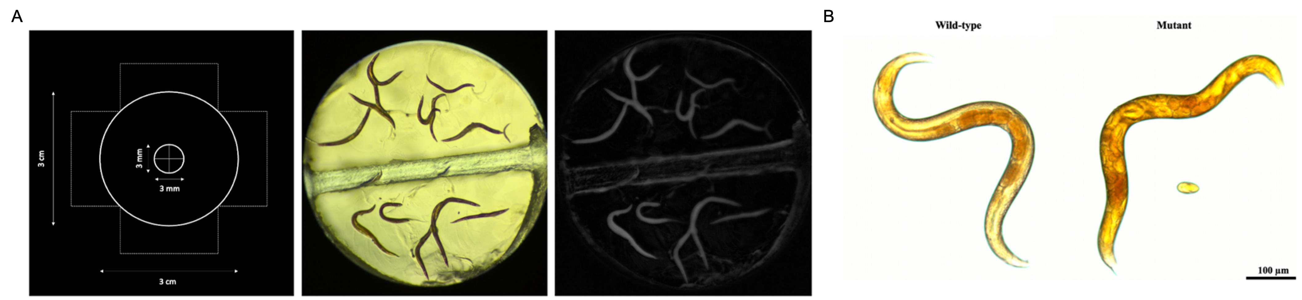
Overview of the imaging protocol for iodine staining. (A) From left to right: schematic of 3D-printed pad used for staining, an example of an acquired image after staining, and image processing using a macro in Fiji software for quantification. Control and test worms are simultaneously exposed to iodine. (B) Representative image of glycogen buildup visualized by iodine staining in a COP2008 mutant compared to a wild-type C. elegans.
Background
Iodine-based staining, most performed using Lugol’s solution, has historically been the primary method for visualizing glycogen in Caenorhabditis elegans, appearing in light-brown to dark-brown color [1]. However, its quantitative reliability is limited because staining intensity is highly sensitive to iodine concentration and rapidly becomes unstable after application. Early work using Lugol’s solution demonstrated that glycogen localizes to distinct anatomical regions in adult worms, including the area anterior to the posterior pharyngeal bulb, the dorso-rectal ganglion region of the tail, and the most proximal oocytes of the gonad arm [2]. Yet subsequent biochemical studies revealed key limitations: mixtures of polysaccharides produce additive colorimetric signals, and the characteristic red–brown iodine–glycogen complex varies in intensity with glycogen concentration, iodine concentration, temperature, and even the biological source of glycogen. These factors collectively introduce substantial variability into Lugol staining [3,4].
To improve reproducibility, we used unfixed worms, which can be rapidly and effectively stained using iodine vapor. We designed a 3D printed pad to improve reproducibility. The vapor method allows multiple populations to be stained simultaneously under identical exposure conditions. Beyond these technical considerations, glycogen itself plays an important physiological role: worms exposed to a high-glucose diet accumulate roughly twice as much glycogen as controls, and this elevation is associated with a reduction in lifespan, potentially mediated by glucose toxicity, increased reactive oxygen species, or glycogen-dependent signaling pathways [5]. Together, these findings emphasize both the central role of glycogen in C. elegans biology and the need for standardized approaches that overcome the inherent variability of traditional staining [3,4].
Materials and reagents
Biological materials
1. C. elegans N2 strain [Caenorhabditis Genetics Center (CGC)]
2. C. elegans COP2008; agl-1(knu864) strain (CGC)
Note: These are the strains presented in this protocol. They can be changed based on the strains of interest.
Reagents
1. Iodine (Sigma-Aldrich, catalog number: 207772-100G)
2. Agarose (Life Technologies, catalog number: 16500-500)
Laboratory supplies
1. Erlenmeyer flask (ThermoFisher Scientific, catalog number: 4103-0125)
2. P1000 pipette (Mandel Scientific, catalog number: GF-F167360)
3. P1000 pipette tips (Mettler & Toledo, catalog number: RC-L1000/10)
Equipment
1. 3D printer (FormLabs, model: Form 3B)
2. Resin (FormLabs, model: Amber Biomed)
3. Leica S6E microscope
4. iPhone 6 (Apple)
5. LabCam Microscope Adapter for iPhone 6/6S Plus (Idu Optics, Directnine)
6. Microscope slides (Cedarlane, catalog number: A547100-5, FisherbrandTM Premium Clipped-Corner Microscope Slides)
7. Coverslips (Mandel Scientific, NEU-GG-15-PRE, #15mm)
8. Micro-dissecting forceps
9. Scissors
10. Worm pick
Software and datasets
1. Fiji software (ImageJ2, version: 2.16.0/1.54p, Build: 26d66057dd, Date: 2014-10-15T19:41:44+0000)
2. Prism 9 software (GraphPad, version: 9.0.2)
Procedure
登录/注册后免费查看全文
文章信息
稿件历史记录
提交日期: Mar 12, 2026
接收日期: May 29, 2026
在线发布日期: Jun 12, 2026
出版日期: Jul 20, 2026
版权信息
© 2026 The Author(s); This is an open access article under the CC BY license (https://creativecommons.org/licenses/by/4.0/).
如何引用
Daghar, H., Samarut, É. and Parker, A. J. (2026). Iodine Staining of Glycogen Storage in Caenorhabditis elegans. Bio-protocol 16(14): e5745. DOI: 10.21769/BioProtoc.5745.

 Download Citation in RIS Format
分类
生物化学 > 糖类 > 糖原
细胞生物学 > 细胞染色 > 糖类
细胞生物学 > 细胞新陈代谢 > 糖类
您对这篇实验方案有问题吗?
在此处发布您的问题,我们将邀请本文作者来回答。同时,我们会将您的问题发布到Bio-protocol Exchange,以便寻求社区成员的帮助。
 发布问题
发布问题


