


Preparation and Characterization of Neutrophil Membrane-Fused Mitochondria (nMITO)
中性粒细胞膜融合线粒体(nMITO)的制备与表征

发布: 2026年07月20日第16卷第14期 DOI: 10.21769/BioProtoc.5744 浏览次数: 258
评审: Elena A. OstrakhovitchAnonymous reviewer(s)
 PDF
PDF  Q&A
Q&A
参见作者原研究论文
The authors used this protocol in:

Mar 2025
Advertisement








Abstract
Mitochondrial transplantation is an emerging strategy for cellular repair, yet its efficiency is often limited by poor targeting and environmental instability. This protocol details the fabrication and comprehensive characterization of neutrophil membrane-fused mitochondria (nMITO), a hybrid organelle platform designed to combine the metabolic vigor of natural mitochondria with the targeting and anti-inflammatory properties of neutrophil membranes. We describe an optimized workflow for mouse heart mitochondrial isolation, lipopolysaccharide (LPS)-activated neutrophil membrane (NEM) extraction, and the subsequent sonication-mediated fusion process. Characterization techniques include dynamic light scattering (DLS) for size and zeta potential, transmission electron microscopy (TEM) for ultrastructural integrity, and bioenergetic assays [ATP synthesis and tetramethylrhodamine methyl ester (TMRM)-based membrane potential] to ensure functional preservation.
Key features
• The protocol provides a methodology for the isolation of neutrophil membranes from mouse bone marrow and mitochondria from the heart.
• The protocol provides a methodology for the fabrication of neutrophil membrane-fused mitochondria (nMITO).
Keywords: nMITO (nMITO) Mitochondria isolation (线粒体分离)
Neutrophil membranes (中性粒细胞膜)
Membrane coating (膜包覆)
Mitochondrial assessment (线粒体评估)
Mitochondria isolation (线粒体分离)
Neutrophil membranes (中性粒细胞膜)
Membrane coating (膜包覆)
Mitochondrial assessment (线粒体评估)
Graphical overview
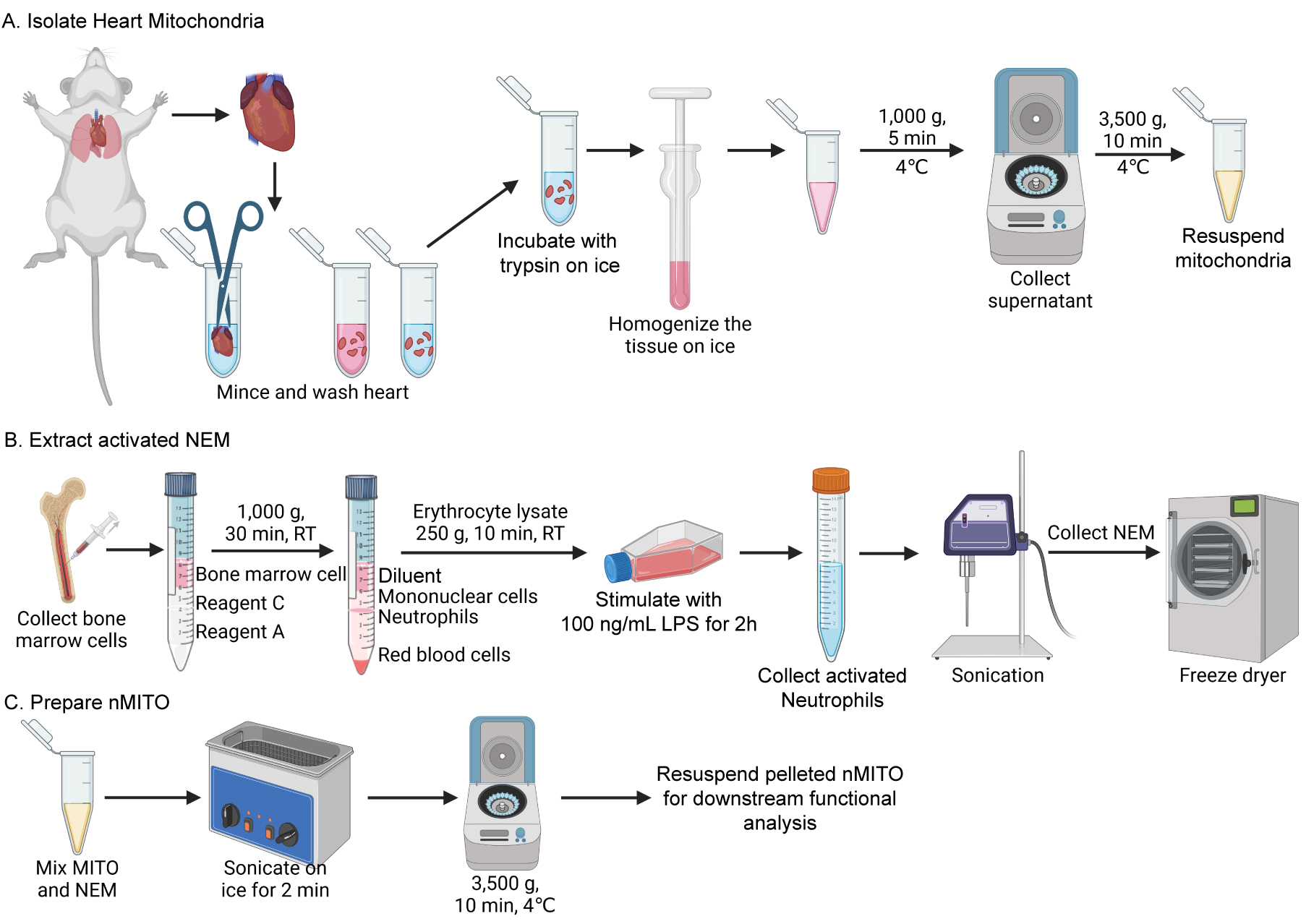
Schematic of the preparation of neutrophil membrane-fused mitochondria (nMITO). Isolation of heart mitochondria and extraction of activated neutrophil membranes from the bone marrow, followed by ice-bath sonication-mediated fusion. Created in BioRender.
Background
Mitochondrial transplantation has emerged as a promising method to restore cellular homeostasis [1,2]; however, “naked” mitochondria lack the ability to target specific inflammatory sites [3,4]. Neutrophils are naturally home to sites of injury via β-integrin/ICAM-1 interactions and carry receptors capable of neutralizing inflammatory cytokines [5,6]. By fusing neutrophil membranes onto isolated mitochondria, neutrophil membrane-fused mitochondria (nMITO) integrates the internal bioenergetic machinery of the mitochondria with the surface-level biological functionality of the neutrophil. In this hybrid system, the mitochondria provide ATP, while the neutrophil membrane facilitates targeted “homing” and “protective” functions. To maximize these capabilities, lipopolysaccharides (LPSs) are utilized in this protocol to activate neutrophils prior to membrane extraction. This activation significantly upregulates the expression of chemokine receptors, inflammatory cytokine receptors, and β-integrins. Consequently, the resulting neutrophil membrane (NEM) can effectively adsorb and neutralize pro-inflammatory cytokines while utilizing the enhanced β-integrin/ICAM-1 interaction for precise site-specific homing.
The preparation of nMITO consists of three primary stages: (i) isolation of mitochondria from mouse heart tissue via differential centrifugation; (ii) extraction of activated neutrophil membranes (NEM) from LPS-stimulated bone marrow neutrophils; (iii) fabrication of nMITO through ice-bath sonication-mediated fusion of the two components. Following the preparation, basic characterization is performed to evaluate the physicochemical properties, bioenergetic functions, and protein marker inheritance of the resulting nMITO.
Materials and reagents
Biological materials
1. Male C57BL/6J mice (6–8 weeks old) for the isolation of heart mitochondria and bone marrow neutrophils
Reagents
1. LPS (Sigma, catalog number: L2630)
2. Tissue Mitochondrial Isolation kit (Beyotime, catalog number: C3606)
3. Mouse Bone Marrow Neutrophil Isolation kit (Solarbio, catalog number: P8550)
4. ATP Assay kit (Beyotime, catalog number: S0027)
5. TMRM (MCE, catalog number: HY-D0984A)
6. CCCP (MCE, catalog number: HY-100941)
7. PBS (Beyotime, catalog number: C0221A)
8. Red blood cell lysis buffer (Beyotime, catalog number: C3702)
9. EDTA (Beyotime, catalog number: ST1305)
10. Mannitol (CHRON CHEMICALS, CAS number: 69-65-8)
11. Sucrose (MCE, catalog number: HY-B1779)
12. 1 M Tris-HCl, PH 7.4 (Beyotime, catalog number: ST786)
13. Protease inhibitor (Beyotime, catalog number: P1005)
14. RPMI 1640 medium (Gibco, catalog number: 11875093)
15. Bradford Protein Assay kit (Beyotime, catalog number: P0006)
16. COX IV monoclonal antibody (Proteintech, catalog number: 66110-1-Ig)
17. Beta actin monoclonal antibody (Proteintech, catalog number: 66009-1-Ig)
18. IL-6R polyclonal antibody (Thermo Fisher Scientific, catalog number: PA5-102425)
19. CXCR2 antibody (Affinity, catalog number: DF7095)
Solutions
1. Neutrophil membrane (NEM) isolation buffer (see Recipes)
Recipes
1. NEM isolation buffer
| Reagent | Final concentration | Quantity or volume |
|---|---|---|
| Mannitol | 225 mM | 2.0494 g |
| Sucrose | 75 mM | 1.2835 g |
| Tris-HCl | 30 mM | 1.5 mL |
| EDTA | 0.5 mM | 7.306 mg |
| Protease inhibitor | 1% (v/v) | 0.5 mL |
Prepare 50 mL of neutrophil membrane isolation buffer according to the recipe. Adjust the final volume to 50 mL with ddH2O and store at 4 °C.
Laboratory supplies
1. Centrifuge tubes (1.5 mL and 15 mL) (Servicebio, catalog numbers: EP-150X-J, EP-1501-J)
2. Culture flask (LABSELECT, catalog number: 13112A)
3. Cell scraper (Beyotime, catalog number: FSCP029)
4. 10 cm scissors and tweezers (Beyotime, catalog numbers: FS001, FS225)
5. 26 G disposable sterile syringe (Beyotime, catalog number: FS801)
Equipment
1. Dounce homogenizer and pestles (glass, 2 mL) (Beyotime, catalog number: FGH002)
2. Centrifuge (angle and swing-out rotor) (CENCE, model: HT165R; HERMLE, model: Z32 HK)
3. Bath sonicator (SCIENTZ, model: SB25-12D)
4. Probe sonicator (LICHEN, model: LC-UP-400)
5. Cell culture incubator (PHCBI, model: MCO-170AICUVDL-PC)
6. Dynamic light scattering analyzer (Brookhaven, model: NanoBrook 90PlusPALS)
7. Flow cytometer (Agilent, model: NovoCyte Advanteon)
8. Vertical electrophoresis system (Bio-Rad, model: PowerPac Basic and mini PROTEAN® Tetra cell)
9. Automatic Cell Analyzer (Countstar, model: Mira BF-S)
10. Freeze dryer (SCIENTZ, model: 10YG/B)
11. Multimode microplate reader (Agilent, model: Synergy H1)
12. Chemiluminescence imaging system (GE, model: Amersham Imager600)
Software and datasets
1. NovoExpress (Agilent Technologies); used for flow cytometry data acquisition and analysis
2. Gen5 Microplate Reader and Imager Software (Agilent Technologies); used for ATP luciferase assay data acquisition and analysis
3. GraphPad Prism 10 (GraphPad Software); used for statistical analysis and data visualization
4. Particle Solutions (Brookhaven Instruments); used for dynamic light scattering (DLS) data acquisition and particle size/zeta potential analysis
5. ImageQuant TL (GE Healthcare); used for chemiluminescence image acquisition of western blots
6. Countstar Software (Countstar); used for automatic cell counting
Procedure
登录/注册后免费查看全文
文章信息
稿件历史记录
提交日期: Mar 30, 2026
接收日期: May 25, 2026
在线发布日期: Jun 14, 2026
出版日期: Jul 20, 2026
版权信息
© 2026 The Author(s); This is an open access article under the CC BY license (https://creativecommons.org/licenses/by/4.0/).
如何引用
Readers should cite both the Bio-protocol article and the original research article where this protocol was used:
- Zhang, Q., Yue, Y. and Zhou, X. (2026). Preparation and Characterization of Neutrophil Membrane-Fused Mitochondria (nMITO). Bio-protocol 16(14): e5744. DOI: 10.21769/BioProtoc.5744.
- Zhang, Q., Shen, Y., Zhang, C., Zhang, H., Li, X., Yang, S., Dai, C., Yu, X., Lou, J., Feng, J., et al. (2025). Immunoengineered mitochondria for efficient therapy of acute organ injuries via modulation of inflammation and cell repair. Sci Adv. 11(12): eadj1896. https://doi.org/10.1126/sciadv.adj1896

 Download Citation in RIS Format
分类
生物工程 > 生物医学工程
免疫学 > 免疫细胞分离 > 嗜中性粒细胞
细胞生物学 > 细胞器分离 > 线粒体
您对这篇实验方案有问题吗?
在此处发布您的问题,我们将邀请本文作者来回答。同时,我们会将您的问题发布到Bio-protocol Exchange,以便寻求社区成员的帮助。
 发布问题
发布问题


