


A Simplified Langendorff-Based Method for Mouse Cardiac Myocyte Isolation
基于Langendorff灌流的小鼠心肌细胞简化分离方法

发布: 2026年07月20日第16卷第14期 DOI: 10.21769/BioProtoc.5743 浏览次数: 365
评审: Olga KopachRishith RavindranKarthik Amudhala Hemanthakumar
 PDF
PDF  Q&A
Q&A
参见作者原研究论文
The authors used this protocol in:

Sep 2025
Advertisement







Abstract
Isolation of adult mouse ventricular myocytes is essential for studying cardiac physiology and cellular function. Traditional methods commonly rely on Langendorff perfusion systems, which provide continuous retrograde coronary perfusion but require specialized equipment and can be complex to operate. Here, we describe a simplified Langendorff-based protocol that uses a syringe pump–driven system to achieve constant-flow retrograde aortic perfusion during enzymatic digestion. The setup incorporates an inline heater for precise temperature control and uses widely available laboratory components, enabling consistent delivery of digestion enzymes. This approach maintains stable perfusion despite changes in coronary resistance and reduces variability associated with conventional gravity-driven systems. The protocol yields high-quality adult ventricular myocytes suitable for downstream functional analyses, including electrophysiology, contractility, and calcium imaging. Compared with traditional systems, this method is more accessible, reduces technical complexity, and improves reproducibility, facilitating adoption in laboratories without dedicated isolated-heart perfusion infrastructure.
Key features
• Accessible cardiomyocyte isolation without dedicated Langendorff apparatus, suitable for laboratories with limited perfusion infrastructure.
• Constant-flow perfusion overcomes enzyme delivery variability caused by changing coronary resistance during tissue digestion.
• Inline heating enables rapid, precise temperature control without water-jacket systems, reducing setup complexity and contamination risk.
• Optimized for producing calcium-tolerant adult ventricular myocytes for electrophysiology, contractility, and calcium imaging studies. Alternative methods may be more appropriate for non-cardiomyocyte populations.
Keywords: Langendorff perfusion (Langendorff灌流) Cardiomyocyte isolation (心肌细胞分离)
Mouse ventricular myocytes (小鼠心室肌细胞)
Retrograde aortic perfusion (主动脉逆行灌流)
Constant-flow perfusion (恒流灌流)
Cardiomyocyte isolation (心肌细胞分离)
Mouse ventricular myocytes (小鼠心室肌细胞)
Retrograde aortic perfusion (主动脉逆行灌流)
Constant-flow perfusion (恒流灌流)
Graphical overview
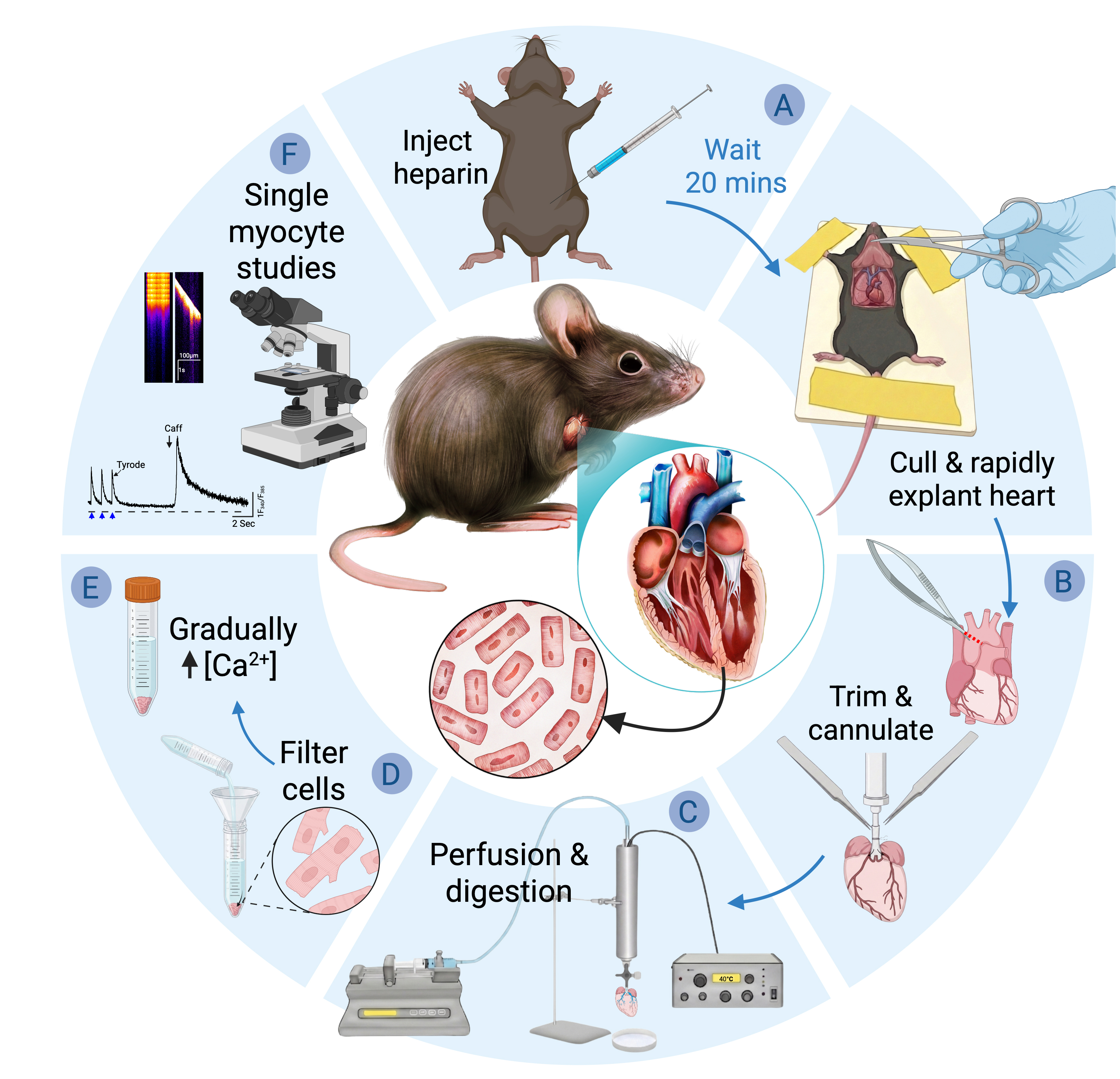
Simplified Langendorff-based mouse cardiac myocyte isolation protocol. Schematic overview of the workflow for isolating adult mouse ventricular cardiomyocytes. (A) Animal preparation and heart excision. (B) Cannulation and setup. (C) Perfusion and enzymatic digestion. (D) Tissue dissociation and cell isolation. (E) Gradual Ca2+ reintroduction. (F) Isolated cardiomyocytes can be used for downstream single-cell functional analyses, including calcium imaging and electrophysiological measurements.
Background
The isolation of intact adult cardiac myocytes is a foundational technique in cardiac research, enabling direct investigation of cellular structure and function under controlled experimental conditions. The isolated heart preparation described by Oscar Langendorff in 1897 [1], and methodologically refined by others [2,3], forms the basis of the modern “Langendorff” method [4]. In this preparation, the excised heart is rapidly cannulated via the ascending aorta and perfused ex vivo while suspended in a temperature-controlled chamber. Retrograde perfusion through the aorta allows controlled delivery of physiological solutions through the intact coronary vasculature while preserving myocardial architecture [1]. Its further adaptation in the 1970s for enzymatic dissociation established the Langendorff method as the standard approach for isolating viable cardiac myocytes [5,6], a status it has maintained for more than five decades [7–9]. Langendorff perfusion enables enzymatic delivery through the coronary circulation while minimizing mechanical trauma to the myocardium by retrograde aortic perfusion. In this configuration, flow delivered through the cannulated proximal aorta fills the aortic root, generating pressure that causes coaptation of the aortic cusps and closure of the valve. Aortic valve closure prevents retrograde ventricular filling and directs perfusate into the coronary circulation via the coronary ostia at the base of the root [4]. This technique is particularly well-suited to rodents and other small-animal models, in which the aorta is sufficiently large for reliable cannulation yet small enough to allow economical use of dissociation enzymes [10]. For larger hearts, perfusing a coronary branch and excising the region is more practical than perfusing the whole heart [10].
Langendorff systems operate under either constant-pressure or constant-flow perfusion regimes [8]. Constant-pressure setups are typically gravity-fed from a reservoir of perfusate and exhibit variable coronary flow as vascular resistance changes during digestion. As enzymatic dissociation reduces resistance, coronary flow increases, providing a useful indicator of digestion-progression that is commonly monitored by changes in drip rate [10,12–15]. Because coronary vascular resistance dynamically influences the pressure–flow relationship [16], variations in flow under constant-pressure perfusion may result in variable delivery of enzyme, particularly as resistance drops during digestion [10,15]. Variability in extracellular matrix composition and fibrosis degree across strain, age, and pathological conditions ostensibly will also influence myocardial resistance and thereby perfusion flow dynamics [15].
In contrast, constant-flow systems employ a fixed volumetric perfusion rate while perfusion pressure varies in response to downstream resistance. Careful selection of the flow rate is important: if set too low, inadequate coronary perfusion may occur; if excessive, supraphysiological perfusion pressures can invert the aortic valve cusps and divert perfusate into the ventricular cavity rather than the coronary circulation. Many traditional constant-flow Langendorff setups rely on peristaltic pumps as well as dedicated perfusion rigs incorporating water-jacketed glassware, bubble traps, recirculation loops, and continuous carbogen or oxygen gassing of bicarbonate-buffered solutions [5,6,9,12]. The complexity of specialized apparatus, combined with the technical expertise required for rapid aortic cannulation, has been criticized as limiting accessibility. In response, simplified “Langendorff-free” methods have been developed [17–20].
Langendorff-free methods, sometimes referred to as “injection methods,” are based on direct injection of perfusate into the ventricular lumen [18–21]. In these approaches, the aorta is clamped, and buffer is forced into the coronary circulation in an antegrade manner, flowing through the aortic valve into the coronary ostia. Although injection-based methods are touted as requiring less technical skill than aortic cannulation, they nonetheless depend on accurate clamp placement and precise ventricular injections into the right ventricular lumen, followed by multiple injections into the left ventricular lumen. These procedures demand operator precision and introduce distinct risks. Repeated ventricular punctures, particularly in the small and thin-walled mouse heart, introduce the risk of rupture or buffer leakage. Preservation of aortic integrity is also critical, as damage can compromise effective coronary perfusion. These risks may be exacerbated in structurally fragile or diseased hearts or those exhibiting pathological remodeling.
Moreover, injection methods rely on non-physiological alkaline buffers (pH ~7.8) for optimal cell yield [17–19], the basis for which is speculative rather than mechanistically established [18]. Cardiomyocyte function is sensitive to pH, and extracellular alkalinization directly influences intracellular pH via established acid–base transport mechanisms, including Na+/H+ exchange and lactate/H+ transport [22,23]. Because intracellular pH must be tightly maintained within a narrow physiological range (~7.15–7.25) to preserve excitation–contraction coupling and calcium homeostasis, even modest deviations can alter Ca2+ handling and myofilament sensitivity [24–27]. Furthermore, extracellular pH fluctuations regulate autophagy, a process central to cellular viability and metabolic control [28]. Exposure to alkaline conditions during isolation may influence post-isolation cellular physiology and subsequent functional readouts. For studies requiring transcriptomic or functional characterization of the full spectrum of cardiac cell types, including fibroblasts, endothelial cells, and immune cells, non-perfusion enzymatic digestion approaches have been developed that enable isolation of viable cells across all major cardiac populations, as demonstrated by single-cell sequencing studies of both healthy and diseased myocardium [29,30].
Taken together, these considerations highlight the need for a method that preserves the physiological fidelity and controlled delivery afforded by constant-flow coronary perfusion while eliminating the specialized equipment and technical expertise required by traditional Langendorff systems. The protocol described here addresses this need through a simplified, syringe pump–driven Langendorff approach. It retains the critical advantages of aortic cannulation and continuous retrograde perfusion while implementing them within a minimal and accessible apparatus optimized for the isolation of adult mouse ventricular myocytes.
Materials and reagents
Biological materials
1. Adult C57BL/6N mice, obtained from Janvier Labs (Le Genest-Saint-Isle, France)
Reagents
1. Collagenase II (Worthington Biochemical, catalog number: LS004177)
Note: Store as a lyophilized powder at 2–8 °C. Stable for long-term storage when kept dry and protected from moisture. Reconstitute fresh prior to use.
2. Collagenase IV (Worthington Biochemical, catalog number: LS004189)
Note: Store as a lyophilized powder at 2–8 °C. Stable for long-term storage when kept dry and protected from moisture. Reconstitute fresh prior to use.
3. Protease XIV (Merck, catalog number: P5147)
Note: Store as a solid at -20 °C. Stable for long-term storage when kept dry and protected from moisture. Reconstitute fresh prior to use.
4. Calcium chloride (CaCl2) (Merck/Sigma-Aldrich, catalog number: 21115)
5. Magnesium chloride hexahydrate (MgCl2·6H2O) (Merck/Sigma-Aldrich, CAS number: 7791-18-6)
6. Bovine serum albumin (BSA) (Merck/Sigma-Aldrich, CAS number: 9048-46-8)
7. Sodium chloride (NaCl) (Merck/Sigma-Aldrich, CAS number: 7647-14-5)
8. HEPES (Merck/Sigma-Aldrich, CAS number: 7365-45-9)
9. MES (Merck/Sigma-Aldrich, CAS number: 4432-31-9)
10. Glucose (Merck/Sigma-Aldrich, CAS number: 50-99-7)
11. Taurine (Merck/Sigma-Aldrich, CAS number: 107-35-7)
12. 2,3-butanedione monoxime (BDM) (Merck/Sigma-Aldrich, CAS number: 57-71-6)
13. EDTA (Merck/Sigma-Aldrich, CAS number: 60-00-4)
14. Potassium chloride (KCl) (Merck/Sigma-Aldrich, CAS number: 7447-40-7)
15. Sodium phosphate monohydrate (NaH2PO4·H2O) (Merck/Sigma-Aldrich, CAS number: 10049-21-5)
16. Sodium hydroxide (NaOH) (Merck/Sigma-Aldrich, CAS number: 1310-73-2)
17. (Optional) Fura-2 AM (Thermo Fisher Scientific, InvitrogenTM, catalog number: F1221); store at -20 °C and protect from light
Solutions
1. EDTA buffer (see Recipes)
2. Perfusion buffer (see Recipes)
3. Enzyme solution (see Recipes)
4. Stop buffer (see Recipes)
5. Ca2+-free Tyrode’s solution (see Recipes)
Recipes
1. EDTA buffer
| Reagent | Final concentration | Quantity for 100 mL |
|---|---|---|
| NaCl | 125 mM | 730.5 mg |
| KCl | 5 mM | 37.3 mg |
| NaH2PO4·H2O | 0.5 mM | 6.9 mg |
| HEPES | 10 mM | 238.3 mg |
| BDM | 10 mM | 101.1 mg |
| Taurine | 10 mM | 125.2 mg |
| EDTA | 5 mM | 146.1 mg (free acid) |
| Glucose | 10 mM | 180.2 mg |
Prepare using ultrapure (Milli-Q) water. Adjust pH to 7.4 at 37 °C using NaOH and bring to a final volume of 100 mL.
Note: Reagent masses are calculated for the specific hydration states listed above. Adjust quantities accordingly if using hydrated salts or alternative chemical forms.
2. Perfusion buffer
| Reagent | Final concentration | Quantity for 100 mL |
|---|---|---|
| NaCl | 130 mM | 759.7 mg |
| KCl | 5 mM | 37.3 mg |
| NaH2PO4·H2O | 0.5 mM | 6.9 mg |
| HEPES | 10 mM | 238.3 mg |
| BDM | 10 mM | 101.1 mg |
| Taurine | 10 mM | 125.2 mg |
| MgCl2·6H2O | 1 mM | 20.3 mg |
| Glucose | 10 mM | 180.2 mg |
Prepare using ultrapure (Milli-Q) water. Adjust pH to 7.4 at 37 °C using NaOH and bring to a final volume of 100 mL.
Note: Reagent masses are calculated for the specific hydration states listed above. Adjust quantities accordingly if using hydrated salts or alternative chemical forms.
3. Enzyme solution
Perfusion buffer supplemented with collagenase II (3,250 U/25 mL), collagenase IV (26,25 U/25 mL), and protease XIV (1.25 mg/25 mL). Prepare fresh immediately prior to use.
4. Stop buffer
Perfusion buffer supplemented with 5% (w/v) BSA. Minimum volume: 10 mL.
5. Ca2+-free Tyrode’s solution
| Reagent | Final concentration | Quantity for 100 mL |
|---|---|---|
| NaCl | 136 mM | 794.8 mg |
| KCl | 4 mM | 29.8 mg |
| HEPES | 5 mM | 119.2 mg |
| MES | 5 mM | 97.6 mg |
| MgCl2·6H2O | 0.8 mM | 16.3 mg |
| Glucose | 10 mM | 180.2 mg |
Prepare using ultrapure (Milli-Q) water. Adjust pH to 7.4 at 37 °C using NaOH and bring to a final volume of 100 mL.
Laboratory supplies
Note: For any item, equivalent products meeting the same specifications are acceptable. Listed brands and catalog numbers are examples only.
1. Beakers (Corning/PYREX®, catalog numbers: 1395-25, 1395-100)
2. Volumetric flask, 50 mL (Corning/PYREX®, catalog number: CLS564050FO)
3. Petri dishes, 60–100 mm (any supplier)
4. Braided silk sutures, USP size 5-0 (Galerie Vömel GmbH, catalog number: 14739)
5. Cell strainer, 300 μm (pluriSelect Life Science, catalog number: 43-50300)
6. Analytical funnel (Semadeni, catalog number: 224)
7. Conical tubes, 15 mL (Sarstedt, catalog number: 62.554.502)
8. Pasteur pipettes, 3 mL (Auxilab, catalog number: DLB009)
9. Syringes, 1 mL (BD Plastipak Luer-Slip, catalog number: 303172)
10. Syringes, 10 mL (B. Braun, catalog number: 4617100V-02)
11. 26G Luer-lock needle (BD, catalog number: 305110)
12. Flexible tubing (compatible with syringe pump and cannula)
Equipment
Note: For any item, equivalent products meeting the same specifications are acceptable. Listed brands and catalog numbers are examples only.
1. Analytical balance (Mettler Toledo, model: AT261 DeltaRange)
2. Magnetic stirrer (IKA, model: COMBIMAG RCO)
3. Water bath (Techne, model: Tempette TE-8A)
4. Syringe pump (KD Scientific, model: 100)
5. Inline heater and temperature controller (Warner Instruments, models: SH-27B and TC-324B)
6. Dissecting microscope (ZEISS, model: Stemi 305)
7. Cannulation/dissection chamber (custom or standard)
8. Benchtop centrifuge (capable of low-speed centrifugation)
9. Aortic metal cannula for mouse heart perfusion (Harvard Apparatus, catalog number: 73-2800; outer diameter 1.3 mm)
Note: Equivalent cannulas compatible with mouse aortic cannulation (approximately 23–25G) may also be used. Cannulas may be modified by creating a shallow circumferential ridge/groove near the tip to improve suture retention and secure the aorta during perfusion.
10. Tissue forceps (Aesculap, catalog number: BD557R)
11. Iris scissors (Aesculap, catalog number: BC110R)
12. Spring scissors (Vannas-Tübingen, catalog number: 15003-08)
13. Operating scissors (KRUUSE, catalog number: 130460)
14. Hemostatic forceps (Aesculap, catalog number: BH111R)
15. Curved iris forceps (Graefe/CHIRU+, catalog number: 19-7013.01)
16. Fine forceps No. 5 (Dumont, catalog number: F6521)
Procedure
登录/注册后免费查看全文
文章信息
稿件历史记录
提交日期: Apr 22, 2026
接收日期: Jun 2, 2026
在线发布日期: Jun 11, 2026
出版日期: Jul 20, 2026
版权信息
© 2026 The Author(s); This is an open access article under the CC BY-NC license (https://creativecommons.org/licenses/by-nc/4.0/).
如何引用
Larsen, M. S., Thomsen, M. B. and Zawadzki, T. (2026). A Simplified Langendorff-Based Method for Mouse Cardiac Myocyte Isolation. Bio-protocol 16(14): e5743. DOI: 10.21769/BioProtoc.5743.

 Download Citation in RIS Format
分类
细胞生物学 > 细胞分离和培养 > 细胞分离
细胞生物学 > 基于细胞的分析方法 > 电生理技术
医学 > 心血管疾病 > 心脏组织培养技术
您对这篇实验方案有问题吗?
在此处发布您的问题,我们将邀请本文作者来回答。同时,我们会将您的问题发布到Bio-protocol Exchange,以便寻求社区成员的帮助。
 发布问题
发布问题


