


CRISPR-PITA: An Imaging-Based CRISPR/dCas9 Assay to Determine Recruitment Directionality of Nuclear Proteins
CRISPR-PITA:一种用于确定核蛋白招募方向性的基于成像的 CRISPR/dCas9 检测方法

(§Technical contact: Idol@gri.org.il) 发布: 2026年07月05日第16卷第13期 DOI: 10.21769/BioProtoc.5742 浏览次数: 296
评审: Sreeja V NairAnonymous reviewer(s)
 PDF
PDF  Q&A
Q&A
参见作者原研究论文
The authors used this protocol in:
Mar 2025
Advertisement







Abstract
Determining the recruitment relationships of nuclear proteins is essential for understanding the mechanisms underlying nuclear complex assembly and gene regulation. A widely used method for studying recruitment is chromatin immunoprecipitation (ChIP), but it requires fixation, chromatin shearing, and specific antibodies and cannot easily resolve recruitment directionality. Other systems like lacO/LacI are restricted to a limited number of specialized cell lines containing this lacO array’s integration. To overcome these limitations, we developed a novel microscopy-based assay, CRISPR-PITA (protein interaction and telomere recruitment assay), to assess whether a nuclear protein can recruit other nuclear factors in living cells. The protein of interest is targeted to repetitive genomic loci (e.g., telomeres) using catalytically inactive Cas9 (dCas9) fused to a SunTag array, resulting in visible nuclear foci. Recruitment of endogenous proteins is evaluated by immunofluorescence. For proof-of-concept, we tested the Kaposi’s sarcoma herpesvirus (KSHV) latency-associated nuclear antigen (LANA). CRISPR-PITA revealed that LANA recruits known interactors, such as ORC2 and SIN3A, but not MeCP2. Conversely, MeCP2 recruits LANA, indicating a unidirectional recruitment relationship. Similarly, MeCP2 could recruit HDAC1, while HDAC1 could not recruit MeCP2, further supporting directional nuclear interactions. Here, we present an easy, straightforward protocol applicable to any transfectable cell line, enabling researchers to dissect recruitment dynamics at high spatial resolution. CRISPR-PITA provides a powerful, flexible, and accessible platform to interrogate recruitment directionality between nuclear proteins in their native cellular context.
Key features
• Enables direct visualization of endogenous protein recruitment in living cells.
• Detects directionality of recruitment.
• Compatible with virtually any transfectable cell line.
• Requires three days to complete.
Keywords: Recruitment (招募) Nuclear proteins (核蛋白)
CRISPR (CRISPR)
dCas9 (dCas9)
SunTag (SunTag)
Transfection (转染)
Immunofluorescence assay (免疫荧光检测)
Confocal microscopy (共聚焦显微镜)
Nuclear proteins (核蛋白)
CRISPR (CRISPR)
dCas9 (dCas9)
SunTag (SunTag)
Transfection (转染)
Immunofluorescence assay (免疫荧光检测)
Confocal microscopy (共聚焦显微镜)
Graphical overview
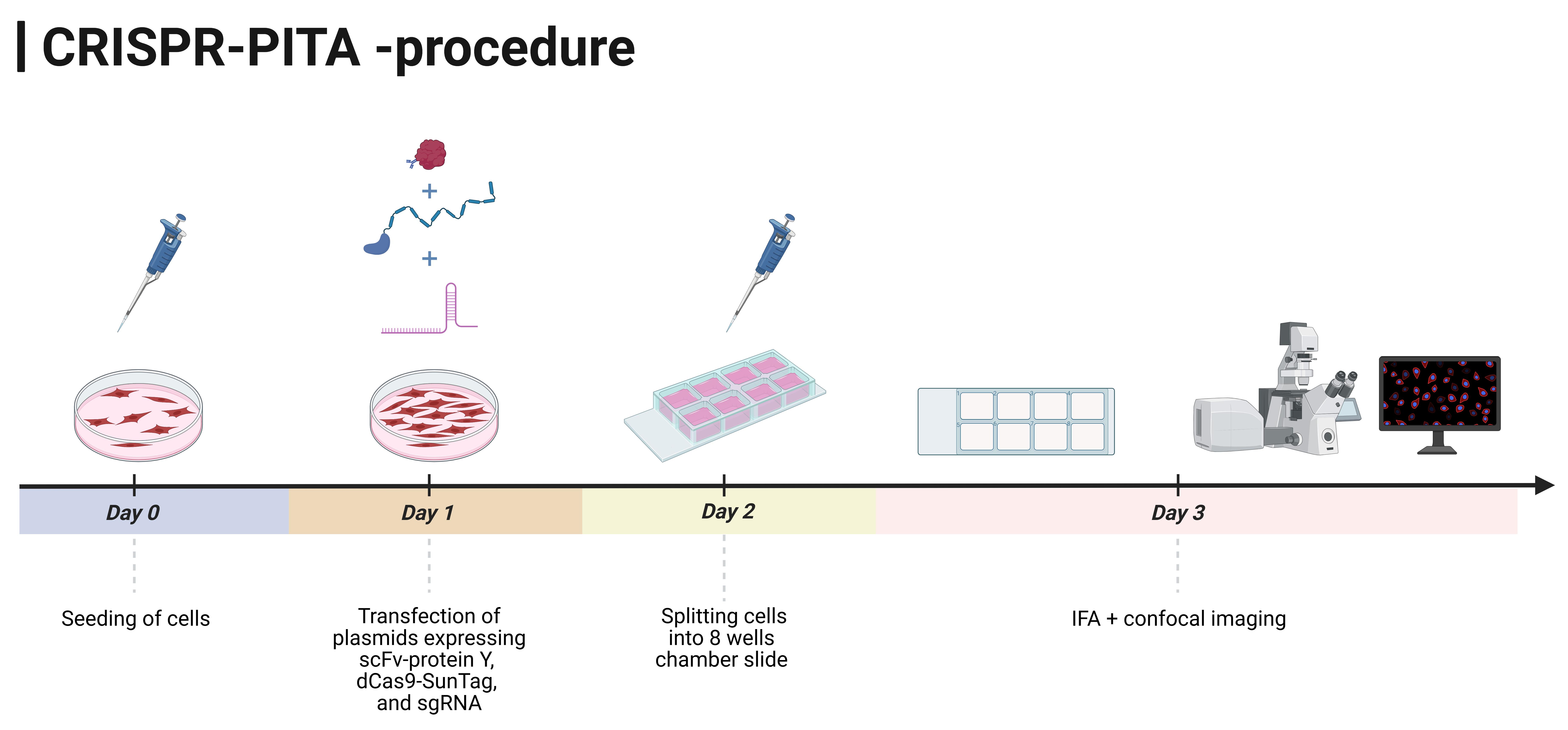
Schematic overview of the CRISPR-PITA procedure
Background
The study of protein–protein and protein–chromatin interactions is fundamental for understanding cellular regulation and nuclear organization. Widely used biochemical approaches, such as co-immunoprecipitation (co-IP), enable the detection of protein complexes by selectively enriching interacting partners via a specific antibody. However, while co-IP is effective for identifying protein interactions, it does not provide spatial or hierarchical information and cannot determine the directionality of recruitment, i.e., which protein acts as the primary recruiter and which proteins are subsequently assembled.
Understanding protein recruitment to specific genomic regions is therefore essential for dissecting mechanisms of chromatin organization and transcriptional regulation. Several assays have been developed to investigate protein–DNA or protein–chromatin interactions, with chromatin immunoprecipitation (ChIP) being one of the most widely used methods [1]. ChIP allows the detection of proteins bound to defined genomic loci, and, in some cases, can provide indirect information on chromatin-associated protein complexes, for example, through ChIP-based proteomic approaches that identify proteins co-associated with specific loci [2]. However, the technique requires fixation, chromatin fragmentation, and immunoprecipitation with high-quality antibodies that can recognize cross-linked proteins. In addition, when multiple proteins are found at the same genomic site, ChIP alone does not resolve the order or directionality of recruitment [3].
To overcome this, ChIP is sometimes combined with genetic perturbations such as knockout (KO) [4] or knockdown (KD) [5] of a suspected recruiter protein. However, these approaches are time-consuming and require extensive validation. An alternative approach relies on the integration of the bacterial lac operator (lacO) array into the host genome and tethering of a protein of interest via fusion to the Lac repressor (LacI) [6]. This system enables recruitment of candidate proteins to a defined genomic locus and allows visualization by microscopy. However, its applicability is limited by the availability of suitable cell lines carrying lacO arrays, and it does not readily support reciprocal or bidirectional recruitment analyses. To address these limitations, we developed a simple, flexible method to test protein recruitment relations in living cells [7]. This approach utilizes dCas9 targeted to repetitive genomic sequences (telomeres) in combination with the SunTag system [8], which amplifies the signal by generating robust, microscopically visible nuclear foci. Briefly, the SunTag system consists of a tandem array of peptide epitopes fused to dCas9, which recruits multiple copies of a cognate single-chain antibody (scFv) fused to the protein of interest, thereby amplifying the local signal at the targeted locus and facilitating the detection of potential interactions. Proteins recruited to these foci can be detected by using antibodies that recognize native epitopes or by tagging the proteins with fluorescent markers (Figure 1). While targeting dCas9 to telomeric repeats has previously been employed to study nuclear dynamics, including heterochromatin organization and liquid–liquid phase separation (LLPS), it has not been adapted to systematically investigate recruitment directionality [9,10].
Our protocol does not require chromatin cross-linking or shearing and does not depend on specialized cell lines or complex genetic manipulations. It allows flexible testing of both direct and reciprocal recruitment relationships, making it broadly applicable to studies of chromatin-associated factors, transcriptional complexes, and virus–host interactions. Using this approach, we have successfully identified unidirectional recruitment dependencies between nuclear proteins, demonstrating the method’s value in dissecting functional hierarchies within protein complexes.
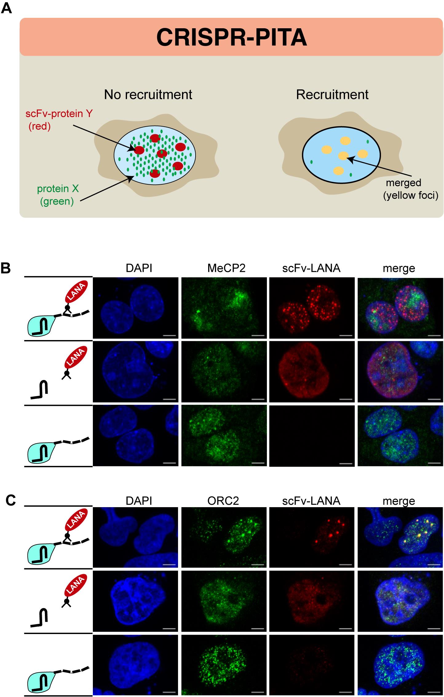
Figure 1. Recruitment phenotypes detected using the CRISPR-PITA assay. (A) Negative recruitment: The recruiter protein (“protein Y,” red) is enriched at repetitive telomeric regions, while the protein of interest (“protein X,” green) remains diffusely distributed throughout the nucleus, indicating the absence of recruitment (left). Positive recruitment: Both the recruiter protein and the protein of interest co-localize at repetitive telomeric regions, resulting in a merged yellow signal, indicative of positive recruitment (right). (B) The recruiter protein LANA is enriched at repetitive telomeric regions (red), while the protein of interest MeCP2 (green) remains diffusely distributed throughout the nucleus, indicating the absence of recruitment. (C) Both the recruiter protein LANA (red) and the protein of interest ORC2 (green) co-localize at repetitive telomeric regions, resulting in a merged yellow signal, indicative of positive recruitment. HCT cells (panel B) and SLK cells (panel C) were transfected with CRISPR-PITA components, as can be seen in the illustrations on the left of each panel. Upper panel: transfection with dCas9-SunTag, Fc-LANA, and sgRNA. Middle panel: transfection with Fc-LANA and sgRNA. Lower panel: transfection with dCas9-SunTag and sgRNA. Immunofluorescence assays were performed to detect mouse anti-LANA antibody (red, Alexa Fluor 594) and rabbit anti-MeCP2 or rabbit anti-ORC2 (green, Alexa Fluor 488) cellular localization. The nucleus was stained with DAPI. Scale bars = 5 μm.
Materials and reagents
Biological materials
1. HEK293, NIH 3T3, SLK, BJAB, BC3, and BCBL1 cells
Reagents
1. Dulbecco’s modified Eagle’s medium (DMEM) (Thermo Fisher Scientific, Gibco, catalog number: 11965092)
2. Roswell Park Memorial Institute (RPMI) 1640 medium (Thermo Fisher Scientific, Gibco, catalog number: 11875093)
3. Fetal bovine serum (Thermo Fisher Scientific, Gibco, catalog number: 16000069)
4. Penicillin-Streptomycin (pen/strep) (5,000 U/mL) (Thermo Fisher Scientific, Gibco, catalog number: 15070063)
5. L-Glutamine (200 mM) (Thermo Fisher Scientific, Gibco, catalog number: 25030024)
6. Sodium pyruvate (100 mM) (Thermo Fisher Scientific, Gibco, catalog number: 11360070)
7. PolyJet In-Vitro DNA transfection reagent (SignaGene Laboratories, catalog number: SL100688)
8. PBS pH 7.4 10× (Invitrogen, catalog number: AM9624)
9. VECTASHIELD® with DAPI (Vector Laboratories, catalog number: H-1200-10)
10. Clear nail polish
11. Plasmids: pHRdSV40-dCas9-10xGCN4_v4-P2A-BFP (Addgene plasmid # 60903; http://n2t.net/addgene:60903; RRID: Addgene_60903), pHR-scFv-GCN4-sfGFP-GB1-dWPRE (Addgene plasmid # 60907; http://n2t.net/addgene:60907; RRID: Addgene_60907), and pSLQ1651-sgTelomere (F+E) (Addgene plasmid # 51024; https://www.addgene.org/51024/; RRID: Addgene_51024)
12. Appropriate antibodies for detecting the proteins of interest and secondary antibodies for IF
13. Paraformaldehyde (Sigma-Aldrich, catalog number: 158127)
14. Triton X-100 (Thermo Fisher Scientific, catalog number: A16046.AE)
15. Tween 20 (Sigma-Aldrich, catalog number: P9416)
16. Bovine serum albumin (BSA) (Sigma-Aldrich, catalog number: A4612)
17. Glycine (Sigma-Aldrich, catalog number: G7126)
Solutions
1. PBS 1× (see Recipes)
2. DMEM (including supplements) (see Recipes)
Recipes
1. PBS 1×
| Reagent | Quantity or volume |
|---|---|
| PBS pH 7.4 10× | 100 mL |
| Double-distilled water | 900 mL |
| Total | 1,000 mL |
2. DMEM (including supplements)
| Reagent | Final concentration | Quantity or volume |
|---|---|---|
| DMEM | 1× | 500 mL |
| Fetal bovine serum | 10% | 50 mL |
| Penicillin 10,000 U/mL | 100 U/mL | 5 mL |
| Streptomycin 10 mg/mL | 100 μg/mL | 5 mL |
| L-glutamine 200 mM | 2 mM | 5 mL |
| Sodium pyruvate 100 mM | 2 mM | 10 mL |
Laboratory supplies
1. Cell culture flasks, 25 cm2, cell-repellent surface (T25 non-adherent flask) (Greiner Bio-One, catalog number: 690980)
2. 100 mm tissue culture-treated dishes (Corning, catalog number: CLS353003)
3. Cell culture multi-well plates (6-well plate) (Greiner Bio-One, catalog number: 657160)
4. 25 mL sterile reservoirs (Thermo Fisher Scientific, catalog number: 95128095) or 50 mL sterile reservoirs (InvitroLab, catalog number: IV-6002)
5. Cell scrapers (TH Geyer, catalog number: 7696760)
6. 15 mL conical tubes (TH Geyer, catalog number: 7696714)
7. 50 mL conical tubes (Greiner Bio-One, catalog number: 227261)
8. 1.7 mL microcentrifuge tube (Axygen, catalog number: MCT-175-C-S)
9. 8-well culture slide (Corning, Falcon, catalog number: CLS354118)
Equipment
1. Refrigerated centrifuge (Eppendorf, model: 5810R)
2. Class II A2 Biological Safety Cabinet (Unicorn, model: BSC-1000IIA2)
3. Inverted confocal microscope (Zeiss, model: LSM780)
Software and datasets
1. Zenn software black edition (Zeiss, Version 2.3)
2. ImageJ (JACoP Plugin, v2.1.4)
3. Prism 9 (GraphPad)
Procedure
登录/注册后免费查看全文
文章信息
稿件历史记录
提交日期: Feb 11, 2026
接收日期: May 21, 2026
在线发布日期: Jun 11, 2026
出版日期: Jul 5, 2026
版权信息
© 2026 The Author(s); This is an open access article under the CC BY-NC license (https://creativecommons.org/licenses/by-nc/4.0/).
如何引用
Lavi, I., Bhattacharya, S., Gurevich, V. and Shamay, M. (2026). CRISPR-PITA: An Imaging-Based CRISPR/dCas9 Assay to Determine Recruitment Directionality of Nuclear Proteins. Bio-protocol 16(13): e5742. DOI: 10.21769/BioProtoc.5742.

 Download Citation in RIS Format
分类
分子生物学 > 蛋白质 > 蛋白质-蛋白质相互作用
细胞生物学 > 细胞成像 > 共聚焦显微镜
您对这篇实验方案有问题吗?
在此处发布您的问题,我们将邀请本文作者来回答。同时,我们会将您的问题发布到Bio-protocol Exchange,以便寻求社区成员的帮助。
 发布问题
发布问题


