


Generation and Characterization of Adaptive Anoikis-Resistant Cells Using Cyclic Attachment-Detachment Culture of Cancer Cells
利用癌细胞周期性贴壁脱附培养构建并表征适应性抗失巢凋亡细胞

发布: 2026年07月05日第16卷第13期 DOI: 10.21769/BioProtoc.5736 浏览次数: 247
评审: Salah BoudjadiSucheta ChopraAnonymous reviewer(s)
 PDF
PDF  Q&A
Q&A
参见作者原研究论文
The authors used this protocol in:
Feb 2026






Abstract
Anoikis resistance, or the ability of cancer cells to evade cell death triggered by immediate detachment from the extracellular matrix, is a critical established hallmark of metastatic cancer. While suspension culture models have been used to study anoikis, most focus on defined single time points or prolonged suspension that may not recapitulate the effects of repeated stress that tumor cells experience during metastatic dissemination. Here, we describe a detailed protocol for generating anoikis-resistant (AnR) cancer cells that have adapted to such stress through exposure to repeated cycles of suspension stress on poly-HEMA-coated plates, followed by recovery under standard attached conditions. The protocol includes methods for determining baseline anoikis sensitivity, generating AnR cells over 7–9 attachment-detachment cycles, assessing the stability and reversion of the anoikis-resistant phenotype, and characterizing AnR cells using Live/Dead staining of spheroids, flow cytometry–based apoptosis assays, and immunofluorescence for proliferation markers. This approach produces a non-genetic, reversible anoikis-resistant state that models the adaptive transcriptional reprogramming underlying metastatic progression, providing a reproducible and physiologically relevant in vitro system for studying anoikis resistance mechanisms and evaluating therapeutic strategies for prevention and reversal of such adaptations.
Key features
• Generates adapted anoikis-resistant cancer cells through cyclic attachment-detachment culture on poly-HEMA-coated plates over 7–9 passages.
• Produces a non-genetic and reversible resistant state that models adaptive transcriptional reprogramming during metastatic dissemination.
• Includes methods for assessing anoikis resistance stability, reversion kinetics, and cryopreservation of cells at defined passages.
• Provides three complementary characterization assays: Live/Dead spheroid imaging, Annexin V/PI flow cytometry, and Ki67 immunofluorescence.
Keywords: Anoikis (失巢凋亡) Matrix detachment (基质脱离)
Cell death (细胞死亡)
Cell survival (细胞存活)
Apoptosis (细胞凋亡)
Spheroid (细胞球)
Adaptation (适应性)
Matrix detachment (基质脱离)
Cell death (细胞死亡)
Cell survival (细胞存活)
Apoptosis (细胞凋亡)
Spheroid (细胞球)
Adaptation (适应性)
Graphical overview
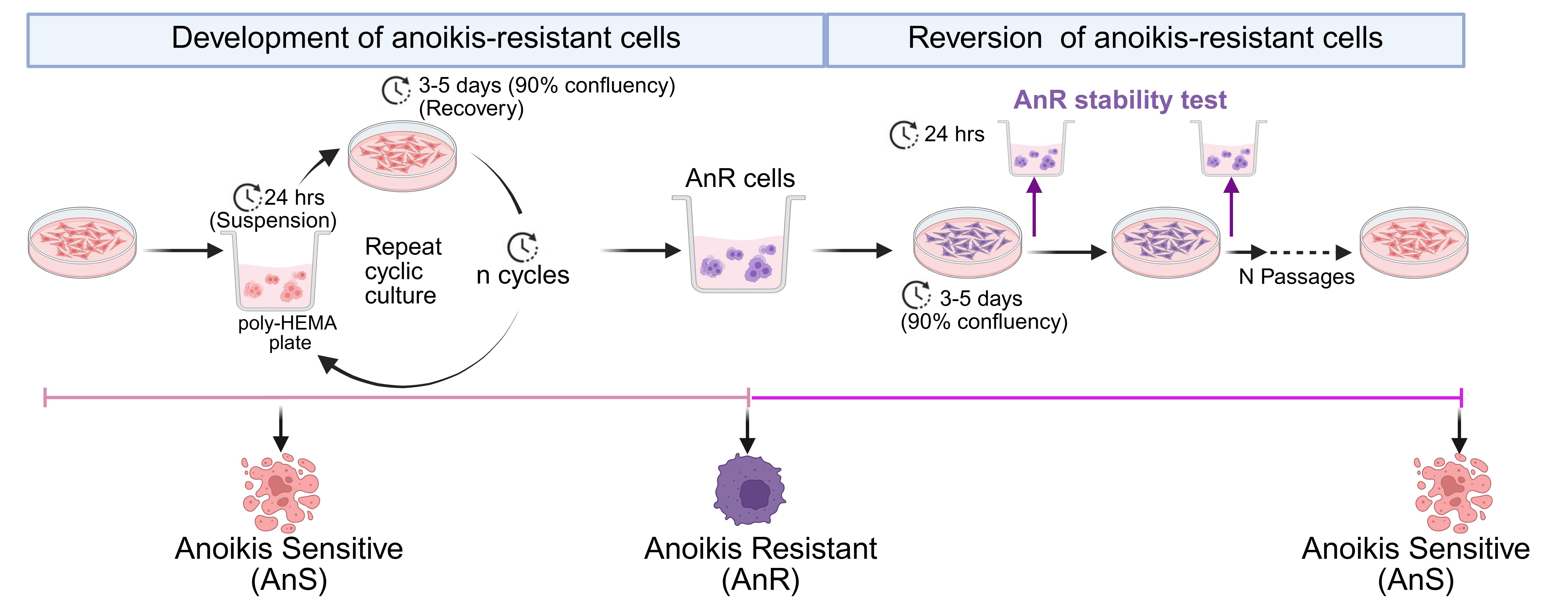
Workflow and timeline for the generation of anoikis-resistant cells through alternating suspension and attached culture, followed by reversion to an anoikis-sensitive state by continuous passaging under attached conditions
Background
Anoikis, a form of programmed cell death triggered by loss of cell–extracellular matrix attachment, can serve as a critical barrier to metastasis. Since cell death is triggered rapidly, cancer cells that acquire anoikis resistance can survive during dissemination, enabling colonization of distant sites. Anoikis resistance is essential across diverse modes of metastatic spread. Circulating tumor cells (CTCs) in the bloodstream must withstand detachment to seed hematogenous metastases [1–3], while in ovarian and other peritoneal cancers, tumor cells must survive transit through the peritoneal cavity within malignant ascites to establish metastatic implants [4–7]. Despite its central role in metastasis across cancer types, methods for modeling the progressive acquisition of anoikis resistance in vitro remain limited.
Several approaches have been used to study anoikis resistance in vitro. These include culturing cells on ultra-low attachment surfaces for single time points, prolonged suspension culture to select for surviving populations, or the use of intrinsically anoikis-resistant cell lines [8–12]. While informative, single-time-point studies capture only the acute response to detachment stress and do not model the progressive adaptation that could occur during metastasis. Prolonged continuous suspension culture may select for preexisting resistant subpopulations rather than recapitulating the adaptive process. Furthermore, comparing intrinsically resistant lines to unrelated sensitive lines introduces confounding variables unrelated to anoikis adaptation.
The protocol described here addresses these limitations by generating adapted anoikis-resistant cells through repeated cycles of suspension stress followed by recovery under attached conditions, mimicking repeated cycles of detachment followed by attached regrowth/survival that tumor cells may experience during metastasis [13]. This approach produces an acquired, non-genetic anoikis-resistant state that is transient and reverts upon prolonged culture without suspension stress, consistent with transcriptional rather than mutational mechanisms of adaptation [13]. The model has been validated across multiple ovarian cancer cell lines with different baseline sensitivities to detachment stress and different mutational backgrounds [13]. The resulting anoikis-adapted cells display enhanced migration, chemoresistance, immune evasion, and metastatic potential in vivo [13], consistent with known associations of anoikis resistance and aggressive metastatic behavior [1,14–17]. The protocol is applicable to any adherent cancer cell lines where baseline anoikis sensitivity can be tested. The paired anoikis-sensitive (AnS) and anoikis-resistant (AnR) derivatives provide an isogenic system for studying the mechanisms underlying metastatic adaptation and for evaluating candidate therapeutics targeting this process.
Materials and reagents
Biological materials
1. OV90 cells (ATCC, catalog number: CRL-11732)
2. CAOV3 cells (ATCC, catalog number: HTB-75)
Reagents
1. Medium 199, Earle’s salts (Thermo Fisher Scientific, Gibco, catalog number: 11150059)
2. MCDB 105 medium (Millipore Sigma, catalog number: 117-500)
3. DMEM (Corning, catalog number: MT10013CV)
4. FBS (Corning, catalog number: 35-011-CV)
5. Penicillin-Streptomycin (pen/strep) (10,000 U/mL) (Thermo Fisher Scientific, Gibco, catalog number: 15140122)
6. Poly (2-hydroxyethyl methacrylate) (poly-HEMA) (Millipore Sigma, catalog number: P3932)
7. Trypsin (2.5%) (Thermo Fisher Scientific, Gibco, catalog number: 15090046)
8. Trypsin EDTA (2.5%) (Millipore Sigma, catalog number: T4174-20ML)
9. Trypan Blue solution, 0.4% (Thermo Fisher Scientific, Gibco, catalog number: 15250061)
10. Dimethyl sulfoxide (DMSO) (Fisher Scientific, catalog number: BP231-100)
11. LIVE/DEADTM Viability/Cytotoxicity kit (Thermo Fisher Scientific, Invitrogen, catalog number: L3224)
12. Annexin V Apoptosis Detection kits (Thermo Fisher Scientific, eBioscience, catalog number: 88-8005-74)
13. Paraformaldehyde (PFA) (Fisher Scientific, J.T. Baker, catalog number: S898-07)
14. Ammonium chloride (NH4Cl) (Fisher Scientific, catalog number: AA11595A1)
15. Sodium hydroxide (NaOH) (Avantor, catalog number: MAL-7708-06)
16. Sodium chloride (NaCl) (Thermo Fisher Scientific, catalog number: S271-3)
17. Potassium chloride (KCl) (Thermo Fisher Scientific, catalog number: P217-500)
18. Sodium phosphate monobasic (NaH2PO4) (Millipore Sigma, catalog number: S0751-100G)
19. Potassium phosphate monobasic (KH2PO4) (Thermo Fisher Scientific, catalog number: P285-500)
20. Triton-X100 (Amresco, catalog number: 0694-1L)
21. Absolute ethanol (Fisher Scientific, catalog number: BP2818-4)
22. Bovine serum albumin (BSA) (Fisher Scientific, catalog number: BP9706100)
23. Ki-67 antibody (Cell Signaling, catalog number: 9449)
24. Goat anti-mouse IgG1 cross-adsorbed secondary antibody Alexa Fluor (Thermo Fisher Scientific, Invitrogen, catalog number: A-21125)
25. ProLongTM Gold Antifade mountant (Thermo Fisher Scientific, Invitrogen, catalog number: P36930)
26. DAPI solution (Thermo Fisher Scientific, Invitrogen, catalog number: 62248)
Solutions
1. OV90 media (see Recipes)
2. CAOV3 media (see Recipes)
3. Poly-HEMA (see Recipes)
4. 4% PFA (see Recipes)
5. 10 mM NH4Cl solution (see Recipes)
6. 0.3% Triton-X 100 solution (see Recipes)
7. BSA (5% and 3%) (see Recipes)
8. 10× PBS (see Recipes)
9. FACS buffer (see Recipes)
Recipes
1. OV90 media
| Reagent | Final concentration | Quantity or volume |
|---|---|---|
| Medium 199, Earle’s salts | 42% | 42 mL |
| MCDB 105 medium | 42% | 42 mL |
| FBS | 15% | 15mL |
| Pen/strep | 1% | 1 mL |
| Total | 100 mL |
2. CAOV3 media
| Reagent | Final concentration | Quantity or volume |
|---|---|---|
| DMEM | 89% | 89 mL |
| FBS | 10% | 10 mL |
| Pen/strep | 1% | 1 mL |
| Total | 100 mL |
3. Poly-HEMA
| Reagent | Final concentration | Quantity or volume |
|---|---|---|
| poly-HEMA | 2% | 2 g |
| Absolute ethanol | 95% | 95 mL |
| Deionized (DI) water | 5% | 5 mL |
| Total | 100 mL |
Place the solution in the oven at 50 °C overnight. The next day, cool the solution to room temperature before using or store it at 4 °C for longer-term storage.
4. 4% PFA
| Reagent | Final concentration | Quantity or volume |
|---|---|---|
| PFA | 4% | 4 g |
| Distilled water | 90 mL | |
| 10 N NaOH | ~5 μL | |
| 10× PBS | 1× | 10 mL |
| Total | 100 mL |
Warm 80 mL of distilled water to 60 °C (do not boil) in a ventilation hood. Dissolve 4 g of PFA in the water by stirring. Slowly add 10 N NaOH (40 g of NaOH in 100 mL of DI H2O) using a dropper until the solution turns clear. Add 10 mL of 10× PBS. Adjust the final pH of the solution to 7.4. Make up the final volume of the solution to 100 mL. Filter the solution and store the excess in -20 °C.
5. 10 mM NH4Cl solution
| Reagent | Final concentration | Quantity or volume |
|---|---|---|
| NH4Cl | 10 mM | 21.4 mg |
| PBS (1×) | 100 mL | |
| Total | 100 mL |
6. 0.3% Triton X-100 solution
| Reagent | Final concentration | Quantity or volume |
|---|---|---|
| Triton X-100 | 0.3% | 300 μL |
| PBS (1×) | 100 mL | |
| Total | 100 mL |
Sonicate the 0.3% Triton X-100 solution with two 15 s pulses. Prepare fresh solutions.
7. BSA solution (5% and 3%)
| Reagent | Final concentration | Quantity or Volume |
|---|---|---|
| BSA | 5% | 5 g |
| BSA | 3% | 3 g |
| PBS (1×) | 100 mL | |
| Total | 100 mL |
8. 10× PBS
| Reagent | Final concentration | Quantity or volume |
|---|---|---|
| NaCl | 1.37 M | 80 g |
| KCl | 27 mM | 2 g |
| Na2HPO4 | 100 mM | 14.2 g |
| KH2PO4 | 18 mM | 2.4 g |
| Total | 1 L |
Add all reagents to 900 mL of DI water. Adjust the pH between 7.2 and 7.4. Make up the total volume to 1,000 mL. For 1× PBS, add 100 mL of 10× PBS to 900 mL of DI water to make a total of 1 L.
9. FACS buffer
| Reagent | Final concentration | Quantity or volume |
|---|---|---|
| 1× PBS | 98 mL | |
| FBS | 2% | 2 mL |
| Total | 100 mL |
Laboratory supplies
1. Tissue culture dishes (100 mm) (Fisher Scientific, catalog number: FB012924)
2. Tissue culture dishes (60 mm) (Fisher Scientific, catalog number: FB012921)
3. Tissue culture plates (6-well) (Fisher Scientific, catalog number: FB012927)
4. Millicell ultra-low attachment plate (96 well) (Millipore Sigma, catalog number: MC96ULA20)
5. 1,250 μL pipette tips (Fisher Scientific, catalog number: 12-111-010)
6. 200 μL pipette tips (Fisher Scientific, catalog number: 12-111-008)
7. 10 μL pipette tips (Fisher Scientific, catalog number: 12-111-006)
8. 10 mL serological pipettes (Fisher Scientific, catalog number: 14-955-234)
9. 25 mL serological pipettes (Fisher Scientific, catalog number: 14-955-235)
10. 15 mL conical tubes (Fisher Scientific, catalog number: 12-565-269)
11. 50 mL conical tubes (Fisher Scientific, catalog number: 12-565-271)
12. Hemocytometer (Reichert bright-line, catalog number: HS-1490)
13. 2 mL cryovial (Fisher Scientific, catalog number: 02-912-729)
14. Freezing container (Fisher Scientific, catalog number: 15-350-50)
15. Falcon round-bottom test tubes (FACS tubes) (Fisher Scientific, catalog number: 14-959-1A)
16. Cytoclip (Fisher Scientific, Epredia, catalog number: 59-910-052)
17. Cytofunnel (Fisher Scientific, Epredia, catalog number: 59-910-40)
18. Microscope slides (Fisher Scientific, catalog number: 22-037-246)
19. Coplin jars (Webber Scientific, catalog number: 2005-19)
20. PAP pen (Fisher Scientific, catalog number: NC9204359)
21. Micro cover glass (VWR, catalog number: 48366067)
Equipment
1. Purifier class II biosafety cabinet (Labconco, catalog number: 3620904)
2. Centrifuge with rotors for 15 and 50 mL conical tubes (Eppendorf, catalog number: 5804)
3. Centrifuge with rotors for 1.5 mL microfuge tubes (Eppendorf, catalog number: 5424R)
4. Revco CO2 incubator (Thermo Electron, catalog number: 29584)
5. Convection oven (Thermo Scientific, catalog number: TS-PR305)
6. Vortex Genie 2 (Thermo Scientific, catalog number: 12-812)
7. Revco Elite Plus freezer (Thermo Scientific, catalog number: ULT1786-6-A43)
8. Liquid nitrogen tank (Thermo Scientific, catalog number: CY50985)
9. Refrigerator (Roper Scientific, model number: RT18DKXFW01)
10. Water bath (Polyscience, catalog number: NC1633292)
11. Inverted microscope (Nikon, catalog number: N-TS2)
12. Confocal microscope (Nikon, model: A1R HD)
13. Flow cytometer (BD LSRFortessa, UAB Flow Cytometry and Single Cell Core Facility)
14. Cytospin III (Shandon, catalog number: 8358-30-0001)
Software and datasets
1. FlowJo software (BD Biosciences, version 10.8.1)
Note: A license is required to use FlowJo. License options are available based on the number of users and frequency of use.
2. ImageJ (NIH, version win64)
Procedure
登录/注册后免费查看全文
文章信息
稿件历史记录
提交日期: Mar 23, 2026
接收日期: May 19, 2026
在线发布日期: Jun 9, 2026
出版日期: Jul 5, 2026
版权信息
© 2026 The Author(s); This is an open access article under the CC BY license (https://creativecommons.org/licenses/by/4.0/).
如何引用
Rajkarnikar, R., Monavarian, M. and Mythreye, K. (2026). Generation and Characterization of Adaptive Anoikis-Resistant Cells Using Cyclic Attachment-Detachment Culture of Cancer Cells. Bio-protocol 16(13): e5736. DOI: 10.21769/BioProtoc.5736.

 Download Citation in RIS Format
分类
癌症生物学 > 细胞死亡 > 细胞生物学试验 > 细胞活性
细胞生物学 > 基于细胞的分析方法 > 非贴壁培养
癌症生物学 > 侵袭和转移 > 细胞生物学试验
您对这篇实验方案有问题吗?
在此处发布您的问题,我们将邀请本文作者来回答。同时,我们会将您的问题发布到Bio-protocol Exchange,以便寻求社区成员的帮助。
 发布问题
发布问题


