


An Optimized Protocol for the Characterization of Zebrafish ApoB-Containing Lipoproteins Using the LipoGlo System
利用 LipoGlo 系统表征斑马鱼含 ApoB 脂蛋白的优化实验方案

发布: 2026年07月05日第16卷第13期 DOI: 10.21769/BioProtoc.5732 浏览次数: 242
评审: Alberto RissoneSukrut Chandrashekhar KamerkarIvonne Sehring
 PDF
PDF  Q&A
Q&A
参见作者原研究论文
The authors used this protocol in:

Jul 2019
Advertisement







Abstract
Apolipoprotein B–containing lipoproteins (ApoB-LPs) transport lipids throughout the circulation and are closely associated with cardiovascular disease in humans. Many aspects of ApoB-LP biology remain elusive, often due to their indirect characterization through the measurement of plasma triglycerides and cholesterol. The conventional approach provides limited information on ApoB-LPs number and size distribution, essential features that influence cardiovascular disease risk. Additionally, drug studies have historically been limited to the use of mammalian research models, which are not suited for high-throughput experiments. Therefore, we generated a reporter system (LipoGlo) utilizing a luciferase enzyme (NanoLuc) fused to the C-terminus of the zebrafish (Danio rerio) ApoBb.1 protein. In metazoans, ranging from insects to humans, each ApoB-LP contains a single ApoB molecule, such that the luminescence emitted from these transgenic fish is proportional to the total number of ApoB-LPs. The LipoGlo zebrafish reporter generates a quantitative chemiluminescent signal that can be used in plate-based assays to measure lipoprotein quantities, a gel-based assay that can measure lipoprotein size distribution, and chemiluminescent microscopy that can, for the first time, visualize lipoprotein localization in a larval zebrafish. LipoGlo, combined with the amenability of zebrafish to genetic approaches, facilitates the rapid assessment of any gene or drug’s role in ApoB-LP molecular and cell biology. This protocol describes three optimized LipoGlo assays that facilitate ApoB-LP characterization with 100× less starting material than prior assays routinely used for mammalian lipoprotein analysis.
Key features
• Improves upon the methods from Thierer et al. [1].
• Lipoprotein profiles from individual zebrafish larvae that can simultaneously be genotyped for specific mutations.
Keywords: Lipoproteins (脂蛋白) ApoB-containing lipoproteins (含 ApoB 脂蛋白)
Zebrafish (斑马鱼)
LipoGlo (LipoGlo)
Cholesterol (胆固醇)
Luciferase (荧光素酶)
ApoB-containing lipoproteins (含 ApoB 脂蛋白)
Zebrafish (斑马鱼)
LipoGlo (LipoGlo)
Cholesterol (胆固醇)
Luciferase (荧光素酶)
Graphical overview
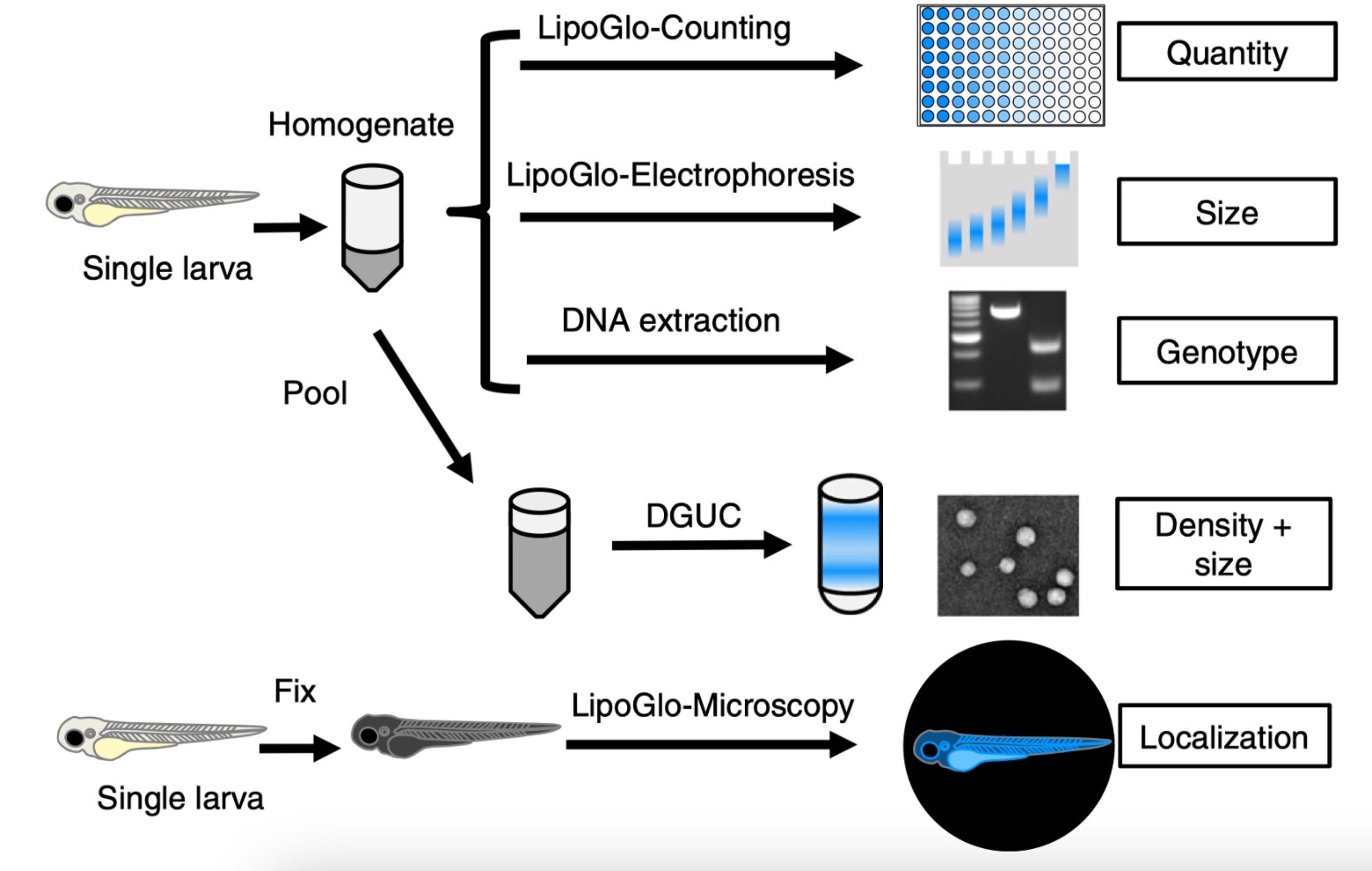
LipoGlo zebrafish tissue homogenate can be used to measure the quantity, size, density, and localization of Apolipoprotein B–containing lipoproteins (ApoB-LPs). Reprinted/adapted from Thierer et al. [1]. © The Authors, some rights reserved; exclusive licensee, Springer Nature. Distributed under a Creative Commons Attribution Non-Commercial License 4.0 (CC BY-NC) http://creativecommons.org/licenses/by-nc/4.0/
Background
Apolipoprotein B–containing lipoproteins (ApoB-LPs) perform the vital role of transporting lipids throughout the circulation, being closely associated with cardiovascular disease, a leading cause of death worldwide [2]. Many fundamental aspects of ApoB-LP basic biology remain elusive [3–5]. Zebrafish and humans have many of the same key lipoprotein-related genes, including the major apolipoprotein genes APOB, APOA1, and APOC2 [6,7]. They also share the conservation of the cholesterol ester transfer protein gene (cetp), which is a transporter of cholesterol esters and triglycerides between high-density lipoproteins (HDLs) and ApoB-LPs [8], a critical gene that is lacking in most rodents [9]. Thus, like humans, the zebrafish carry most of their cholesterol in their ApoB-LP fraction, whereas mice use HDL. The larval zebrafish are also ideally suited for high-throughput drug screens due to their small size (~5 mm in length), optically transparent development, and rapid organ development being completed by 5 days post fertilization (dpf) [10]. Despite the many advantages of using larval zebrafish, previous assays lack in sensitivity to profile ApoB-LP size and number in individual larvae [6,11,12].
In 2019, Thierer et al. described the LipoGlo reporter zebrafish system, in which genomic engineering was used to insert the coding sequence of a nanoluciferase (NanoLuc) reporter fused to the C-terminus of the zebrafish ApoBb.1 gene [1]. The small (19.1 kDa) NanoLuc luciferase reporter generates a quantitative chemiluminescent signal through processing of its substrate, furimazine [13]. NanoLuc is ~100 times brighter than firefly luciferase and provides a robust signal-to-noise ratio that enables accurate detection at femtomolar concentrations [13]. Each lipoprotein contains one ApoB molecule, making the luminescence emitted proportional to the total number of ApoB-LPs [14].
In our original publication, we found that homozygous LipoGlo zebrafish emitted twice as much luminescence as heterozygous animals; however, in the years since, we found that this 2-fold increase is no longer consistently observed. We tested the hypothesis that the attenuated assay performance was due to premature depletion of LipoGlo substrate during the assay. In support of this hypothesis, we found that using 10× less tissue homogenate restores assay performance. ApoB-LPs come in a variety of sizes, with chylomicrons being the largest, and low-density lipoproteins (LDLs) being the smallest. The LipoGlo system can differentiate and quantify the levels of the different ApoB-LP size subclasses by subjecting tissue homogenates to a 3% native polyacrylamide gel electrophoresis (PAGE) followed by bioluminescent imaging. In our original publication, we used a commercially available DiI-labeled human LDL as a migration marker, but over time, we observed variations in the migration of the DiI-LDL, perhaps due to batch variations in LDL labeling. To better benchmark the LipoGlo Electrophoresis assay, we sought to develop a more consistent and validated migration marker. To achieve this, we used tissue homogenates from ldlrasd52 mutants [15] that are enriched in small LDL particles and apoC2sd38 mutants that have high levels of large very low density lipoprotein (VLDL) particles, confirmed by an orthogonal density gradient ultra-centrifugation (DGUC) assay to differentiate small and large particle fractions. Importantly, the DiI-LDL marker and the ldlrasd52 ApoB-LPs consistently appear in the same DGUC fraction, indicating that, while the DiI label causes the resulting LDL to migrate at a slower rate on the PAGE gel, making it appear larger, it remains a useful standardization marker to address gel-to-gel variation [1]. In our original deployment of the LipoGlo electrophoresis assay using a PAGE gel to describe the sizes of ApoB-LPs, we included the signal from the loading well (zero mobility; ZM), thinking that it represented extremely large particles (e.g., chylomicrons). The protocol presented here suggests excluding the ZM signal because we now know from the DGUC analysis that it reflects aggregation of particles from a range of sizes that occurs when samples are loaded and run in the PAGE gel. In sum, we describe a series of LipoGlo protocol improvements to provide a more robust platform for studying ApoB-LPs in individual zebrafish larvae.
Materials and reagents
Biological materials
Zebrafish stocks were maintained at 27 °C in a circulating aquarium facility. Adult zebrafish were fed once daily with ~2% body weight Gemma 300 (Skretting USA). Natural spawnings were used to obtain embryos, which were kept in embryo medium at 28 °C on a 14/10 h light/dark cycle until 7 dpf, unless noted otherwise. All experiments in this work were performed in larvae between 1 and 7 dpf. However, it should be noted that this protocol is optimized for 1–15 dpf [1] and has not been tested on earlier or later time points.
1. Zebrafish mutant line ldlrasd52, generated by the Yury Miller Lab, ZFIN ID ZDB-ALT-170913–3 [15]
2. Zebrafish mutant line apoC2sd38, generated by the Yury Miller Lab, ZFIN ID ZDB-ALT-151110–1 [6]
3. Zebrafish reporter line Fus(ApoBb.1-nluc), generated in the Farber Lab, ZFIN ID ZDB-ALT-191218–4 [1]
Reagents
1. Ammonium persulfate (APS) (Bio-Rad, catalog number: 1610700)
2. Anhydrous calcium chloride (CaCl2) (Sigma, catalog number: C5670)
3. Boric acid (Sigma, catalog number: B0394)
4. Bromophenol blue (Sigma, catalog number: 114391)
5. Complete mini EDTA-free protease inhibitor cocktail (Sigma, catalog number: 11836170001)
6. Complete protease inhibitor cocktail (Sigma, catalog number: 11697498001)
7. Ethylene glycol-bis(2-amino-ethylether)-N,N,N,N’-tetraacetic acid (EGTA) (Sigma, catalog number: E3889)
8. Ethylenediaminetetraacetic acid (EDTA) (Sigma, catalog number: E9884)
9. Ethly 3-aminobenzonate methanesulfonate (tricaine, ms-222) (Sigma, catalog number: E10521)
10. HEPES (Goldbio, catalog number: H-400-100)
11. Low-density lipoprotein from human plasma, DiI complex (DiI-LDL) (Thermo Fischer, catalog number: L3482)
12. Low-melt agarose (Thermo Fischer, catalog number: BP165)
13. Magnesium sulfate anhydrous (MgSO4) (Sigma, catalog number: M7506)
14. Nano-Glo luciferase assay system (contains Nano-Glo® luciferase assay substrate and Nano-Glo® luciferase assay buffer) (Promega, catalog number: N1110)
15. OptiPrep density gradient medium (Sigma, catalog number: D1556)
16. Phosphate buffer saline (PBS) (Sigma, catalog number: 08057-12TAB-F)
17. Potassium chloride (KCl) (Sigma, catalog number: 7447-40-7)
18. Potassium phosphate monobasic (KH2PO4) (Sigma, catalog number: P8709)
19. Rain X original treatment (Walmart, catalog number: 800002242W)
20. Sodium bicarbonate (NaHCO3) (Sigma, catalog number: 1613655)
21. Sodium chloride (NaCl) (Sigma, catalog number: 1064040500)
22. Sodium hydroxide (NaOH) (Sigma, catalog number: 221465)
23. Sodium phosphate dibasic anhydrous (Na2HPO4) (Sigma, catalog number: S9763)
24. Sucrose (VWR, catalog number: BDH9308)
25. Tetramethylethylenediamine (TEMED) (Thermo Fisher, catalog number: 17919)
26. Tris Bas, (Thermo Fisher, catalog number: BP152-500)
27. Tris hydrochloride (Roche, catalog number: 10812846001)
28. Tween-20 (Sigma, catalog number: P1379)
29. 32% paraformaldehyde aqueous solution (PFA) (Electron Microscopy Sciences, catalog number: 15714)
30. 40% Acrylamide:Bis solution 19:1 (Bio-Rad, catalog number: 1610144)
Solutions
1. 20× embryo medium (see Recipes)
2. 1× embryo medium (see Recipes)
3. Tricaine (see Recipes)
4. 2× ApoB-LP stabilization buffer (see Recipes)
5. 2× ApoB-LP stabilization buffer (no sucrose) (see Recipes)
6. 0.5 M EGTA (see Recipes)
7. LipoGlo reaction buffer (see Recipes)
8. DNA lysis buffer (see Recipes)
9. 3% Native-PAGE gel (see Recipes)
10. 5× TBE (see Recipes)
11. 1× TBE (see Recipes)
12. Native-PAGE gel loading dye (see Recipes)
13. Native-PAGE gel imaging solution (see Recipes)
14. Low-density lipoprotein from human plasma (DiI-LDL) (see Recipes)
15. HEPES-buffered saline (HBS) (see Recipes)
16. 1 M HEPES buffer pH 7.4 (see Recipes)
17. 9% iodixanol solution (see Recipes)
18. 12% iodixanol solution (see Recipes)
19. 4% PFA (see Recipes)
Recipes
1. 20× embryo medium
| Reagent | Final concentration | Quantity or volume |
|---|---|---|
| NaCl | 0.299 M | 17.5 g |
| KCl | 10.1 mM | 0.75 g |
| CaCl2 | 19.6 mM | 2.18 g |
| KH2PO4 | 3.01 mM | 0.41 g |
| Na2HPO4 | 1.0 mM | 0.142 g |
| MgSO4 | 19.8 mM | 2.39 g |
| Milli-Q H2O | n/a | 1 L |
| Total | n/a | 1 L |
Store at 4 °C.
2. 1× embryo medium
| Reagent | Final concentration | Quantity or volume |
|---|---|---|
| 20× embryo medium | n/a | 1 L |
| NaHCO3 | 0.83 mM | 1.4 g |
| Milli-Q H2O | n/a | 20 L |
Store at room temperature.
3. Tricaine
| Reagent | Final concentration | Quantity or volume |
|---|---|---|
| Tricaine | 19.5 mM | 400 mg |
| 1 M Tris | 21 mM | 2.1 mL |
| Milli-Q H2O | n/a | 100 mL |
Adjust pH to 7.0 with sodium bicarbonate and store at room temperature.
4. 2× ApoB-LP stabilization buffer
| Reagent | Final concentration | Quantity or volume |
|---|---|---|
| EGTA | 40 mM | 400 μL |
| Sucrose | 0.58 M | 1 g |
| Milli-Q H2O | n/a | Top up to 5 mL |
| Complete mini EDTA-free protease inhibitor | n/a | 1 tablet |
When making a 10 mL total volume, use one complete EDTA-free protease inhibitor tablet.
5. 2× ApoB-LP stabilization buffer (no sucrose)
| Reagent | Final concentration | Quantity or volume |
|---|---|---|
| EGTA | 40 mM | 400 μL |
| Milli-Q H2O | n/a | Top up to 5 mL |
| Complete mini EDTA-free protease inhibitor | n/a | 1 tablet |
When making a 10 mL total volume, use one complete EDTA-free protease inhibitor tablet.
6. 0.5 M EGTA
| Reagent | Final concentration | Quantity or volume |
|---|---|---|
| EGTA | 0.5 M | 9.3 g |
| Milli-Q H2O | n/a | 50 mL |
| Total | n/a | 50 mL |
Store at room temperature.
7. LipoGlo reaction buffer
| Reagent | Final concentration | Quantity or volume |
|---|---|---|
| 1× Phosphate Buffer Saline (PBS) | n/a | 6.68 mL |
| Nano-Glo luciferase buffer* | n/a | 1.1 mL |
| Nano-Glo luciferase substrate* | n/a | 22 μL |
| Total | n/a | 8 mL |
*Reagents from Nano-Glo luciferase assay system.
Make fresh for each experiment.
8. DNA lysis buffer
| Reagent | Final concentration | Quantity or volume |
|---|---|---|
| NaOH | 50 mM | 2 g |
| Milli-Q H2O | n/a | 500 mL |
| Total | n/a | 500 mL |
Store at room temperature.
9. 3% Native-PAGE gel
| Reagent | Final concentration | Quantity or volume |
|---|---|---|
| 5× TBE | n/a | 1.6 mL |
| Milli-Q H2O | n/a | 5.7 mL |
| Acrylamide:Bis solution 19:1 | 40% | 0.6 mL |
| Ammonium persulfate (APS) | 10% | 62.5 μL |
| TEMED | n/a | 5 μL |
| Total | n/a | 8.5 mL |
10. 5× TBE
| Reagent | Final concentration | Quantity or volume |
|---|---|---|
| Tris base | 446 mM | 5.4 g |
| Boric acid | 445 mM | 2.75 g |
| 0.5M EDTA pH 8.0 | 0.01 mM | 2 mL |
| Milli-Q H2O | n/a | 98 mL |
| Total | n/a | 100 mL |
Filter-sterilize and store at 4 °C.
11. 1× TBE
| Reagent | Final concentration | Quantity or volume |
|---|---|---|
| 5× TBE | n/a | 10 mL |
| Milli-Q H2O | n/a | 40 mL |
| Total | n/a | 50 mL |
12. Native-PAGE gel loading dye
| Reagent | Final concentration | Quantity or volume |
|---|---|---|
| Sucrose | 1.17 M | 4 g |
| Bromophenol blue | 3.7 mM | 25 mg |
| 1× TBE | n/a | 10 mL |
| Total | n/a | 10 mL |
Store at -20 °C.
13. Native-PAGE gel imaging solution
| Reagent | Final concentration | Quantity or volume |
|---|---|---|
| 5× TBE | n/a | 1 mL |
| Nano-Glo luciferase substrate | n/a | 2 μL |
| Total | n/a | 1 mL |
Make fresh for each experiment.
14. DiI-LDL
| Reagent | Final concentration | Quantity or volume |
|---|---|---|
| 1× TBE | n/a | 4 mL |
| Sucrose | 0.1 g/mL | 0.48 g |
| Human DiI-LDL | 0.04 mg/mL | 200 μL |
| Total | n/a | 4.8 mL |
Store aliquots at -80 °C.
15. HBS
| Reagent | Final concentration | Quantity or volume |
|---|---|---|
| NaCl | 0.145 M | 0.85 g |
| 1 M HEPES buffer (pH 7.4) | 0.1 M | 10.0 mL |
| Milli-Q H2O | n/a | 90 mL |
16. 1 M HEPES buffer (pH 7.4)
| Reagent | Final concentration | Quantity or volume |
|---|---|---|
| HEPES | 1 M | 238.3 g |
| Milli-Q H2O | n/a | 1 L |
Adjust pH to 7.4 and store at room temperature.
17. 9% iodixanol solution
| Reagent | Final concentration | Quantity or volume |
|---|---|---|
| OptiPrep density gradient medium | n/a | 1.5 mL |
| HBS | n/a | 8.5 mL |
18. 12% iodixanol solution
| Reagent | Final concentration | Quantity or volume |
|---|---|---|
| OptiPrep density gradient medium | n/a | 2.0 mL |
| HBS | n/a | 8.0 mL |
19. 4% PFA
| Reagent | Final concentration | Quantity or volume |
|---|---|---|
| 32% PFA aqueous solution | 4.0% | 12.5 mL |
| PBS | n/a | 87.5 mL |
Laboratory supplies
1. 96-well black OptiPlate (Perkin-Elmer, catalog number: 6005290)
2. Microseal B PCR plate sealing film (Bio-Rad, catalog number: MSB1001)
3. Template 96-well non-skirted PCR plate (USA Scientific, catalog number: 1402-9598)
4. 50 mL conical sterile polypropylene centrifuge tubes (Thermo Fisher, catalog number: 339653)
5. 15 mL conical sterile polypropylene centrifuge tubes (Thermo Fisher, catalog number: 339650)
6. 1.5 mL tubes (Thermo Fischer, catalog number: 3401-DLB)
7. 100 mm × 20 mm polystyrene Petri dishes (Sigma, catalog number: P5606-400EA)
8. Staples high-capacity heavyweight sheet protectors (Staples, catalog number: 15944)
9. Dumont No. 5 forceps (Sigma, catalog number: F6521-1EA)
Equipment
1. Microplate-horn system (QSONICA, model: Q700MPXC)
2. Labnet Mini PCR plate spinner centrifuge (Spectra Services, model: C1000)
3. 4.9 mL OptiSealTM polypropylene tube, 13 mm × 51 mm (Beckman Coulter, catalog number: 362185)
4. 200 μL tapered round gel loading tip (USA Scientific, catalog number: 1252-0610)
5. Mini-PROTEAN® Tetra cell casting module (Bio-Rad, catalog number: 1658021)
6. Mini-PROTEAN® Tetra vertical electrophoresis cell (Bio-Rad, catalog number: 1658004)
7. Bausch & Lomb benchtop refractometer (Wazobia Scientific, catalog number: 33.467.10)
8. Beckman Coulter Optima XL 80K ultracentrifuge (Beckman Coulter, catalog number: A99833)
9. Rotor VTi65.2 (Beckman Coulter, catalog number: 362754)
10. Petri dishes 100 mm × 20 mm (Fisher Scientific, catalog number: FB0875711Z)
11. Zeiss AxioZoom microscope (Zeiss, model: V16)
12. Zeiss AxioCam MRm (Coastal Microscopes, catalog number: 51322K)
13. Zeiss BG40 IR blocking filter (Edmund Optics, catalog number: 16-367)
14. BioTek Syngery H1 microplate reader (Agilent, model: H1MF-SI)
15. Odyssey Fc imager (LI-COR Bioscience, model: Fc)
Software and datasets
1. FIJI imaging processing software (NIH/http://fiji.sc/Fiji) [16]
2. Excel (Microsoft)
3. Prism (version 10.4.1 (532) GraphPad)
Procedure
登录/注册后免费查看全文
文章信息
稿件历史记录
提交日期: Feb 9, 2026
接收日期: May 7, 2026
在线发布日期: Jun 10, 2026
出版日期: Jul 5, 2026
版权信息
© 2026 The Author(s); This is an open access article under the CC BY license (https://creativecommons.org/licenses/by/4.0/).
如何引用
Hensley, M. R. and Farber, S. A. (2026). An Optimized Protocol for the Characterization of Zebrafish ApoB-Containing Lipoproteins Using the LipoGlo System. Bio-protocol 16(13): e5732. DOI: 10.21769/BioProtoc.5732.

 Download Citation in RIS Format
分类
生物化学 > 脂质 > 脂质测定
生物化学 > 脂质 > 脂质转运
您对这篇实验方案有问题吗?
在此处发布您的问题,我们将邀请本文作者来回答。同时,我们会将您的问题发布到Bio-protocol Exchange,以便寻求社区成员的帮助。
 发布问题
发布问题


