


4D Imaging of Brown Algal Cells
褐藻细胞的四维成像

发布: 2026年07月05日第16卷第13期 DOI: 10.21769/BioProtoc.5730 浏览次数: 370
评审: Favour Chukwudumebi OnoniwuAnonymous reviewer(s)
 PDF
PDF  Q&A
Q&A
参见作者原研究论文
The authors used this protocol in:
Oct 2025
Advertisement







Abstract
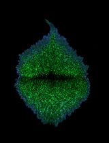
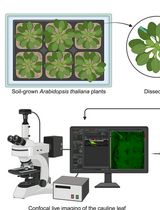
In vivo imaging of brown algal cells in 3D is extremely challenging because of the presence of pigments, such as fucoxanthin and chlorophyll, that diffract light. Moreover, brown algae live in seawater, a high ionic environment that can change the fluorochrome behavior or cause aggregates. Despite the importance of in vivo monitoring the developmental process of brown algal tissues, 4D imaging (x, y, z, t) on a conventional fluorescence microscope is limited. Here, we propose a detailed protocol using a new orange-emitting fluorochrome, styryl benzoindoleninium sulfonate (SBIS), suitable for labeling the plasma membrane of brown algal cells and multicolor in vivo imaging in 3D using confocal and light sheet microscopy. Unlike calcofluor white (CFW), SBIS enables the observation of brown algal cells at thicknesses up to 25 μm and over periods up to 7 days on brown algae such as Ectocarpus sp., Sphacelaria rigidula, and Saccharina latissima. This step-by-step protocol includes labeling of brown algal tissues, mounting for 3D confocal time-lapse microscopy, and mounting for 3D time-lapse light sheet microscopy. The imaging setup and parameters have been optimized for minimizing toxicity for brown algal tissues, improving signal-to-noise ratio, and enabling detailed visualization of cell shape. Therefore, this protocol provides robust and multiplexed imaging with 4D visualization of brown algal cell shape throughout the brown algae growth, offering broad applications to brown algae study at the cellular level.
Key features
• Introducing a new orange-emitting fluorochrome, styryl benzoindolenium sulfonate (SBIS), labeling the plasma membrane of brown algae.
• A step-by-step protocol to visualize brown algal cell shape in 3D using confocal and light sheet microscopy, suitable for live in vivo imaging.
• Live 4D imaging without interfering with the brown algae growth.
• Adaptable to different brown algae, including Ectocarpus sp., Sphacelaria rigidula, and Saccharina latissima.
Keywords: Brown algae (褐藻) Plasma membrane probe (质膜探针)
In vivo imaging (活体成像)
3D (三维成像)
Confocal microscope (共聚焦显微镜)
Light sheet microscope (光片显微镜)
Plasma membrane probe (质膜探针)
In vivo imaging (活体成像)
3D (三维成像)
Confocal microscope (共聚焦显微镜)
Light sheet microscope (光片显微镜)
Background
The study of cell shapes and sizes in 3D during embryogenesis and growth is a widely studied topic in metazoans, plants, and brown algae. Brown algae (Phaeophyceae) are photosynthetic organisms and one of the five groups of eukaryotes that have acquired complex multicellularity and evolved independently of red algae, green plants (including green algae and land plants), animals, and fungi. In multicellular organisms, 3D growth is considered an intricate morphogenetic process. In metazoans, cellular migration commonly drives the establishment of growth axes in multicellular eukaryotes growing in 3D. However, brown algal cells, like plant cells, are surrounded by a semi-rigid cell wall that cements them together. Consequently, the orientation of 3D growth relies on changes in the cell division planes at specific locations. The abundant natural carotenoid and chlorophyll a pigments present in the thylakoid membranes of brown algal chloroplasts [1] absorb blue-green light (excitation wavelength, 480–500 nm) and emit red light (emission wavelength, 646–655 nm for carotenoid and 677–680 nm for chlorophyll a [2]); this characteristic makes in vivo imaging challenging. Moreover, despite the emergence of genome editing tools in brown algae, which have enabled a knockout to be achieved in Ectocarpus sp. [3], knock-ins are not yet feasible. Therefore, along with the development of microscopy techniques enabling time-lapse studies of these organisms in 3D, research aiming at developing fluorochromes labeling specific compartments of brown algal cells is essential. Several fluorescent dyes have been used to label cellular structures in brown algal tissues, but each presents limitations for long-term 4D imaging. Calcofluor white (CFW), a UV-absorbing and blue-emitting fluorochrome, is capable of forming hydrogen bonds with β (1→4) and β (1→3) polysaccharides, thus binding mainly to the chitin and cellulose present in the cell wall of brown algal cells [4,5]. However, the use of CFW reaches a limit when imaging living organisms in 3D over a long period of time, as UV increases the generation of reactive oxygen species that are toxic to living organisms. Furthermore, blue light is less penetrating into the living tissues than longer wavelengths. Fluorochromes labeling the plasma membrane, like FM1-43 [N-(3-Triethylammoniumpropyl)-4-(4-(Dibutylamino) Styryl) Pyridinium Dibromide] and FM4-64 [dibromure de N-(3-triethylammoniumpropyl)-4-(6-(4-(diethylamino) phenyl) hexatrienyl) pyridinium] are internalized by endocytosis within minutes after labeling Ectocarpus sp., S. rigidula, and S. latissima cells. As a result, they are not suitable for a 4D study of brown algae embryogenesis and growth. The aim of this study is to propose a protocol for visualizing brown algal cells in 4D by optimizing mounting for confocal and light sheet microscopy, adjusting acquisition parameters to minimize phototoxicity, and carefully selecting a fluorochrome that allows clear visualization of the plasma membrane.
Materials and reagents
Biological materials
1. Ectocarpus sp. produced from fragmentation of the wild-type male strain Ec32 (CCAP 1310/335; origin: San Jan de Marcona, Peru) parthenosporophytes
2. Embryos of S. latissima, produced from fertilization of the female gametophyte strain F1 with the male strain M1 as described by [6]
3. Female gametophytes of S. rigidula, grown from the fragmentation of adult cultures
Note: All alga thalli were grown in full-strength Provasoli-enriched [7] autoclaved natural or artificial seawater (pH 7.8) (Tropic Marin®) in a culture cabinet at 13 °C with a 12:12 light:dark cycle (light intensity, 29 μmol photon/m2/s) [8].
Reagents
1. Sea salt (Tropic Marin® classic sea salt)
2. Low-melting agarose (Sigma-Aldrich, catalog number: A9045)
Solutions
1. Artificial sea water (ASW) (see Recipes)
2. Low-melting agarose (see Recipes)
3. styryl benzoindoleninium sulfonate (SBIS) (see Recipes)
Recipes
1. Artificial sea water (ASW)
| Reagent | Final concentration | Quantity or volume |
|---|---|---|
| Tropic Marin® classic sea salt | 35 g/L | 35 g |
| MilliQ water | n/a | 1 L |
Filter ASW with a 0.2 μm filter to remove big salt crystals and other particles, then autoclave and store at 4 °C.
2. Low-melting agarose
| Reagent | Final concentration | Quantity or volume |
|---|---|---|
| Low-melting agarose Sigma-Aldrich #A9045 | 1% | 1 g |
| ASW | n/a | 100 mL |
Store at 4 °C.
3. SBIS
| Reagent | Final concentration | Quantity or volume |
|---|---|---|
| SBIS | 10 μM | 10 μL |
| ASW | n/a | 990 μL |
Store at 4 °C.
Laboratory supplies
1. 20 mm diameter glass-bottom cell culture dish, with glass of standard thickness 0.16–0.19 mm (NEST Biotech, catalog number: 801001)
2. Petri dish (Greiner, Standard, sterile, 20 PCS/BAG, catalog number: 632181)
3. Parafilm (Amcor, Parafilm “M” All-Purpose Laboratory film, catalog number: PM-996)
4. Glass capillary (Brand, transferpettor caps 10 μL, catalog number: 701902)
5. Glue (Leroy Merlin®, catalog number: 62928642)
Equipment
1. Confocal microscope (Zeiss, model: LSM 880)
2. Light sheet microscope (Zeiss, model: Light sheet 7)
Software and datasets
1. Zen Blue, Carl Zeiss Microscopy, LLC software
2. Fiji (open source, available at http://fiji.sc/Fiji; [9])
Procedure
登录/注册后免费查看全文
文章信息
稿件历史记录
提交日期: Mar 11, 2026
接收日期: May 5, 2026
在线发布日期: Jun 10, 2026
出版日期: Jul 5, 2026
版权信息
© 2026 The Author(s); This is an open access article under the CC BY-NC license (https://creativecommons.org/licenses/by-nc/4.0/).
如何引用
Zilliox, M. and Charrier, B. (2026). 4D Imaging of Brown Algal Cells. Bio-protocol 16(13): e5730. DOI: 10.21769/BioProtoc.5730.

 Download Citation in RIS Format
分类
植物科学 > 藻类学 > 细胞分析
细胞生物学 > 细胞成像 > 活细胞成像
细胞生物学 > 细胞成像 > 共聚焦显微镜
您对这篇实验方案有问题吗?
在此处发布您的问题,我们将邀请本文作者来回答。同时,我们会将您的问题发布到Bio-protocol Exchange,以便寻求社区成员的帮助。
 发布问题
发布问题


