


Visualizing Membrane Nanotube Dynamics in Drosophila Oocyte Using Live-Cell Imaging
利用活细胞成像观察果蝇卵母细胞中膜纳米管的动态变化

(*contributed equally to this work) 发布: 2026年07月05日第16卷第13期 DOI: 10.21769/BioProtoc.5728 浏览次数: 319
评审: Sreesankar EaswaranAnonymous reviewer(s)
 PDF
PDF  Q&A
Q&A
参见作者原研究论文
The authors used this protocol in:
Dec 2025
Advertisement







Abstract
Thin membrane protrusions in cells help them communicate, create traction forces during their movement, and coordinate complex development in multicellular organisms. These structures include cytonemes, tunneling nanotubes, and microtubule-based nanotubes (MT-nanotubes), each with a different cytoskeletal constitution and function. Actin-based cytonemes help deliver signaling molecules, while microtubule-based nanotubes assist with transporting vesicles and organelles. Despite their physiological role, we still do not fully understand how these thin membrane protrusions form and function. In this study, we introduce an improved live-cell imaging method to observe polar cell protrusions during micropyle morphogenesis in developing Drosophila eggs. This technique combines precise developmental staging, careful dissection, and optimized ex vivo culture conditions to maintain tissue health during extended imaging. We also fine-tuned the imaging settings to reduce phototoxicity and thermal stress. This allows for continuous, high-resolution tracking of protrusion dynamics in real time. Our protocol addresses major drawbacks of fixed-tissue methods by capturing the entire process of protrusion formation, extension, and remodeling in intact living tissue. Additionally, it works well with drugs, making it a useful tool for functional studies. Overall, this approach builds a strong foundation for exploring membrane protrusion biology. It can also be applied to investigate similar developmental processes in other systems, aiding our understanding of normal development and diseases.
Key features
• We optimized a live-imaging protocol ensuring accurate staging and fine dissection of Drosophila egg chambers for reproducible polar cell nanotube visualization.
• The enhanced culture conditions maintain egg chamber viability for extended periods, enabling the continuous, real-time observation of dynamic membrane protrusion formation.
• The optimized image acquisition settings minimize phototoxicity and sample heating, preventing imaging-induced artifacts and ensuring the acquisition of high-resolution, reproducible datasets while preserving tissue health.
• This protocol supports pharmacological treatments for functional perturbation experiments and can be adapted to study similar developmental processes across various other insect systems.
Keywords: MT-Nanotube (MT-纳米管) Drosophila (果蝇)
Oogenesis (卵子发生)
Micropyle (卵孔)
Polar cells (极细胞)
Germline development (生殖系发育)
Arthropoda (节肢动物门)
Drosophila (果蝇)
Oogenesis (卵子发生)
Micropyle (卵孔)
Polar cells (极细胞)
Germline development (生殖系发育)
Arthropoda (节肢动物门)
Graphical overview
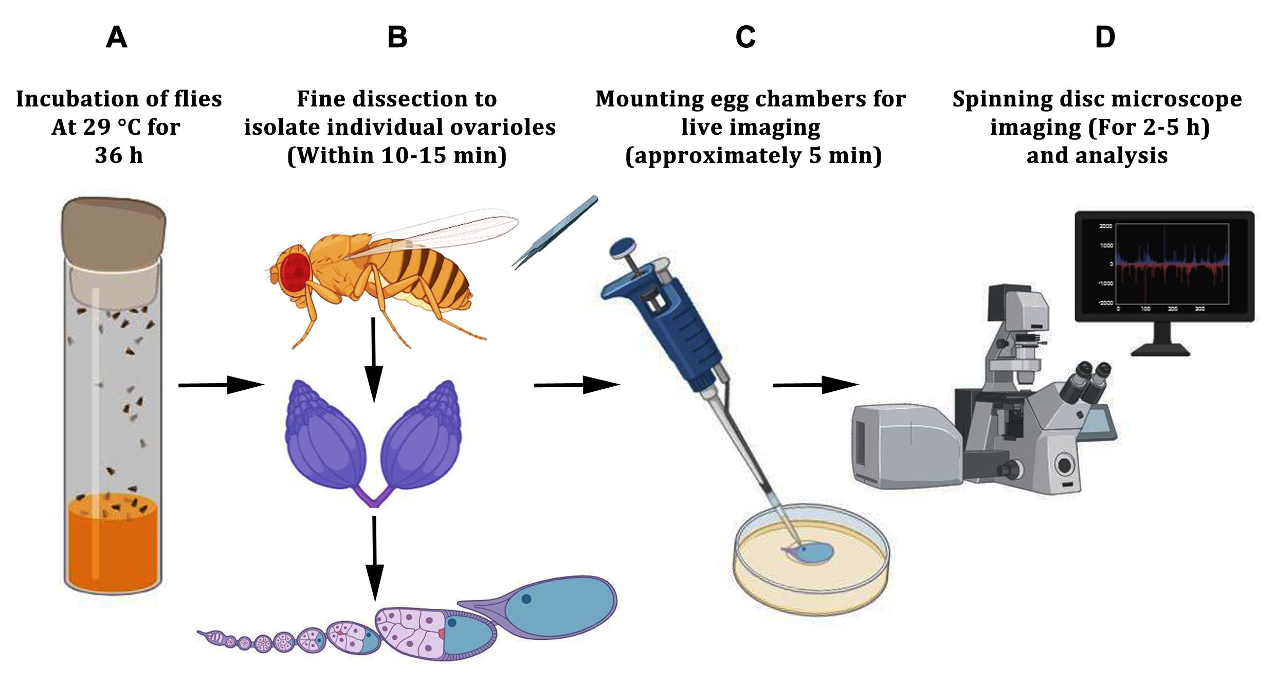
Schematic representation of the workflow for live-cell imaging of desired egg chambers. (A) Five to six females, along with 2–3 males, are incubated in a vial containing fly food supplemented with dry yeast. The flies are incubated at 29 °C to induce the development of egg chambers (termed as fly fattening). (B) The fattened female flies are dissected into live imaging media. The ovaries are taken out of the abdomen using a pair of forceps, followed by gently pulling out the individual egg chambers from the sheath of the ovarioles. (C) Using a 200 μL tip, the desired egg chambers, along with dissection media, are carefully aspirated and evenly distributed onto a poly-D-Lysine-coated coverslip in the imaging dish. (D) Imaging is carried out using a spinning disk confocal microscope. The images are analyzed using Fiji software.
Background
Membrane protrusions are specialized cellular structures that perform diverse functions, including environmental sensing, generation of traction forces during cell migration, and mediation of intercellular communication. Beyond their roles in normal development and cellular homeostasis, these structures are also implicated in disease progression [1]. In developing multicellular organisms, membrane extensions enable long-range communication between cells, which is crucial for coordinating complex morphogenetic processes.
Membrane protrusions exhibit substantial structural and functional diversity and are described by various terms, such as cytonemes, tunneling nanotubes (TNTs), membrane nanotubes, and microtubule-based nanotubes (MT-nanotubes) [2,3]. While these structures can connect distant cells, they differ in cytoskeletal composition and function. Cytonemes are predominantly actin-based extensions that facilitate the targeted delivery of signaling molecules to neighboring cells [4]. In contrast, nanotubes are generally microtubule-based and support a broader range of cellular activities, including the transport of vesicles, organelles, signaling molecules, and survival factors [5,6]. Long, narrow protrusions (typically less than 1.5 μm wide) that span several cell diameters are commonly classified as MT-nanotubes [7]. When these nanotubes form open connections between cells, they enable direct cytoplasmic exchange. While this connectivity can support normal physiological communication, it can also be exploited by pathogens or cancer cells to promote survival and disease progression.
Closed membrane nanotubes, on the other hand, often function in paracrine or juxtacrine signaling and play essential roles in tissue morphogenesis [8]. For example, in the Drosophila male germline, closed nanotubes mediate communication between germline stem cells and hub cells, maintaining stem cell identity [9]. Similarly, actin-based protrusions coordinate myoblast fusion during muscle development, and cytonemes facilitate long-range morphogen signaling during Drosophila wing and eye development [10,11]. Despite the diversity, the mechanisms underlying nanotube formation and function remain poorly understood. Drosophila micropyle development provides a useful model to study how thin membrane protrusions contribute to organ morphogenesis. The micropyle is a tubular structure at the anterior end of the eggshell that allows sperm entry during fertilization [12]. From stage 12 onward, a thin protrusion from the polar cells extends into the developing micropyle and generates an open channel in the eggshell that serves as an entry point for fertilizing sperm (Figure 1A–E) [13].
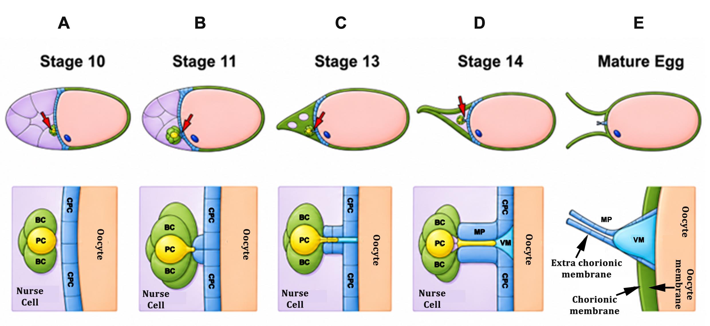
Figure 1. Schematic of stage-wise tubular morphogenesis of the micropyle at late oogenesis. (A) By stage 10, the border cell (BC) cluster has completed its migration and reaches the oocyte, while the centripetal follicle cells (CFCs) begin migrating inward from both the dorsal and ventral sides of the egg chamber. These CFCs move to enclose the anterior region of the oocyte, creating a partition between the nurse cells (NCs) and the oocyte. (B) At stage 11, CFC migration is completed, and they initiate the early steps of tubular morphogenesis of the micropyle. Concurrently, polar cells (PCs), located centrally within the BC cluster, begin to form a protrusion directed toward the oocyte surface, marking the tip of the developing micropyle. (C–E). From stage 13 onward, this protrusion elongates into a narrow, thin extension that pierces the forming tubular structure, contributing to the creation of an open channel that serves as the conduit for fertilizing sperm. In the schematic representation, NCs are depicted in pastel purple, the oocyte in pastel magenta, BCs in green, PCs in yellow, CFCs in blue, the vitelline membrane (VM) in sky blue, and the complete extra chorionic structure known as micropyle (MP). (Images were modified from Acharjee et al. [13].)
A major challenge in studying these tubular structures is that they are extremely thin, fragile, and dynamic. Fixed-tissue imaging provides useful structural information but captures only static snapshots, making it difficult to resolve protrusion formation, extension, remodeling, and interactions with surrounding cells. In addition, delicate membrane structures may be distorted or lost during fixation. Thus, live-cell imaging offers a key advantage by enabling direct visualization of protrusion dynamics in real time and revealing the cellular events that drive morphogenesis.
Here, we describe an optimized live-cell imaging protocol for visualizing the polar cell protrusion during micropyle morphogenesis in Drosophila egg chambers. The protocol includes methods for accurate staging and fine dissection of egg chambers, which are essential for isolating intact samples at the appropriate developmental window. We also optimized the ex vivo culture conditions to maintain egg chamber viability for extended imaging periods, allowing continuous observation of this dynamic event. In addition, imaging parameters were refined to minimize phototoxicity and sample heating during prolonged acquisition, preserve tissue health, and prevent imaging-induced artifacts. These optimizations improve sample stability and image quality, enabling reproducible acquisition of high-resolution datasets across the complete morphogenetic process. The protocol is also compatible with pharmacological treatments, providing a practical platform for functional perturbation experiments in isolated egg chambers. Overall, this method offers a robust and accessible approach for studying dynamic membrane protrusions in living tissue and, with minor modifications, may be adapted to investigate related developmental processes in other insect systems.
Materials and reagents
Biological materials
1. Drosophila stocks: Flies with the fluorescent reporter UAS mCD8::GFP (BL-5237), UAS GFP (BL-6874) were employed; upd-GAL4 (gifted by Dr. Harrison) was used for driving the overexpression of fluorescent reporter in the polar cells
Reagents
1. Non-absorbent cotton wool (Bengal Surgicals Limited)
2. Local sugar
3. Agar powder (SRL, catalog number: 19661)
4. Yeast (AB-MAURI)
5. Cornflour (Ganesh)
6. Malt (SRL, catalog number: 83358)
7. Propionic acid (Merck, catalog number: DH3D-731323)
8. Orthophosphoric acid (Merck, catalog number: CL9C690802)
9. Benzyl-benzoate (LOBA Chemie, catalog number: 0004700500)
10. Ethanol (Merck, catalog number: 1.00983.0511)
11. Phosphate buffered saline (PBS) (Sigma, catalog number: P3813)
12. Schneider's media (Life Technologies, catalog number: 21720-024)
13. Insulin, 10 mg/mL (Sigma, catalog number: I5550) dissolved in acidified water
14. Fetal bovine serum (FBS) (Gibco, catalog number: A5256801)
15. poly-D-Lysine (Sigma-Aldrich, catalog number: P7280)
16. Halocarbon oil 27 (Sigma-Aldrich, catalog number: H8773)
17. Pen-strep (Life Technologies, catalog number: 15140-122)
18. Fungizone (HyClone, catalog number: SV30078.01)
19. Sodium hydroxide pellets purified (Merck, catalog number: 1064980500)
20. Acetic acid glacial, 90%–100% (Merck, catalog number: 1.93402.0521)
Solutions
1. Live imaging Schneider’s medium cocktail (see Recipes)
2. Fly food media (see Recipes)
3. Acidified water (see Recipes)
Recipes
1. Live imaging Schneider’s medium cocktail (for 15 mL) [14]
| Component | Concentration | Composition (in 15 mL) |
|---|---|---|
| Schneider’s medium | - | 10.8 mL |
| FBS | 15% | 2.25 mL |
| Pen/strep | 0.01% | 1.5 μL |
| Fungizone | 0.001% | 0.15 μL |
| Insulin | 0.2 mg/mL | 300 μL (add fresh before use) |
2. Fly food media (for 1 L)
| Component | Composition (in 1 L) |
|---|---|
| Cornflour | 75 g |
| Sugar | 80 g |
| Dry yeast | 15 g |
| Malt extract (3%) | 30 g |
| Agar-agar | 10 g |
| Water | Up to 1 L |
| Propanoic acid | 5 mL |
| Orthophosphoric acid | 1 mL |
| Benzyl-benzoate | 5 mL (from 5% solution in EtOH) |
3. Acidified water
Add 1 μL of concentrated 37% HCl to 1 mL of water.
Laboratory supplies
1. Fly culture vials (Crystal make)
Equipment
1. Incubator at 25 and 29 °C for Drosophila stock maintenance and cross-incubation
2. Laminar flow hood (BIOAIR, model: s@feflow0.9)
3. Stereo microscope (Olympus, model: SZX16)
4. Epi fluorescence microscope (Olympus, models: IX81, IX83)
5. Confocal microscope (Nikon, model: AX, with NIS Elements software)
6. Spinning disk microscope (Olympus, model: IX83 spinning disk microscope)
7. Greiner Lumox culture dish hydrophilic (50 mm) (Sigma, catalog number: Z376744)
8. Paintbrush (1–5 size range) (Dayal Series-68 Taklon Round P)
9. Grooved slide (Samtech Instruments)
10. Dumont #5 forceps (Fine Science Tools, catalog number: 11251-20)
11. Micropipette with disposable tips (1,000 μL, 200 μL, 20 μL, 10 μL) (Gilson)
12. No. 1 coverslips (Fisher, catalog number: 12-542-B)
13. pH meter (BR Biochem)
Software and databases
1. ImageJ/Fiji (ImageJ 1.53f51)
2. Cell sense dimension (V 4.4)
3. NIS-elements (AR.20.00)
4. Microsoft Excel (Microsoft Office 365)
5. GraphPad Prism 9 and 6
Procedure
登录/注册后免费查看全文
文章信息
稿件历史记录
提交日期: Mar 25, 2026
接收日期: May 13, 2026
在线发布日期: Jun 3, 2026
出版日期: Jul 5, 2026
版权信息
© 2026 The Author(s); This is an open access article under the CC BY-NC license (https://creativecommons.org/licenses/by-nc/4.0/).
如何引用
Saha, B., Acharjee, S., Nandi, J. and Prasad, M. (2026). Visualizing Membrane Nanotube Dynamics in Drosophila Oocyte Using Live-Cell Imaging. Bio-protocol 16(13): e5728. DOI: 10.21769/BioProtoc.5728.

 Download Citation in RIS Format
分类
发育生物学 > 形态建成 > 细胞结构
发育生物学 > 细胞生长和命运决定 > 卵母细胞
细胞生物学 > 细胞成像 > 活细胞成像
您对这篇实验方案有问题吗?
在此处发布您的问题,我们将邀请本文作者来回答。同时,我们会将您的问题发布到Bio-protocol Exchange,以便寻求社区成员的帮助。
 发布问题
发布问题


