


Stepwise Differentiation of Mouse Embryonic Stem Cells Into Murine Blood Vessel Organoids With Endothelial Lineage Tracing for Quality Control
将小鼠胚胎干细胞逐步分化为小鼠血管类器官并通过内皮细胞谱系追踪进行质量控制

(§ Technical contact) 发布: 2026年07月05日第16卷第13期 DOI: 10.21769/BioProtoc.5727 浏览次数: 804
评审: Jessica DavisThirupugal GovindarajanNeha Saxena
 PDF
PDF  Q&A
Q&A
参见作者原研究论文
The authors used this protocol in:

Oct 2025
Advertisement






Abstract
In vitro vascular models are most informative when they recapitulate endothelial assembly within a 3D microenvironment. Blood vessel organoids (BVOs) enable the study of vascular heterogeneity, function, and organ-instructive cues in development, homeostasis, and disease. Here, we present a robust stepwise method to generate murine blood vessel organoids (mBVOs) from feeder-dependent mouse embryonic stem cells (mESCs) of common genetic backgrounds. Embryoid bodies (EBs) are formed using strain-specific seeding densities (day 0–3), followed by mesoderm induction (day 3–6) and vascular induction (day 6–8). Induced EBs are embedded in collagen I with Geltrex to drive sprouting and network formation (day 8–13). Vascular networks are microdissected and grown in suspension to yield mature mBVOs (day 21–30). The inclusion of a Cre-inducible VE-cadherin-GFP reporter line enables a quantitative quality control, reducing variability by excluding poorly differentiated organoids. The protocol reliably produces ~100 mBVOs per differentiation and is compatible with engineered mouse strains for gain- and loss-of-function studies, functional assays of vascular plasticity, and syngeneic grafting to assess perfusion. Thus, mBVOs provide a scalable and traceable 3D platform that bridges endothelial assays, mouse models, and human organoid systems.
Key features
• A detailed timeline to differentiate feeder-dependent mESCs into mBVOs, with key success readouts and troubleshooting.
• Efficient across three genetic backgrounds with strain-specific EB seeding densities and typical yields of ~100 mBVOs per differentiation.
• The inducible VE-cadherin-GFP lineage tracing/reporter system provides an endothelial quality control to quantify efficiency and exclude poorly differentiated organoids.
• Compatible with engineered mouse strains for gain/loss-of-function, with in vitro assays of vascular plasticity/remodeling, and with syngeneic in vivo validation.
Keywords: Murine blood vessel organoids (mBVOs) (小鼠血管类器官) Mouse embryonic stem cells (mESCs) (小鼠胚胎干细胞)
Feeder-dependent culture (饲养层依赖性培养)
Embryoid bodies (EBs) (胚状体)
Vascular differentiation (血管分化)
Sprouting angiogenesis (出芽性血管生成)
VE-cadherin (Cdh5) lineage tracing (VE-钙黏蛋白谱系追踪)
Quality control of differentiation (分化质量控制)
Mouse embryonic stem cells (mESCs) (小鼠胚胎干细胞)
Feeder-dependent culture (饲养层依赖性培养)
Embryoid bodies (EBs) (胚状体)
Vascular differentiation (血管分化)
Sprouting angiogenesis (出芽性血管生成)
VE-cadherin (Cdh5) lineage tracing (VE-钙黏蛋白谱系追踪)
Quality control of differentiation (分化质量控制)
Graphical overview
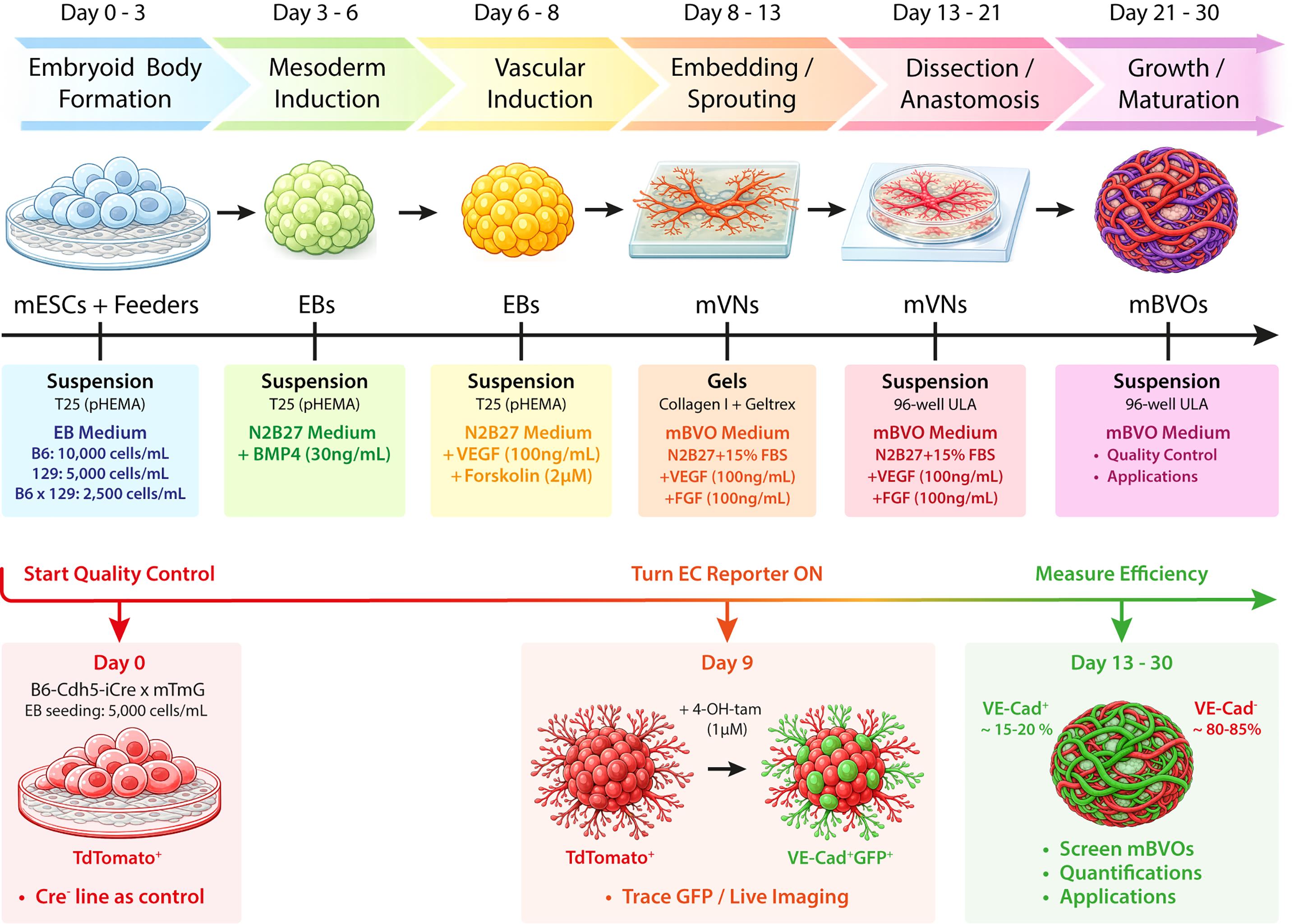
Workflow for the differentiation of mESCs into mBVOs, including the B6-Cdh5-iCre × mTmG reporter-based quality control. 129, 129S6 wild-type mouse strain; 4-OH-tam, 4-hydroxytamoxifen; B6, C57BL/6 wild-type mouse strain; B6x129, C57BL/6x129S6 F1 hybrid mouse strain; BMP, bone morphogenic protein 4; EB, embryoid body; EC, endothelial cell; FBS, fetal bovine serum; FGF, fibroblast growth factor; GFP, green fluorescent protein; mBVO, murine blood vessel organoid; mESC, murine embryonic stem cell; mVN, murine vascular network; pHEMA, poly(2-hydroxyethyl methacrylate); T25, cell culture flasks (25 cm2); ULA, ultra-low attachment; VE-Cad, vascular endothelial cadherin; VEGF, vascular endothelial growth factor.
Background
Modeling blood vessels in vitro has long been technically challenging because a functional vasculature is not solely defined by endothelial cells. In vivo, endothelial tubes are supported by mural/perivascular cells, a basement membrane, and the surrounding stroma, altogether maintaining barrier function and stability [1–3].
As a result, 2D endothelial cultures or simple sprouting assays are well-suited for endothelial-centered studies, but they provide limited insight into how multicellular vascular units self-organize and adapt within vascular beds [4]. More complex co-culture and matrix-based systems increase physiological relevance, yet they are typically optimized to answer narrow questions rather than capturing a broader, self-organizing vascular architecture that can be compared across experiments [5,6].
Human blood vessel organoids (hBVOs), initially described by Wimmer and colleagues, marked a major step forward. Taking advantage of the multilineage potential of pluripotent stem cells, they self-assemble into complex vascular structures in 3D, including endothelial, mural, mesenchymal, and immune compartments [7–9]. Since the original reports, hBVOs have been incorporated into a variety of organ-instructed assembloid systems. Notable examples include their integration with neural tissues [10,11] and their combination with pancreatic islets [12]. Beyond these applications, hBVOs have been widely used for disease modeling, including studies of diabetes [7], CNS malformations [13], neurodegeneration [14,15], aging [16], sepsis [17], and cancer metastasis [18].
Like other organoid systems [19], hBVOs offer clear advantages but also carry important limitations. A major strength is their ability to recapitulate human vascular organization and tissue-context effects. However, the system has practical limits. Human pluripotent stem cells are not always readily available, and hBVOs lack a substantial immune component, which restricts their use in vascular-immune and inflammation-focused studies [20]. Moreover, although perfusion can be engineered in vitro [21], many long-term perfusion and remodeling assays rely on transplantation into immunocompromised mice, which complicates the interpretation of immune-related processes [7,22].
Vascular studies have long been performed in mice because of their genetic traceability and physiological relevance [23]. Yet, there are significant differences between mouse and human vascular gene-expression programs [24,25]. Importantly, there is a strong push to replace, reduce, and refine animal experiments when possible [26], in accordance with European legislation (Directive 2010/63/EU; available at https://eur-lex.europa.eu/eli/dir/2010/63/oj/eng).
In this protocol, we describe the stepwise differentiation of mouse embryonic stem cells (mESCs) into murine blood vessel organoids (mBVOs) following a defined timeline [27]. We adapted the hBVO method logic to the requirements of feeder-dependent mESC cultures grown on mitotically inactivated mouse embryonic fibroblasts (MEFs). The protocol is reproducible with distinct genetic backgrounds and uses strain-specific embryoid body (EB) seeding densities to enhance reproducibility across lines. During routine differentiations, we typically obtain a consequent number of mBVOs (~100), enabling sufficient replicates for quantitative assays and statistical analyses [27].
A common challenge in organoid differentiation is achieving—and accurately quantifying—differentiation efficiency. To address this, we include an endothelial lineage-tracing module using B6-Cdh5-iCre × mTmG mESCs. After 4-hydroxytamoxifen (4-OH-tam) induction in vitro, VE-cadherin-expressing cells switch from membrane tdTomato to membrane GFP, allowing identification and exclusion of poorly differentiated organoids. Cre-negative controls define the tomato-only baseline [27,28]. This approach offers a practical advantage over strategies that rely mostly on endpoint marker staining or morphology, and it provides a standardized method for batch selection [27].
Because the system is based on mESCs, it is compatible with genetically engineered mouse strains, enabling gain- or loss-of-function studies targeting the endothelial compartment, as well as mural and immune cells, if suitable Cre tools are available. Finally, mBVOs provide a platform to study vascular plasticity and remodeling in vitro. In our original study, we used mBVOs to assess responses to pro-inflammatory cues, anti-angiogenic perturbations, and tumor–vasculature interactions using glioblastoma assembloids. We also demonstrated perfusion after syngeneic implantation in mice [27].
Together, mBVOs bridge the gap between mouse models, endothelial assays, and human BVO platforms. They provide a scalable murine 3D vascular system that is genetically traceable, with a quantitative quality control step to measure differentiation efficiency. We expect this protocol to enhance the reproducibility of BVO generation and to facilitate mechanistic studies that connect murine genetics with questions rooted in human vascular biology and disease.
Materials and reagents
Biological materials
1. Mouse embryonic stem cells (mESCs), C57BL/6 × 129S6 F1 hybrid (B6x129)
2. mESCs, C57BL/6 wild type (B6)
3. mESCs, 129S6 wild type (129)
4. mESCs, B6-Cdh5-iCre × mTmG (VE-cadherin lineage tracing line)
5. mESCs, B6-Cdh5-iCre × mTmG Cre-negative (tdTomato-only control)
6. Mouse embryonic fibroblasts (MEFs), isolated from mouse embryos and expanded as primary cultures before mitomycin C (MMC) inactivation and cryo-storage as feeder stocks
Note: mESC strains and primary MEFs were previously isolated and provided by the laboratories of Prof. Pieter Carmeliet and Prof. Kian Peng Koh, following previously described procedures [29–37].
Reagents
mESC/MEF culture
1. KnockOut DMEM (Thermo Fisher Scientific, catalog number: 10829-018)
2. ESC-qualified fetal bovine serum (FBS) (Thermo Fisher Scientific, catalog number: 10270106)
3. L-glutamine (Thermo Fisher Scientific, catalog number: 25030-024)
4. Sodium pyruvate (Thermo Fisher Scientific, catalog number: 11360-039)
5. Non-essential amino acids (Thermo Fisher Scientific, catalog number: 11140-035)
6. 2-mercaptoethanol (Sigma-Aldrich, catalog number: M6250); store at 4 °C
7. Penicillin-streptomycin (Thermo Fisher Scientific, catalog number: 15140-122)
8. Mouse leukemia inhibitory factor (mLIF) (PeproTech, catalog number: 250-02)
9. DMEM high glucose (Thermo Fisher Scientific, catalog number: 41965-062)
10. Trypsin-EDTA 0.25% (Thermo Fisher Scientific, catalog number: 25200056)
11. Gelatin, from porcine skin (Sigma-Aldrich, catalog number: G1890)
12. Distilled water, cell culture grade (Thermo Fisher Scientific, catalog number: 15230162)
13. D-PBS without calcium and magnesium (Thermo Fisher Scientific, catalog number: 14190094)
14. Mitomycin C (MMC) (Sigma-Aldrich, catalog number: M0503)
Differentiation (N2B27 medium + growth factors)
15. DMEM/F-12 (Thermo Fisher Scientific, catalog number: 11320-033)
16. Neurobasal medium (Thermo Fisher Scientific, catalog number: 21103-049)
17. N-2 supplement (Thermo Fisher Scientific, catalog number: 17502-048)
18. B-27 supplement (Thermo Fisher Scientific, catalog number: 17504-044)
19. Bovine albumin fraction V, 7.5% (Thermo Fisher Scientific, catalog number: 15260-037)
20. Monothioglycerol (Sigma-Aldrich, catalog number: D6145); store at 4 °C
21. Recombinant murine BMP4 (PeproTech, catalog number: 315-27)
22. Recombinant murine VEGF165 (PeproTech, catalog number: 450-32)
23. Forskolin (Bio-Techne, catalog number: 1099)
Embedding/matrices
24. MEM (10×), no glutamine (Thermo Fisher Scientific, catalog number: 21430020)
25. Ham’s F-12 (Thermo Fisher Scientific, catalog number: 11765054)
26. HEPES, 1 M (Thermo Fisher Scientific, catalog number: 15630080)
27. NaHCO3, 7.5% (Sigma-Aldrich, catalog number: S8761)
28. NaOH, 1.0 N (Sigma-Aldrich, catalog number: S2770)
29. Bovine collagen I (PureCol, 3 mg/mL) (Advanced BioMatrix, catalog number: 5005)
30. Geltrex LDEV-free reduced growth factor basement membrane matrix (Gibco, catalog number: A1413202)
mBVO maturation
31. Recombinant bFGF (PeproTech, catalog number: 450-33)
32. Fetal bovine serum (FBS), batch-tested (Biowest, catalog number: S1810)
Endothelial tracing for quality control
32. 4-hydroxytamoxifen (4-OH-tamoxifen) (Sigma-Aldrich, catalog number: SML1666)
Coating for embryoid body (EB) culture
33. Poly(2-hydroxyethyl methacrylate) (polyHEMA) (Sigma-Aldrich, catalog number: P3932)
34. Ethanol, 95% (VWR Chemicals, catalog number: 85829.360)
Solutions
1. ESC medium (see Recipes)
2. MEF medium (see Recipes)
3. Mitomycin C (MMC) (see Recipes)
4. Embryoid body (EB) medium (see Recipes)
5. N2B27 medium (N2B27) (see Recipes)
6. Mesoderm induction medium (N2B27 + BMP4) (see Recipes)
7. Vascular induction medium (N2B27 + VEGF + forskolin) (see Recipes)
8. Collagen I/Geltrex embedding mix (Embedding mix) (see Recipes)
9. mBVO maturation medium (mBVO medium) (see Recipes)
10. 0.1% gelatin coating solution (see Recipes)
11. 4-hydroxytamoxifen (4-OH-tamoxifen) working solution (QC module) (see Recipes)
12. polyHEMA coating solution (see Recipes)
Recipes
1. ESC medium (for feeder-dependent mESC maintenance)
| Reagent | Final concentration | Quantity or volume |
|---|---|---|
| KnockOut DMEM | n/a | to 500 mL |
| ESC-qualified FBS | 20% (v/v) | 100 mL |
| L-glutamine | 2 mM | 5 mL of 200 mM stock |
| Sodium pyruvate | 1 mM | 5 mL of 100 mM stock |
| Non-essential amino acids | 0.1 mM each | 5 mL of 100× stock |
| 2-mercaptoethanol | 0.1 mM | 6 μL |
| Penicillin-streptomycin | 100 U/mL (or 100 μg/mL) | 5 mL of 100× stock |
| mLIF | 20 U/mL (or 10 ng/mL) | 100 μL |
Prepare to a 500 mL total volume, filter-sterilize using a 0.22 μm PES membrane vacuum filter, and store at 4 °C for up to 2 weeks. Store mLIF stocks at -80 °C following your lab practices. ESC-qualified FBS has been previously batch tested and validated for mESC maintenance.
2. MEF medium
| Reagent | Final concentration | Quantity or volume |
|---|---|---|
| DMEM high glucose | n/a | to 500 mL |
| ESC-qualified FBS | 10% (v/v) | 50 mL |
| L-glutamine | 2 mM | 5 mL of 200 mM stock |
| Penicillin-streptomycin | 100 U/mL (or 100 μg/mL) | 5 mL of 100× stock |
Store at 4 °C for up to 4 weeks. Use the same batch of ESC-qualified FBS for this medium.
3. Mitomycin C (MMC)
Resuspend one vial of MMC powder in 4 mL of sterile D-PBS without calcium and magnesium to prepare a 0.5 mg/mL stock solution. Using a sterile 5 mL syringe, filter-sterilize the solution through a 0.22 μm PES syringe filter into a sterile 15 mL tube. Aliquot the filtered MMC stock, protect from light, and store at 4 °C for up to 1 week once resuspended.
Safety note: MMC is toxic. Prepare the stock solution under a biological safety cabinet, wear appropriate protection, and discard liquid and solid waste according to institutional chemical waste procedures.
4. EB medium
Prepare 250 mL of ESC medium (see Recipe 1) without mLIF, filter-sterilize using a 0.22 μm PES membrane vacuum filter, and store at 4 °C up to 2 weeks.
5. N2B27 medium (base medium used throughout differentiation)
| Reagent | Final concentration | Quantity or volume |
|---|---|---|
| DMEM/F-12 | 50% (v/v) | to 250 mL |
| Neurobasal medium | 50% (v/v) | to 250 mL |
| N-2 supplement | 0.5% (v/v) | 2.5 mL |
| B-27 supplement | 1% (v/v) | 5 mL |
| Bovine albumin fraction V, 7.5% | 0.5 mg/mL | 3.33 mL |
| L-glutamine | 2 mM | 5 mL of 200 mM stock |
| Penicillin-streptomycin | 100 U/mL (or 100 μg/mL) | 5 mL of 100× stock |
| Monothioglycerol | 150 μM | 6.3 μL |
Prepare to a 500 mL total volume, filter-sterilize using a 0.22 μm PES membrane vacuum filter, and store at 4 °C for up to 2 weeks.
6. Mesoderm induction medium
| Reagent | Final concentration | Quantity or volume |
|---|---|---|
| N2B27 medium | n/a | 30 mL |
| Recombinant BMP4 | 30 ng/mL | 9 μL (100 μg/mL stock) |
Prepare fresh on the day of use. Store BMP4 stocks at -80 °C following your lab practices.
7. Vascular induction medium
| Reagent | Final concentration | Quantity or volume |
|---|---|---|
| N2B27 medium | n/a | 50 mL |
| Recombinant VEGF165 | 100 ng/mL | 50 μL (100 μg/mL stock) |
| Forskolin | 2 μM | 10 μL (10 mM stock) |
Prepare fresh on the day of use. Store VEGF165 and forskolin stocks at -80 °C following your lab practices.
8. Collagen I/Geltrex embedding mix, pH 7.4
| Reagent | Final concentration | Quantity or volume |
|---|---|---|
| MEM (10×), no glutamine | n/a | 313 μL |
| Ham’s F-12 | n/a | 460 μL |
| HEPES, 1 M | n/a | 63 μL |
| NaHCO3, 7.5% | n/a | 49 μL |
| L-glutamine | n/a | 31 μL |
| Penicillin-streptomycin | n/a | 25 μL |
| Bovine collagen I, 3 mg/mL | n/a | 3.33 mL |
| NaOH, 1.0 N | adjust pH to 7.4 | 500 μL |
| Geltrex | n/a | 750 μL |
Prepare fresh on ice and use immediately. Mix by gentle inversion and slow pipetting to minimize bubble formation and to prevent premature gelation/precipitation.
Note: The first components listed (up to and excluding collagen I) can be combined in any order. After adding collagen I, adjust the pH to ~7.4 with 1.0 N NaOH, and add Geltrex last. This mix is used to prepare layer 1 and then layer 2 (~5.5 mL per layer) and is sufficient to cover an entire 12-well plate during the embedding step.
9. mBVO maturation medium
| Reagent | Final concentration | Quantity or volume |
|---|---|---|
| N2B27 medium | n/a | to 100 mL (84.8 mL) |
| FBS | 15% (v/v) | 15 mL |
| Recombinant VEGF165 | 100 ng/mL | 100 μL (100 μg/mL stock) |
| Recombinant bFGF | 100 ng/mL | 100 μL (100 μg/mL stock) |
Prepare fresh on the day of use. Store bFGF stocks at -80 °C following your lab practices.
Note: Use batch-tested FBS. ESC-qualified FBS is required for feeder-dependent mESC maintenance but is not required at this stage.
10. 0.1% gelatin coating solution
| Reagent | Final concentration | Quantity or Volume |
|---|---|---|
| Gelatin (porcine skin) | 0.1% (w/v) | 0.5 g |
| Distilled water | n/a | to 500 mL |
Dissolve, filter-sterilize using a 0.22 μm PES membrane vacuum filter, and store at 4 °C up to 1 month.
11. 4-hydroxytamoxifen (4-OH-tamoxifen) working solution
Prepare a 1 mM working solution by diluting the commercial 13 mM stock in ethanol. Aliquot (5 μL) and store at -20 °C protected from light (≤1 month). Avoid repeated freeze-thaw cycles and use fresh aliquots for each induction. Dilute in mBVO medium to a 1 μM final concentration and treat overnight on day 9.
12. polyHEMA 1% coating solution for EB suspension culture
| Reagent | Final concentration | Quantity or volume |
|---|---|---|
| polyHEMA | 1% (w/v) | 5 g |
| Ethanol, 95% | n/a | to 500 mL |
Dissolve with stirring and warming at 75 °C, filter-sterilize using a 0.22 μm PES membrane vacuum filter, and store at 4 °C for up to 1 month.
Laboratory supplies
1. Cell culture flasks, T25 (Corning, catalog number: 430639)
2. Cell culture plates, 6-well (Corning, catalog number: 3516)
3. Cell culture plates, 12-well (Corning, catalog number: 3513)
4. Cell culture dishes, 60 mm (Corning, catalog number: 430166)
5. Cell culture plates, 96-well round-bottom ultra-low attachment (ULA) (Corning, catalog number: 7007)
6. 15 mL conical sterile tubes (Corning, catalog number: 430790)
7. 50 mL conical sterile tubes (Corning, catalog number: 430828)
8. 1.5 mL microcentrifuge tubes (Eppendorf, catalog number: 0030120086)
9. Serological pipettes (5 mL) (Corning, catalog number: 4487)
10. Serological pipettes (10 mL) (Corning, catalog number: 4492)
11. Serological pipettes (25 mL) (Corning, catalog number: 4251)
12. Filter pipette tips, sterile, 10/20 μL (Corning Axygen, catalog number: TF-420-L-R-S)
13. Filter pipette tips, sterile 200 μL (Corning Axygen, catalog number: TF-200-L-R-S)
14. Filter pipette tips, sterile 1,000 μL (Corning Axygen, catalog number: TF-1000-L-R-S)
15. Pipette tips, non-filtered, sterile, 200 μL (Corning Axygen, catalog number: T-200-C-R-S
16. pH indicator strips (Merck, catalog number: 1.09543)
17. Vacuum filter bottles [500 mL, 0.22 μm, polyethersulfone (PES)] (Corning, catalog number: 431097)
18. Syringes, 5 mL, sterile, disposable (VWR, catalog number: 613-0917)
19. Sterile syringe filters (0.22 μm, PES) (VWR, catalog number: 514-4122)
20. Reagent reservoirs, sterile (25 mL) (VWR Collection, catalog number: 613-1175)
21. Plastic forceps (Fine Science Tools, catalog number: 11700-00)
22. Noyes spring scissors (straight) (Fine Science Tools, catalog number: 15012-12)
23. Double spatula (straight, flat/rounded ends, 9 mm blade width) (VWR, catalog number: 231-1034)
Equipment
1. Class II biological safety cabinet (Clean Air, model: EF/S 4)
2. Open biological cabinet with horizontal laminar flow (Telstar, model: Aeolus H5)
3. CO2 incubator, humidified, 37 °C, 5% CO2 (Binder, model: C170-230V-R)
4. Inverted phase-contrast microscope for routine culture (Leica, model: DM IL)
5. Inverted phase-contrast microscope for imaging (Zeiss, model: Axiovert S100)
6. Hemocytometer for cell counting (Blaubrand, model: Neubauer Improved Bright-Line)
7. Fluorescence microscope (for mTmG tdTomato/GFP readout) (Leica, model: Thunder)
8. Stereomicroscope (2×–10×) (Zeiss, model: Stemi 508)
9. LED Lamp, cold light source (Schott, model: KL1600 LED)
10. Refrigerated centrifuge with swinging-bucket rotor (Eppendorf, model: 5702R)
11. Water bath (room temperature to 37 °C) (Grant, model: SUB Aqua Pro 12L)
12. Analytical balance (Sartorius, model: Entris 124-1S)
13. Pipette, P10, 1–10 μL (Gilson, catalog number: F144802)
14. Pipette, P20, 2–20 μL (Gilson, catalog number: F123600)
15. Pipette, P200, 20–200 μL (Gilson, catalog number: F123601)
16. Pipette, P1000, 100–1,000 μL (Gilson, catalog number: F123602)
17. Multichannel pipette (30–300 μL) (Eppendorf, model: 3125000052)
18. Vacuum aspiration system with disinfectant trap (Integra Biosciences, Vacusafe 158320)
19. Aspiration pipette system with accessories (Integra Biosciences, Vacuboy hand operator set 155500)
20. Refrigerator (4 °C) and freezer (-20 °C) (Liebherr)
21. -80 °C freezer (for long-term reagent storage) (Thermo, model: UXF70086 ULT)
22. Dry incubator (room temperature to 60 °C) (VWR, model: VWI1296B03)
Software and datasets
1. Fiji (ImageJ) (NIH, version 1.54p), free
2. GraphPad Prism (GraphPad Software, version 10), license required
3. LasX Office (Leica, version 1.4.7.28982), license required
Procedure
登录/注册后免费查看全文
文章信息
稿件历史记录
提交日期: Feb 25, 2026
接收日期: May 6, 2026
在线发布日期: May 28, 2026
出版日期: Jul 5, 2026
版权信息
© 2026 The Author(s); This is an open access article under the CC BY-NC license (https://creativecommons.org/licenses/by-nc/4.0/).
如何引用
Readers should cite both the Bio-protocol article and the original research article where this protocol was used:
- Guelfi, S., Bopp, S. and Bergers, G. (2026). Stepwise Differentiation of Mouse Embryonic Stem Cells Into Murine Blood Vessel Organoids With Endothelial Lineage Tracing for Quality Control. Bio-protocol 16(13): e5727. DOI: 10.21769/BioProtoc.5727.
- Guelfi, S., Gopalan, K. S., Maene, P., Robbert, D. J., Pallarés-Moratalla, C., Vella, G., Nobis, M., Mourao, L., Bourdely, P., van der Veer, B. K., et al. (2025). Murine vascular organoids are responsive and adaptable 3D systems with cellular heterogeneity and dynamic plasticity. Sci Adv. 11(43): eady4738. https://doi.org/10.1126/sciadv.ady4738

 Download Citation in RIS Format
分类
干细胞 > 类器官培养
干细胞 > 胚胎干细胞 > 细胞分化
您对这篇实验方案有问题吗?
在此处发布您的问题,我们将邀请本文作者来回答。同时,我们会将您的问题发布到Bio-protocol Exchange,以便寻求社区成员的帮助。
 发布问题
发布问题


