


A Flow Cytometry–Based Assay to Quantify the Binding of Transmembrane Ligands to Their Cognate Receptors Using Fluorescent Virus-Like Particles
一种基于流式细胞术利用荧光病毒样颗粒定量检测跨膜配体与其相应受体结合的方法

发布: 2026年07月05日第16卷第13期 DOI: 10.21769/BioProtoc.5726 浏览次数: 559
评审: David PaulSukrut Chandrashekhar KamerkarAnonymous reviewer(s)
 PDF
PDF  Q&A
Q&A
参见作者原研究论文
The authors used this protocol in:
Feb 2026
Advertisement








Abstract
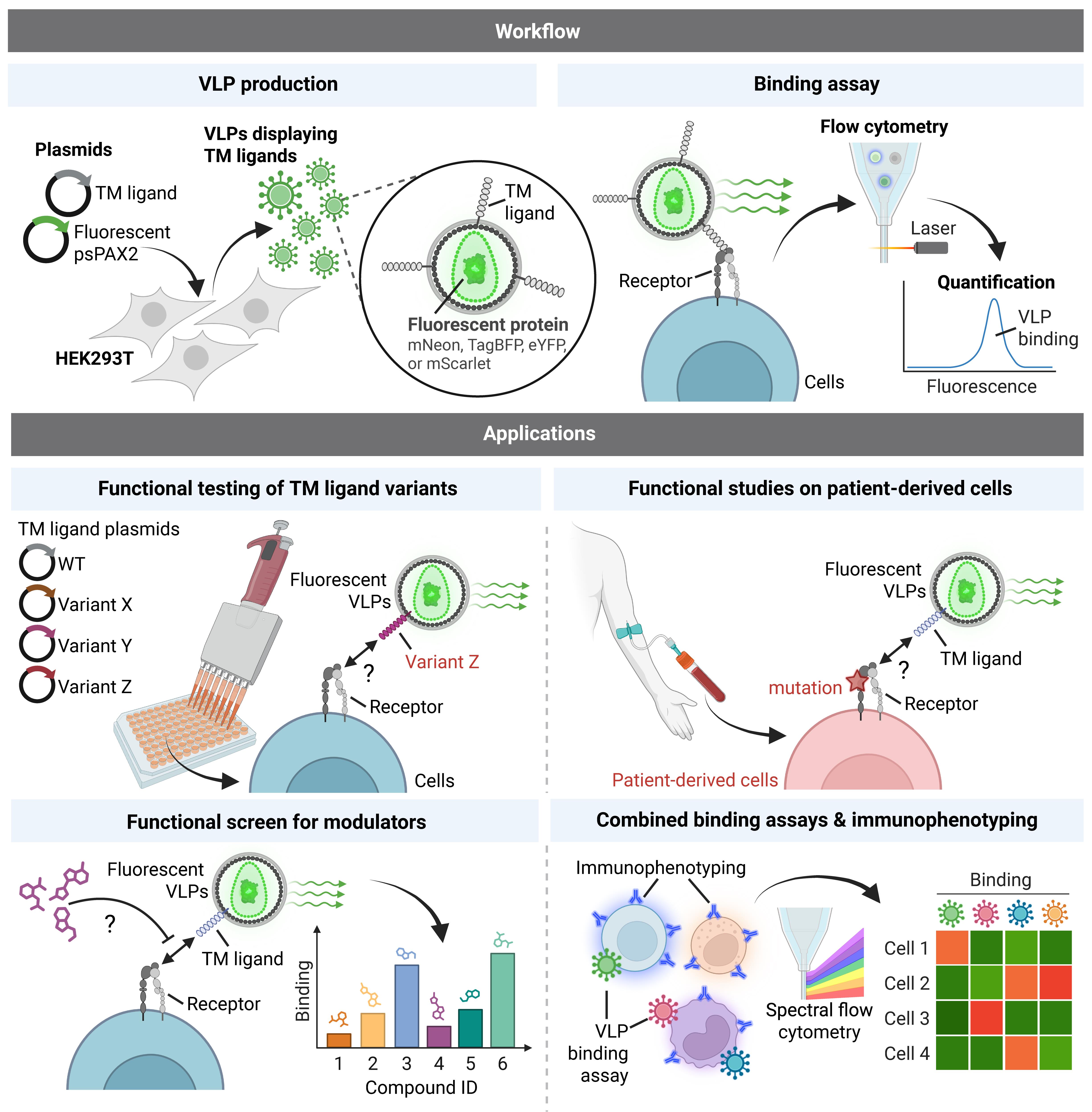
The binding of transmembrane (TM) ligands to their cognate TM receptors on neighboring cells governs intercellular adhesion and direct cell–cell communication. However, these interactions are difficult to study in vitro because they depend on membrane presentation, ligand orientation, receptor clustering, and avidity, features often not captured by soluble recombinant ligands or cell-free assays. Here, we describe a flow cytometry–based assay using fluorescent, lentiviral virus-like particles (VLPs) displaying TM ligands to quantify binding to their receptors on target cells. Fluorescent VLPs are generated in-house by plasmid transfection in HEK293T cells and enable direct fluorescent detection without fluorochrome-conjugated secondary antibodies. The system is modular and readily accommodates engineered ligand constructs, including patient-derived variants. We applied this platform to generate ICAM-1-displaying fluorescent VLPs and to study human LFA-1 function in patient-derived leukocytes. This protocol provides a detailed workflow for VLP production and in vitro binding assays, offering a simple, quantitative, and cost-effective approach for studying TM ligand–receptor interactions in a membrane context. The system is well-suited for mechanistic studies, functional assessment of patient-derived variants, and direct binding assays using patient-derived cells. Integrating the assay into multicolor flow cytometry panels enables simultaneous immunophenotyping and quantification of up to four ligand–receptor interactions at single-cell resolution.
Key features
• Quantifies TM ligand–receptor binding in a membrane context using fluorescent VLPs and flow cytometry.
• Fully in-house, modular system based on plasmid transfection in HEK293T cells, without reliance on recombinant ligands or fluorochrome-conjugated secondary antibodies.
• Supports testing of engineered ligand variants, including patient-derived alleles, and direct functional studies on patient-derived cells.
• Compatible with multicolor flow cytometry panels, enabling simultaneous immunophenotyping and quantification of up to four ligand–receptor interactions at single-cell resolution.
Keywords: LFA-1 (LFA-1) ICAM-1 (ICAM-1)
Virus-like particle (病毒样颗粒)
Transmembrane ligand (跨膜配体)
Ligand binding assay (配体结合实验)
Ligand–receptor interaction (配体–受体相互作用)
Flow cytometry (流式细胞术)
Integrin (整合素)
ICAM-1 (ICAM-1)
Virus-like particle (病毒样颗粒)
Transmembrane ligand (跨膜配体)
Ligand binding assay (配体结合实验)
Ligand–receptor interaction (配体–受体相互作用)
Flow cytometry (流式细胞术)
Integrin (整合素)
Graphical overview
Background
Transmembrane (TM) ligand–receptor interactions are essential mediators of direct cell–cell communication. Their membrane-bound, contact-dependent nature underlies fundamental biological processes, ranging from tissue patterning and morphogenesis to leukocyte trafficking and activation. For example, leukocyte integrins such as LFA-1 (αLβ2), VLA-4 (α4β1), and α4β7 mediate transendothelial migration through binding to endothelial TM ligands ICAM-1, VCAM-1, and MAdCAM-1, respectively [1]. Likewise, T-cell receptors (TCRs) and co-stimulatory receptors such as CD28, OX40, 4-1BB, or ICOS must engage their cognate TM ligands on antigen-presenting cells for productive T-cell priming [2]. Accordingly, genetic variants in genes encoding TM ligands or their receptors underlie a broad range of inherited human diseases, including congenital developmental disorders [3,4] and inborn errors of immunity [5]. Notable examples include defects in key lymphocyte costimulatory and coinhibitory molecules—such as CD28 [6], ICOS/ICOSL [7–9], CTLA-4 [10,11], PD-1/PD-L1 [12–14], OX40 [15], 4-1BB [16], CD40/CD40L [17–21], and CD27/CD70 [22]—as well as essential leukocyte adhesion molecules, such as LFA-1 [1] and other β2-integrins [23–25], highlighting the need for simple assays to evaluate the functional consequences of patient variants in a membrane context. Yet, TM ligand–receptor interactions remain challenging to study because they depend on membrane presentation, ligand orientation, receptor clustering, and avidity—features often poorly captured by soluble recombinant proteins or cell-free assays, while cell-based coculture assays can be more difficult to quantify, scale, and standardize. As a result, there is a need for experimental systems that preserve the membrane-associated nature of these interactions while remaining quantitative, scalable, and accessible.
Here, we describe a flow cytometry–based assay using fluorescent virus-like particles (VLPs) displaying TM ligands to quantify receptor binding on target cells. Building on a previously reported lentiviral packaging vector (psPAX2) encoding mNeon fused to the nucleocapsid protein [26], we generated additional psPAX2-derived constructs carrying distinct fluorescent proteins, enabling the production of VLPs labeled with mNeon (Green), eYFP (Yellow), TagBFP (Blue), or mScarlet (Red), and displaying a TM ligand of interest. This platform combines the advantages of membrane-context presentation, experimental flexibility, and robust flow cytometric readout, without the need for fluorochrome-conjugated secondary antibodies. Because VLPs are generated in-house by plasmid transfection in HEK293T cells, the method does not depend on the availability of commercial recombinant ligands and can be implemented in a cost-effective and highly modular manner. Moreover, ligand constructs can be readily engineered to introduce point mutations, domain swaps, or other modifications, making this system particularly well-suited for mapping structure–function relationships, assessing the impact of patient variants, and performing direct binding studies on patient-derived cells. We successfully applied this platform to study human LFA-1 using fluorescent VLPs displaying its ligand ICAM-1. The assay enabled quantitative analysis of LFA-1 function on patient-derived leukocytes and contributed to the characterization of a novel inborn error of immunity caused by selective LFA-1 deficiency due to loss-of-function variants in its αL subunit [1].
Materials and reagents
Biological materials
1. HEK 293T cells (RRID:CVCL_0063)
2. Jurkat cells (RRID:CVCL_0367)
3. THP-1 cells (RRID:CVCL_0006)
Plasmids
1. psPAX2-D64V-NC-mNeon (Addgene, Plasmid #196509, a gift from Howard Chang)
2. psPAX2-D64V-NC-eYFP (this study; Addgene, Plasmid #257940)
3. psPAX2-D64V-NC-TagBFP (this study; Addgene, Plasmid #257941)
4. psPAX2-D64V-NC-mScarlet (this study; Addgene, Plasmid #257942)
5. pCMV6-ICAM-1 (Addgene, Plasmid #257960)
6. pCMV6-ICAM-1 E34A (Addgene, Plasmid #257961)
Reagents
1. OmniPur BSA, fraction V (Sigma-Aldrich, catalog number: 2960-500GM), store at 4 °C
2. X-tremeGENE 9 transfection reagent (Roche, catalog number: 6365787001), store at 4 °C
3. Lenti-X concentrator (Takara Biosciences, catalog number: 631231), store at 4 °C
4. Fetal bovine serum (FBS) (Gibco), store at -20 °C
Note: FBS should be heat-inactivated at 56 °C for 30 min and filter-sterilized through a 0.22 μm filter before use.
5. DMEM + GlutaMAX (Gibco, catalog number: 10566-016), store at 4 °C
6. RPMI + GlutaMAX (Gibco, catalog number: 61870-036), store at 4 °C
7. OptiMEM serum-free medium (Thermo Fisher Scientific, catalog number: 31985-070), store at 4 °C
8. 1 M HEPES buffer (Gibco, catalog number: 15630080), store at 4 °C
9. Phosphate-buffered saline (PBS) (Corning, catalog number: 21-031-CV), store at room temperature
10. 1 M MgCl2 (Ambion, catalog number: AM9530G), store at room temperature
11. 0.5 M EGTA (bioWORLD, catalog number: 40520008-1), store at room temperature
12. Paraformaldehyde (PFA) solution 4% in PBS (ChemCruz, catalog number: sc-281692), store at 4 °C and protect from light.
Solutions
1. DMEM complete medium (see Recipes)
2. RPMI complete medium (see Recipes)
3. Binding buffer (see Recipes)
4. 2× Mg2+/EGTA solution (see Recipes)
5. FACS buffer (see Recipes)
Recipes
1. DMEM complete medium
| Reagent | Final concentration | Quantity or volume |
|---|---|---|
| DMEM + GlutaMAX | 445 mL | |
| Heat-inactivated, filtered FBS | 10% v/v | 50 mL |
| HEPES 1 M | 10 mM | 5 mL |
| Total | 500 mL |
Store at 4 °C.
2. RPMI complete medium
| Reagent | Final concentration | Quantity or volume |
|---|---|---|
| RPMI + GlutaMAX | 445 mL | |
| Heat-inactivated, filtered FBS | 10% v/v | 50 mL |
| HEPES 1 M | 10 mM | 5 mL |
| Total | 500 mL |
Store at 4 °C.
3. Binding buffer
| Reagent | Final concentration | Quantity or volume |
|---|---|---|
| RPMI + GlutaMAX (without FBS) | 50 mL | |
| BSA fraction V | 0.1% w/v | 50 mg |
| Total | 50 mL |
Combine all components in a 50 mL conical tube and mix by inversion until solid BSA is dissolved. Filter-sterilize the solution using a 0.22 μm vacuum filtration unit. Store at 4 °C.
4. 2× Mg2+/EGTA solution
| Reagent | Final concentration | Quantity or volume |
|---|---|---|
| Binding buffer | 9.82 mL | |
| MgCl2 1 M | 10 mM | 100 μL |
| EGTA 0.5 M | 4 mM | 80 μL |
| Total | 10 mL |
Combine all components in a 15 mL conical tube and mix by inversion. Filter-sterilize the solution using a 0.22 μm vacuum filtration unit. Store at 4 °C.
5. FACS buffer
| Reagent | Final concentration | Quantity or volume |
|---|---|---|
| PBS | 500 mL | |
| BSA fraction V | 0.2% w/v | 1 g |
| Total | 500 mL |
Combine all components and mix by inversion until solid BSA is dissolved. Filter-sterilize the solution using a 0.22 μm vacuum filtration unit. Store at 4 °C.
Laboratory supplies
1. 10 cm tissue culture-treated dishes (Falcon, catalog number: 353003)
2. Vacuum filtration unit, Nalgene Rapid-Flow, 0.22 μm, 50 mL (Thermo Fisher Scientific, catalog number: 564-0020)
3. Vacuum filtration unit, Nalgene Rapid-Flow, 0.22 μm, 500 mL (Thermo Fisher Scientific, catalog number: 566-0020)
4. Microcentrifuge tubes, low-binding, 1.5 mL, autoclaved before use (VWR, catalog number: 76332-068)
5. Conical centrifuge tubes, polypropylene, 15 mL, 50 mL (Falcon, catalog numbers: 352096, 352070)
6. Syringes with Luer lock, 10 mL (BD, catalog number: 302995)
7. Acrodisc syringe filters, 0.45 μm (Pall, catalog number: 4614)
8. Serological pipettes, individually wrapped, 5 mL, 10 mL (Falcon, catalog numbers: 356543, 356551)
9. 96-well polypropylene V-bottom plates (Greiner, catalog number: 651201)
Equipment
1. Single-channel pipettes 1–10, 20–200, and 100–1000 μL
2. Multichannel pipettes 1–50 and 20–300 μL
3. Serological pipette controller
4. Hemocytometer or automated cell counter Countess II FL (Thermo Fisher Scientific, catalog number: AMQAF1000)
5. Class II biological safety cabinet
6. Humidified tissue culture incubator, 37 °C and 5% CO2
7. Centrifuge with temperature control and speeds up to 1500× g
8. Flow cytometer: Attune NxT (Thermo Fisher Scientific, RRID:SCR_019590) equipped with violet (405 nm), blue (488 nm), and yellow (561 nm) lasers
Software and datasets
1. FlowJo Software (v10.10.0, RRID:SCR_008520); requires a license
Procedure
登录/注册后免费查看全文
文章信息
稿件历史记录
提交日期: Mar 14, 2026
接收日期: May 10, 2026
在线发布日期: Jun 3, 2026
出版日期: Jul 5, 2026
版权信息
© 2026 The Author(s); This is an open access article under the CC BY license (https://creativecommons.org/licenses/by/4.0/).
如何引用
Readers should cite both the Bio-protocol article and the original research article where this protocol was used:
- Kim, C. M., Gaballa, M., Lee, D., Jouanguy, E., Zhang, S., Casanova, J. and Yatim, A. (2026). A Flow Cytometry–Based Assay to Quantify the Binding of Transmembrane Ligands to Their Cognate Receptors Using Fluorescent Virus-Like Particles. Bio-protocol 16(13): e5726. DOI: 10.21769/BioProtoc.5726.
- Yatim, A., Youssefian, L., Idani, A., Mourelatos, H., Alipour Olyaei, N., Habibi, L., Peel, J. N., Gaballa, M., Kim, C., Lee, D., et al. (2026). Human LFA-1 governs T cell immune surveillance of the skin. Sci Immunol. 11(116): eadz8360. https://doi.org/10.1126/sciimmunol.adz8360

 Download Citation in RIS Format
分类
免疫学 > 免疫细胞功能 > 淋巴细胞
细胞生物学 > 基于细胞的分析方法 > 流式细胞术
免疫学 > 免疫机理
您对这篇实验方案有问题吗?
在此处发布您的问题,我们将邀请本文作者来回答。同时,我们会将您的问题发布到Bio-protocol Exchange,以便寻求社区成员的帮助。
 发布问题
发布问题


