


Whole-Mount Immunostaining of Tyrosine Hydroxylase for Dopaminergic Neuron Analysis in Zebrafish Larvae
斑马鱼幼体酪氨酸羟化酶整体免疫染色,用于多巴胺能神经元分析

发布: 2026年07月05日第16卷第13期 DOI: 10.21769/BioProtoc.5725 浏览次数: 458
评审: Zihan LingAnonymous reviewer(s)
 PDF
PDF  Q&A
Q&A
参见作者原研究论文
The authors used this protocol in:
Mar 2026
Advertisement






Abstract
Whole-mount techniques are widely used in medical and biological research to analyze protein expression and tissue organization in intact specimens. Traditional approaches for protein localization include section-based immunohistochemistry and in situ hybridization; however, these methods can be limited by tissue disruption and loss of spatial context. Whole-mount protocols generally involve tissue fixation, permeabilization, and staining with specific probes, but their effectiveness varies depending on the antigen–antibody combination and the specimen type. Consequently, no universal protocol is suitable for all experimental conditions. This protocol presents a detailed whole-mount immunostaining protocol for evaluating tyrosine hydroxylase (TH) expression, a key marker of dopaminergic neurons, in zebrafish (Danio rerio) larvae. The procedure outlines critical steps from sample preparation to staining optimization to ensure reproducible and specific signal detection. This approach enables accurate visualization and analysis of dopaminergic neuron distribution in intact larvae. The protocol offers a reliable and adaptable approach that preserves tissue integrity, enables three-dimensional visualization, and is particularly well-suited for developmental and neurobiological studies using zebrafish larvae.
Key features
• Optimized whole-mount immunofluorescence protocol for detecting tyrosine hydroxylase in intact zebrafish larvae.
• Permeabilization and bleaching steps were included to improve antibody penetration and reduce pigmentation-related signal interference
• Three-dimensional brain architecture is preserved, enabling spatial analysis of dopaminergic neuron distribution without tissue sectioning, including region-specific fluorescence quantification using defined regions of interest (ROIs).
• Provides a straightforward approach for assessing dopaminergic neuron distribution through fluorescence imaging and quantitative analysis.
• Suitable for neurodevelopmental and neurotoxicity studies using zebrafish larvae.
Keywords: Dopaminergic neurons (多巴胺能神经元) Zebrafish (斑马鱼)
Larvae (幼体)
Whole-mount (整体染色)
Tyrosine hydroxylase (酪氨酸羟化酶)
Zebrafish (斑马鱼)
Larvae (幼体)
Whole-mount (整体染色)
Tyrosine hydroxylase (酪氨酸羟化酶)
Graphical overview
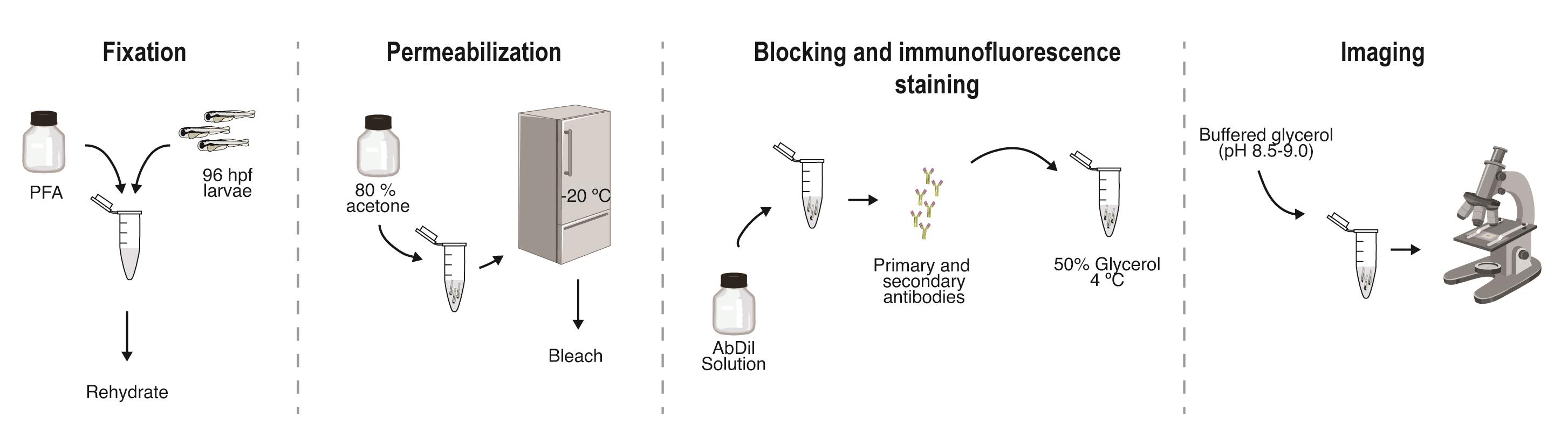
Background
Whole-mount immunohistochemistry, a technique that combines the visualization of tissue structures with the specific detection of proteins using antibodies, can be traced back to the studies by Albert Coons and colleagues in 1941 [1]. Over the years, this technique has been widely used in many research laboratories (especially in developmental biology and neuroscience studies) as well as in clinical diagnostics [2,3]. In general, it can be applied to whole zygotic embryos as well as to dissected tissues, with fixation and preservation of tissue integrity [4], permeabilization to allow antibody access [5], and staining with specific probes [6,7] playing key roles in the successful application of this technique. Yet, despite significant developments and refinements, particularly in sample preparation and staining procedures, the selection of the most suitable method must be based on parameters such as the type of specimen under investigation and the degree of sensitivity required.
In different research areas, zebrafish are increasingly employed as model systems to investigate molecular, genetic, and cellular aspects of developmental and neurobiological processes due to several advantages over other animal models [8,9]. However, although zebrafish represent an excellent experimental system, protein detection methods must be carefully adjusted to account for factors such as tissue permeability, pigmentation, and developmental stage [10,11]. Indeed, there is no single protocol suitable for all experimental conditions, as the effective application of a specific working procedure depends strongly on the antigen–antibody combination [12,13].
Several alternative methodologies have been used to assess protein expression, including section-based immunohistochemistry, in situ hybridization, and tissue-clearing approaches coupled with advanced imaging techniques [14–16]. In addition, tissue- and cell-specific visualization in zebrafish is commonly achieved using transgenic reporter lines or chemical labeling techniques [17,18]. However, their use is limited by the availability of suitable promoters, variability in expression levels, and the need for specialized genetic tools and breeding strategies. Similarly, chemical approaches are often restricted to specific tissues or structures and may lack cellular resolution or specificity for particular protein targets. In contrast, whole-mount immunohistochemistry allows direct detection of endogenous proteins with cellular resolution, without the need for genetic modification. However, these techniques generally require specialized reagents, longer processing times, and access to advanced microscopy platforms. Therefore, in this chapter, a step-by-step protocol for the localization of tyrosine hydroxylase (TH), a standard marker of dopaminergic neurons in the central nervous system [19], in zebrafish larvae is presented. This protocol is adapted from previously published work [20] and includes critical steps and practical considerations to ensure reliable and reproducible staining. Compared with other published whole-mount approaches, the protocol described here emphasizes optimized permeabilization and bleaching steps to improve antibody penetration and minimize pigmentation-related background, making it particularly suitable for early larval stages. However, limitations include dependence on antibody quality and reduced applicability to later developmental stages or adult tissues without further protocol modification. Furthermore, despite recent advancements, the availability of validated antibodies in zebrafish remains limited, particularly for specific cell types or developmental stages, which can constrain immunohistochemical applications.
Beyond the assessment of dopaminergic neurons, this protocol can be readily adapted for the detection of additional neuronal or non-neuronal markers and applied in neurodevelopmental, neurotoxicity, and pharmacological studies using zebrafish larvae.
Materials and reagents
Biological materials
1. 96 h post-fertilization (hpf) AB wild-type zebrafish larvae
Reagents
1. Sodium chloride (NaCl) (Sigma-Aldrich, catalog number: S7653)
2. Potassium chloride (KCl) (Sigma-Aldrich, catalog number: P3911)
3. Sodium phosphate dibasic (Na2HPO4) (Sigma-Aldrich, catalog number: S7907)
4. Potassium phosphate monobasic (KH2PO4) (Sigma-Aldrich, catalog number: P0662)
5. Sodium hydroxide (NaOH) (Sigma-Aldrich, catalog number: 221465)
6. Paraformaldehyde (PFA) (Sigma-Aldrich, catalog number: P6148)
7. Methanol (MeOH) (Sigma-Aldrich, catalog number: 34860)
8. Ethanol (EtOH) (Sigma-Aldrich, catalog number: E7023)
9. Acetone (Sigma-Aldrich, catalog number: 179124)
10. Triton X-100 (Sigma-Aldrich, catalog number: T8787)
11. Tween-20 (MP Biomedicals, catalog number: TWEEN201)
12. Potassium hydroxide (KOH) (Sigma-Aldrich, catalog number: 221473)
13. Hydrogen peroxide (H2O2) (Sigma-Aldrich, catalog number: 216763)
14. Bovine serum albumin (BSA) (Fisher Scientific, catalog number: BP9702)
15. Tyrosine hydroxylase antibody (Merck, catalog number: MAB318)
16. Alexa Fluor 488 secondary conjugated antibody (Jackson ImmunoResearch Europe Ltd., catalog number: 115-545-003)
17. Glycerol (Sigma-Aldrich, catalog number: 356352-M)
18. Sodium carbonate (Na2CO3) (Sigma-Aldrich, catalog number: S7795)
19. Sodium bicarbonate (NaHCO3) (Sigma-Aldrich, catalog number: S6297)
20. MilliQ water
21. Xylol (Sigma-Aldrich, catalog number: 1.08298)
Solutions
1. 10× phosphate-buffered saline (PBS) (see Recipes)
2. 1 N NaOH (see Recipes)
3. 4% PFA (see Recipes)
4. Ethanol (EtOH)/xylol solution (1:1 v/v) (see Recipes)
5. 80% acetone (see Recipes)
6. PTwx (see Recipes)
7. 100 mM KOH
8. 3% H2O2 (see Recipes)
9. Bleaching solution (see Recipes)
10. AbDil solution (see Recipes)
11. Glycerol solution (see Recipes)
12. 0.5 M carbonate buffer (pH 9.0) (see Recipes)
13. Buffered glycerol (see Recipes)
Recipes
1. 10× PBS
| Reagent | Final concentration | Quantity or volume |
|---|---|---|
| NaCl | 1.37 M | 40.0 g |
| KCl | 27 mM | 1.0 g |
| Na2HPO4 | 100 mM | 8.9 g |
| KH2PO4 | 18 mM | 1.2 g |
| MilliQ water | up to 500 mL | |
| Total | n/a | 500 mL |
Note: Adjust to neutral pH by adding acid or base as appropriate until the solution is pH 7.2–7.4. Sterilize by autoclaving for 20 min and store at 4 °C. Dilute in MilliQ water to 1× (v/v) before use.
2. 1 N NaOH
| Reagent | Final concentration | Quantity or volume |
|---|---|---|
| NaOH | 1 N | 4.0 g |
| MilliQ water | 100 mL | |
| Total | n/a | 100 mL |
3. 4% PFA
| Reagent | Final concentration | Quantity or volume |
|---|---|---|
| Paraformaldehyde | 4% | 4.0 g |
| 1× PBS | Up to 100 mL | |
| Total | n/a | 100 mL |
Note: Heat the solution to approximately 60 °C under stirring to facilitate dissolution. Avoid overheating and do not exceed 60 °C, as higher temperatures may lead to paraformaldehyde degradation. Slowly add 1 N NaOH until the solution clears. After, adjust pH to around 7.4 with diluted HCl and adjust the volume of the solution to 100 mL.
4. EtOH/xylol solution (1:1 v/v)
| Reagent | Final concentration | Quantity or volume |
|---|---|---|
| Ethanol | 50% | 25.0 mL |
| Xylol | 50% | 25.0 mL |
| Total | n/a | 50.0 mL |
5. 80% acetone
| Reagent | Final concentration | Quantity or volume |
|---|---|---|
| Acetone | 80% | 80.0 mL |
| MilliQ water | 20.0 mL | |
| Total | n/a | 100 mL |
6. PTwx
| Reagent | Final concentration | Quantity or volume |
|---|---|---|
| Tween20 | 0.1% | 100.0 μL |
| Triton X-100 | 0.1% | 100.0 μL |
| PBS | 99.8 mL | |
| Total | n/a | 100 mL |
7. 100 mM KOH
| Reagent | Final concentration | Quantity or volume |
|---|---|---|
| KOH | 100 mM | 281 mg |
| MilliQ water | 50.0 mL | |
| Total | n/a | 50.0 mL |
8. 3% H2O2
| Reagent | Final concentration | Quantity or volume |
|---|---|---|
| H2O2 | 3% | 5.0 mL |
| MilliQ water | 45.0 mL | |
| Total | n/a | 50.0 mL |
9. Bleaching solution
| Reagent | Final concentration | Quantity or volume |
|---|---|---|
| 3% H2O2 | 1.5% | 25.0 mL |
| 100 mM KOH | 50 mM | 25.0 mL |
| Total | n/a | 50.0 mL |
10. AbDil solution
| Reagent | Final concentration | Quantity or volume |
|---|---|---|
| BSA | 5% | 0.5 g |
| Triton X-100 | 1% | 100.0 μL |
| 1× PBS | 9.9 mL | |
| Total | n/a | 10.0 mL |
11. Glycerol solution
| Reagent | Final concentration | Quantity or volume |
|---|---|---|
| Glycerol | 50%–70% | 50–70 mL |
| 1× PBS | 50–30 mL | |
| Total | n/a | 100 mL |
12. 0.5 M carbonate buffer (pH 9)
| Reagent | Final concentration | Quantity or volume |
|---|---|---|
| Na2CO3 | 13 mM | 0.138 g |
| NaHCO3 | 87 mM | 0.731 g |
| NaCl | 150 mM | 0.877 g |
| MilliQ water | 100.0 mL | |
| Total | n/a | 100 mL |
13. Buffered glycerol
| Reagent | Final concentration | Quantity or volume |
|---|---|---|
| Glycerol | 9 mL | |
| 0.5 M carbonate buffer | 0.05 M | 1 mL |
| Total | n/a | 10 mL |
Laboratory supplies
1. 1.5 mL microcentrifuge tubes
2. 2.0 mL microcentrifuge tubes
3. Routine glassware and consumables (e.g., pipette tips, conical tubes, etc.)
4. Glass microcapillaries (Merck, catalog number: P2049)
Equipment
1. Rotator (e.g., Labnet Mini LabRoller Rotator)
2. Variable volume micropipettes
3. Stir plate
4. pH meter
5. Stereomicroscope (e.g., Olympus, model: SZX7)
6. Fluorescence microscope (e.g., Olympus, model: IX51 inverted fluorescence microscope)
Software and datasets
1. Image analysis software (e.g., ImageJ [21])
2. Statistical software (e.g., GraphPad Prism [22])
Procedure
登录/注册后免费查看全文
文章信息
稿件历史记录
提交日期: Feb 3, 2026
接收日期: May 11, 2026
在线发布日期: May 28, 2026
出版日期: Jul 5, 2026
版权信息
© 2026 The Author(s); This is an open access article under the CC BY license (https://creativecommons.org/licenses/by/4.0/).
如何引用
Félix, L. (2026). Whole-Mount Immunostaining of Tyrosine Hydroxylase for Dopaminergic Neuron Analysis in Zebrafish Larvae. Bio-protocol 16(13): e5725. DOI: 10.21769/BioProtoc.5725.

 Download Citation in RIS Format
分类
神经科学 > 发育 > 神经元
细胞生物学 > 组织分析 > 组织形态学
发育生物学 > 细胞生长和命运决定 > 神经元
您对这篇实验方案有问题吗?
在此处发布您的问题,我们将邀请本文作者来回答。同时,我们会将您的问题发布到Bio-protocol Exchange,以便寻求社区成员的帮助。
 发布问题
发布问题


