


An Immunoprecipitation-Based Nonradioactive Kinase Assay to Measure Akt Kinase Activity in Mammalian Cell Lines
基于免疫沉淀法检测哺乳动物细胞系中Akt激酶活性的非放射性激酶实验

发布: 2026年07月05日第16卷第13期 DOI: 10.21769/BioProtoc.5724 浏览次数: 519
评审: Vipin K. YadavPrashanth N SuravajhalaAnonymous reviewer(s)
 PDF
PDF  Q&A
Q&A
参见作者原研究论文
The authors used this protocol in:

Mar 2024
Advertisement






Abstract
Protein kinase B, more commonly known as Akt, is a family of three serine/threonine kinases (Akt1, Akt2, and Akt3) that play a central role in regulating processes such as proliferation, survival, metabolism, and migration through phosphorylation of downstream targets. Given its involvement in numerous cellular processes, aberrant Akt signaling is prevalent across multiple cancer types, underscoring the need for Akt kinase assays to assess activity, regulatory mechanisms, and the efficacy of targeted interventions. Most existing Akt kinase assays rely on expensive commercial kits, some of which employ pre-purified, constitutively active Akt expressed in insect cells, bypassing physiologic autoinhibition of Akt; therefore, they are not suitable for evaluating allosteric inhibitors or context-dependent regulation. Here, we describe a detailed, step-by-step protocol for a nonradioactive Akt kinase assay using epitope-tagged, recombinant Akt1 expressed in a mammalian cell line and isolated by immunoprecipitation. This method eliminates the need to co-express Akt with upstream regulatory kinases or to purify active enzyme from insect cells, a time-consuming and technically demanding process, particularly when analyzing multiple Akt mutants. Because Akt is assayed in a regulated, autoinhibited state, this protocol enables direct evaluation of allosteric inhibitors that cannot be assessed using active Akt purified from insect cells. We note, however, that Akt1 kinase activity in this assay is measured from epitope-tagged, transiently overexpressed protein, which could influence cellular signaling dynamics. Despite this limitation, the cellular context preserves key regulatory features of Akt1 autoinhibition and membrane-dependent activation that are absent in assays using purified, pre-activated kinase. Together, this protocol supports analysis of Akt kinase activity under diverse experimental conditions, including receptor stimulation, pharmacologic treatment, allosteric inhibitor exposure, and mutations, using an accessible, economical, and physiologically relevant approach.
Key features
• This protocol is broadly accessible, requiring only standard laboratory equipment and commonly used techniques without specialized instrumentation or purified kinase preparations.
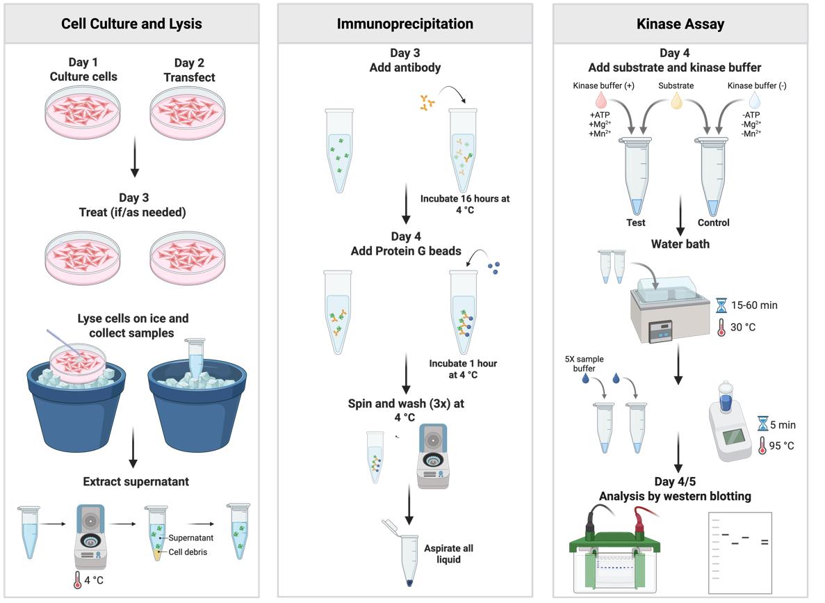
• This protocol measures Akt1 catalytic output by assessing substrate phosphorylation following immunoprecipitation of transiently expressed, epitope-tagged Akt1 from cells.
• The assay is performed in a low-throughput format and provides a semiquantitative readout.
• This protocol can be adapted to other mammalian cell lines and optimized for other protein kinases of choice.
Keywords: Akt (Akt) Phosphorylation (磷酸化)
Nonradioactive kinase assay (非放射性激酶实验)
Immunoprecipitation (免疫沉淀)
Allosteric inhibitors (变构抑制剂)
Phosphorylation (磷酸化)
Nonradioactive kinase assay (非放射性激酶实验)
Immunoprecipitation (免疫沉淀)
Allosteric inhibitors (变构抑制剂)
Graphical overview
Background
Protein kinase B, commonly referred to as Akt, is a serine/threonine kinase that serves as a central integrator of signaling pathways in mammalian cells. Akt regulates a broad spectrum of cellular functions, including cell growth, survival, migration, and metabolism, through phosphorylation of more than 100 identified substrates [1]. Activation of Akt is initiated by growth factor–stimulated phosphoinositide 3-kinase–mediated production of phosphatidylinositol 3,4,5-trisphosphate (PIP3) at the plasma membrane [2]. Binding of PIP3 to the N-terminal pleckstrin homology (PH) domain of Akt promotes its translocation to the plasma membrane [3]. Once localized at the membrane, Akt is sequentially phosphorylated at two critical regulatory sites: a threonine residue within the kinase domain by phosphoinositide-dependent kinase 1 (PDK1) [4,5] and a serine residue within the C-terminal hydrophobic motif primarily by mammalian target of rapamycin complex 2 [6]. Dual phosphorylation at these sites is required for full enzymatic activation, enabling Akt to phosphorylate downstream effectors and propagate signaling responses [2,7]. In the absence of receptor stimulation, intramolecular interactions between the PH and kinase domains enforce AKT autoinhibition [8]; stabilization of this conformation has informed the development of multiple allosteric inhibitors [9].
In cellular studies, Akt activation is most commonly inferred by immunoblot detection of phosphorylation at the two regulatory residues, which serve as widely used surrogate markers of Akt activation. While this approach provides a convenient measure of upstream signaling, Akt phosphorylation status alone does not directly assess catalytic activity toward substrates and may not fully reflect functional output under all experimental conditions. Direct measurement of substrate phosphorylation is therefore necessary to evaluate Akt enzymatic activity more accurately.
While historically kinase activity assays have relied on radioactivity, more recently, assay kits employing colorimetric readouts have been introduced (e.g., Abcam, catalog number: ab139436). This approach is suited for high-throughput in vitro screening and identification of potential Akt substrates. In addition to endpoint assays, lentivirus-based real-time kinetic methods have been developed to monitor Akt activity dynamically in living cells (e.g., Sartorius, catalog number: BA-04868). Although these approaches provide valuable quantitative determination and/or temporal resolution, they are expensive and require specialized instrumentation and expertise, restricting their use to laboratories with dedicated infrastructure and limiting broader accessibility. Also, some of the current assay kits to detect substrate phosphorylation rely on Akt expressed and purified from insect cells, often following co-expression with PDK1 to generate a constitutively active enzyme (e.g., Promega, catalog number: V1911). As a result, these methods are limited in their ability to capture physiologically relevant regulation or to evaluate allosteric inhibitors that depend on intact regulatory interactions.
The protocol described here provides a nonradioactive, immunoprecipitation-based Akt kinase assay that can be readily implemented in laboratories equipped for standard cell biology and biochemistry techniques. By measuring Akt1 kinase activity from mammalian cells transiently expressing an epitope-tagged Akt1, this approach directly assesses substrate phosphorylation while preserving autoinhibitory control and endogenous regulatory mechanisms. Limitations of this assay are that it is low-throughput and semiquantitative, but it is well-suited for initial investigation and hypothesis testing, enabling assessment of Akt regulation, mutant function, and allosteric inhibitor responses under physiologically relevant conditions. In addition to measuring basal and stimulus-dependent Akt activity, this assay can be applied to compare isoform-specific activity among the three Akt isoforms [10] and to evaluate the functional impact of disease-associated Akt mutations. With appropriate optimization, this approach may also be adapted to study other protein kinases whose activity is governed by autoinhibition or complex regulatory interactions.
Materials and reagents
Biological materials
1. HEK293-T (ATCC, catalog number: CRL-3216)
2. NEB5α competent E. coli high efficiency (New England Biolabs, catalog number: C2987H)
Reagents
1. Dulbecco's high glucose modified eagles medium (DMEM) (Cytiva, catalog number: SH30022.02)
2. Dulbecco's phosphate-buffered saline (DPBS) (Cytiva, catalog number: SH30028.02)
3. 0.25% trypsin, 0.1% EDTA in HBSS w/o calcium, magnesium and sodium bicarbonate (Corning, catalog number: 25-053-CI)
4. Fetal bovine serum (FBS) characterized (Cytiva, catalog number: SH30071.03)
5. Penicillin/streptomycin/glutamine (PSG), 100× (Gibco, catalog number: 10378-016)
6. OPTI-MEM I (1×) reduced serum medium (Gibco, catalog number: 31985-062)
7. TransIT®-LT1 transfection reagent (Mirus Bio, catalog number: MIR 2300)
8. pcDNA3.1-HA-Akt1 WT construct (Addgene plasmid #78778) (a gift from Dr. Jaewhan Song)
9. pcDNA3.1-HA-Akt1 K179M construct (Addgene plasmid #73409) (a gift from Dr. Jie Chen)
10. ZymoPURE II Plasmid Midiprep kit (ZYMO RESEARCH, catalog number: D4200)
11. Dimethyl sulfoxide (DMSO) (Corning, catalog number: 25950CQC)
12. MK-2206 hydrochloride (Cayman Chemical Company, catalog number: 11593)
13. ATP solution (100 mM) (Thermo Scientific, catalog number: R0441)
14. PRAS40 recombinant protein antigen (Novus Biologicals, catalog number: NBP2-57165PEP)
15. Dithiothreitol (DTT) (Thermo Fisher Scientific, catalog number: R0861)
16. HEPES (Fisher Scientific, catalog number: BP310-1)
17. Manganese chloride tetrahydrate (Fisher Scientific, catalog number: M87-100)
18. Magnesium chloride hexahydrate (Fisher Scientific, catalog number: M33-500)
19. Phosphatase inhibitor cocktail I (Thermo Fisher Scientific, catalog number: J63907.AA)
20. Phosphatase inhibitor cocktail II (Thermo Fisher Scientific, catalog number: J61022.AA)
21. Protease inhibitor tablets (Thermo Fisher Scientific, catalog number: A32955)
22. Triton X-100 (TX-100) (Fisher Scientific, catalog number: BP151-500)
23. HA-Tag mouse monoclonal antibody (0.2 μg/μL) (Santa Cruz Biotechnology, catalog number: SC-7392)
24. Protein G PLUS-agarose (Santa Cruz Biotechnology, catalog number: SC-2002)
25. PRAS40 rabbit monoclonal antibody (Cell Signaling, catalog number: 2691)
26. Phospho-PRAS40 (Thr246) rabbit monoclonal antibody (Cell Signaling, catalog number: 2997)
27. IRDye® 680RD goat anti-mouse IgG (H+L), 0.1 mg (Li-COR Biosciences, catalog number: 925-68070)
28. IRDye® 680RD goat anti-rabbit IgG (H+L), 0.1 mg (Li-COR Biosciences, catalog number: 925-68071)
29. β-Mercaptoethanol (β-ME) (Millipore Sigma, catalog number: 444203)
30. Bromophenol blue (Fisher Scientific, catalog number: BP115-25)
31. Glycerol (Thermo Fisher Scientific, catalog number: J61059AP)
32. TRIS base (Fisher Scientific, catalog number: BP152-10)
33. HCl (Millipore Sigma, catalog number: HX06034)
34. Glycine (Fisher Scientific, catalog number: BP381-5)
35. Ammonium persulfate (APS) (Fisher Scientific, catalog number: BP179-100)
36. Sodium dodecyl sulfate (SDS) (Fisher Scientific, catalog number: BP166-500)
37. N,N,N',N'-Tetramethylethylenediamine (TEMED) (Thermo Fisher Scientific, catalog number: 17919)
38. Sodium phosphate dibasic anhydrous (Na2HPO4) (Fisher Scientific, catalog number: BP332-500)
39. Potassium phosphate monobasic (KH2PO4) (Fisher Scientific, catalog number: BP362-500)
40. Potassium chloride (KCl) (Fisher Scientific, catalog number: P217-500)
41. Sodium chloride (NaCl) (Fisher Scientific, catalog number: S271-10)
42. TWEEN-20 (Fisher Scientific, catalog number: BP337-500)
43. 30% Acrylamide/Bis solution 29:1 (Bio-Rad, catalog number: 1610156)
44. Precision Plus Protein Dual Color Standards (Bio-Rad, catalog number: 1610374)
45. Methanol (Fisher Scientific, catalog number: A412-4)
46. Non-fat dry milk (Lab Scientific, catalog number: M0841)
Solutions
1. Complete DMEM medium (see Recipes)
2. Lysis buffer (see Recipes)
3. Kinase assay buffer (+) (see Recipes)
4. Kinase assay buffer (-) (see Recipes)
5. 5× sample buffer (see Recipes)
6. 10× running buffer (see Recipes)
7. 1× running buffer (see Recipes)
8. 10× transfer buffer (see Recipes)
9. 1× transfer buffer (see Recipes)
10. 10× PBS (see Recipes)
11. 1× PBS (see Recipes)
12. 1× PBS with Tween 20 (PBS-T) (see Recipes)
13. SDS-PAGE resolving gel (see Recipes)
14. SDS-PAGE stacking gel (see Recipes)
15. 5% non-fat dry milk (see Recipes)
Recipes
1. Complete DMEM medium
| Reagent | Final concentration | Quantity or volume |
|---|---|---|
| DMEM | n/a | 445 mL |
| FBS | 10% (v/v) | 50 mL |
| 100× PSG | 1× | 5 mL |
Store at 4 °C.
2. Lysis buffer
| Reagent | Final concentration | Quantity or volume |
|---|---|---|
| 1 M HEPES, pH 7.4 | 25 mM | 162.5 μL |
| 1 M MgCl2 | 1 mM | 6.5 μL |
| 1 M NaCl | 150 mM | 975 μL |
| 20% Triton-X-100 | 1% | 325 μL |
| 1 M DTT | 1 mM | 6.5 μL |
| 50× Protease inhibitor | 1× | 130 μL |
| 50× Phosphatase inhibitor cocktail I | 1× | 130 μL |
| 100× Phosphatase inhibitor cocktail II | 1× | 60 μL |
| Autoclaved MilliQ water | - | 4704.5 μL |
| Total | N/A | 6.5 mL |
The calculations and volumes shown above are sufficient for 10 dishes. Make the lysis buffer fresh for same-day usage only and keep it on ice. The storage conditions for the stock solutions used to make lysis buffer are as follows: 1 M HEPES at 4 °C; autoclaved MilliQ water, 1 M MgCl2, 1 M NaCl, and 20% TX-100 at room temperature; 1 M DTT, 50× protease inhibitor cocktail, 50× phosphatase inhibitor cocktail I (PIC-I), and 100× phosphatase inhibitor cocktail II (PIC-II) at -20 °C. The stock solutions are stable at these storage conditions for at least 6 months.
3. Kinase assay buffer (+)
| Reagent | Final concentration | Quantity or volume |
|---|---|---|
| 1 M HEPES, pH 7.4 | 25 mM | 3.25 μL |
| 0.1 M MgCl2 | 5 mM | 6.5 μL |
| 0.1 M MnCl2 | 5 mM | 6.5 μL |
| 0.1 M DTT | 1 mM | 1.3 μL |
| 50× protease inhibitor | 1× | 2.6 μL |
| 50× phosphatase inhibitor cocktail I | 1× | 2.6 μL |
| 100× phosphatase inhibitor cocktail II | 1× | 1.3 μL |
| 100 mM ATP | 5 mM | 6.5 μL |
| PRAS40 (0.5 mg/mL) | 0.5 μg | 5.2 μL |
| Autoclaved MilliQ water | N/A | 94.25 μL |
| Total | N/A | 130 μL |
The calculations and volumes shown above are sufficient for five reactions. Make the kinase assay buffer fresh for same-day usage only and keep it on ice. The storage conditions for the solutions used to make the kinase assay buffer are as follows: 1 M HEPES at 4 °C; autoclaved MilliQ water, 0.1 M MgCl2, and 0.1 M MnCl2 at room temperature; 1 M DTT, 100 mM ATP, 50× protease inhibitor cocktail, 50× PIC-I, and 100× PIC-II at -20 °C. The stock solutions are stable at these storage conditions for at least 1 year. The PRAS40 antigen should be stored in small aliquots (10 μL) at -80 °C.
4. Kinase assay buffer (-)
| Reagent | Final concentration | Quantity or volume |
|---|---|---|
| 1 M HEPES, pH 7.4 | 25 mM | 3.25 μL |
| 0.1 M DTT | 1 mM | 1.3 μL |
| 50× protease inhibitor | 1× | 2.6 μL |
| 50× phosphatase inhibitor cocktail I | 1× | 2.6 μL |
| 100× phosphatase inhibitor cocktail II | 1× | 1.3 μL |
| PRAS40 (0.5 μg/μL) | 0.5 μg | 5.2 μL |
| Autoclaved MilliQ water | N/A | 113.75 μL |
| Total | N/A | 60 μL |
The calculations and volumes shown above are sufficient for five reactions. Make the kinase assay buffer fresh for same-day usage only and keep it on ice. The storage conditions for the solutions used to make the kinase assay buffer are as follows: 1 M HEPES at 4 °C; autoclaved MilliQ water at room temperature; 1 M DTT, 50× protease inhibitor cocktail, 50× PIC-I, and 100× PIC-II at -20 °C. The stock solutions are stable at these storage conditions for at least 1 year. The PRAS40 antigen should be stored in small aliquots (10 μL) at -80 °C.
5. 5× sample buffer
| Reagent | Final concentration | Quantity or volume |
|---|---|---|
| 1 M Tris pH 6.8 | 312.5 mM | 3.125 mL |
| Glycerol | 50% (v/v) | 5 mL |
| SDS | 0.35 M | 1 g |
| β-ME | 5% (v/v) | 0.5 mL |
| Bromophenol blue | 0.01% (w/v) | 1 mg |
| Autoclaved MilliQ water | N/A | ~0.375 mL |
| Total | N/A | 10 mL |
Save in aliquots (100–500 μL) at -20 °C. Thaw before use.
6. 10× running buffer (pH ~8.3)
| Reagent | Final concentration | Quantity or volume |
|---|---|---|
| Tris base | 0.25 M | 30 g |
| Glycine | 1.9 M | 144 g |
| SDS | 1% | 10 g |
| MilliQ water | N/A | ~816 mL |
| Total | N/A | 1 L |
Store at room temperature.
7. 1× running buffer (pH ~8.3)
| Reagent | Final concentration | Quantity or volume |
|---|---|---|
| 10× running buffer | 1× | 100 mL |
| MilliQ water | N/A | 900 mL |
| Total | N/A | 1 L |
Store at room temperature.
8. 10× transfer buffer (pH ~8.3)
| Reagent | Final concentration | Quantity or volume |
|---|---|---|
| Tris base | 0.25 M | 30 g |
| Glycine | 1.9 M | 144 g |
| MilliQ water | N/A | ~826 mL |
| Total | N/A | 1 L |
Store at room temperature.
9. 1× transfer buffer (pH ~8.3)
| Reagent | Final concentration | Quantity or volume |
|---|---|---|
| 10× transfer buffer | 1× | 100 mL |
| Methanol | 20% (v/v) | 200 mL |
| MilliQ water | N/A | 700 mL |
| Total | N/A | 1 L |
Store at 4 °C.
10. 10× PBS
| Reagent | Final concentration | Quantity or volume |
|---|---|---|
| NaCl | 1.4 M | 81.82 g |
| KCl | 2.68 mM | 199.8 mg |
| Na2HPO4 | 0.1 M | 14.2 g |
| KH2PO4 | 0.02 M | 2.72 g |
| MilliQ water | N/A | ~900 mL |
| Total | N/A | 1 L |
Store at room temperature.
11. 1× PBS
| Reagent | Final concentration | Quantity or volume |
|---|---|---|
| 10× PBS | 1× | 100 mL |
| MilliQ water | N/A | 900 mL |
| Total | N/A | 1 L |
Store at room temperature.
12. 1× PBS-T
| Reagent | Final concentration | Quantity or volume |
|---|---|---|
| 10× PBS | 1× | 100 mL |
| Tween 20 | 0.1% (v/v) | 0.1 mL |
| MilliQ water | N/A | 899.1 mL |
| Total | N/A | 1 L |
Store at room temperature.
13. SDS-PAGE resolving gel
| Reagent | Final concentration | Quantity or volume |
|---|---|---|
| 30% Acrylamide/Bis solution, 29:1 | 10% | 13.34 mL |
| 1.5 M TRIS pH 8.8 | 0.375 M | 10 mL |
| 10% SDS | 0.1% | 400 μL |
| 10% APS | 0.1% | 400 μL |
| TEMED | 0.1% | 40 μL |
| MilliQ water | N/A | 15.82 mL |
| Total | N/A | 40 mL |
Store the acrylamide solution, 1.5 M Tris pH 8.8, and 10% APS in aliquots at 4 °C. APS stock solution should be made fresh weekly. Store 10% SDS and TEMED at room temperature.
14. SDS-PAGE stacking gel
| Reagent | Final concentration | Quantity or volume |
|---|---|---|
| 30% Acrylamide/Bis solution, 29:1 | 5% | 3.34 mL |
| 1.0 M TRIS pH 6.8 | 0.127 M | 2.54 mL |
| 10% SDS | 0.1% | 200 μL |
| 10% APS | 0.1% | 200 μL |
| TEMED | 0.1% | 20 μL |
| MilliQ water | N/A | 13.7 mL |
| Total | N/A | 20 mL |
Store the acrylamide solution, 1.0 M Tris pH 6.8, and 10% APS in aliquots at 4 °C. APS stock solution should be made fresh weekly. Store 10%SDS and TEMED at room temperature.
15. 5% dry non-fat milk
| Reagent | Final concentration | Quantity or volume |
|---|---|---|
| Dry non-fat milk | 5% | 2.5 g |
| 1× PBS | n/a | 50 mL |
Make it fresh when needed. It should be ok to store at 4 °C for 1–2 days.
Laboratory supplies
1. 100 mm sterile tissue culture dish (Fisher Scientific, catalog number: FB012924)
2. 15 mL centrifuge tubes (Olympus, purchased from Genesee Scientific, catalog number: 28-103)
3. 1.7 mL microcentrifuge tubes (Olympus, purchased from Genesee Scientific, catalog number: 24-282LR)
4. 50 mL centrifuge tubes (Olympus, purchased from Genesee Scientific, catalog number: 28-108)
5. 10 μL barrier tips sterile (Olympus, purchased from Genesee Scientific, catalog number: 24-400)
6. 200 μL barrier tips sterile (Olympus, purchased from Genesee Scientific, catalog number: 26-412)
7. 1,000 μL barrier tips sterile (Olympus, purchased from Genesee Scientific, catalog number: 24-430)
8. 10 μL non-filter pipette tips (Fisherbrand SureOne, catalog number: 02-707-455)
9. 200 μL yellow non-filter pipette tips (Fisherbrand SureOne, catalog number: 02-707-451)
10. 1,000 μL non-filter pipette tips (Olympus, purchased from Genesee Scientific, catalog number: 23-615 R)
11. 200 μL wide-bore universal pipette tips (Axygen, catalog number: T205WBCRS)
12. Gel loading tips (1–200 μL) (Fisherbrand, catalog numer: 02-707-182)
13. 5 mL serological pipette sterile (Genclone, purchased from Genesee Scientific, catalog number: 12-102)
14. 10 mL serological pipette sterile (Genclone, purchased from Genesee Scientific, catalog number: 12-104)
15. 9” disposable Pasteur pipettes (Fisher Scientific, catalog number: 13-678-20D)
16. Cell scrapers (Fisher Scientific, catalog number: 08-100-241)
17. Kimwipes (Kimtech Science Brand, catalog number: 34155)
18. Short plates Mini-Protean (Bio-Rad, catalog number: 1653308)
19. Glass plates Mini-Protean (Bio-Rad, catalog number: 1653312)
20. Blotting paper 703 (VWR International, catalog number: 28298-020)
21. Immobilon FL transfer membrane (Merck Millipore, catalog number: IPFL00010)
22. Black western blot incubation boxes (Li-COR Bioscience, catalog number: 929-97201)
23. CountessTM cell counting chamber slides (Thermo Fisher Scientific, Invitrogen, catalog number: C10283)
24. Parafilm (Genesee Scientific, catalog number: 16-100)
25. Kirkland Stretch-Tite plastic wrap
26. Liquid nitrogen dewar
27. Tube racks
28. Lab coat
29. Gloves
30. Safety glasses
Equipment
1. Biosafety cabinet (The Baker Company, model: RND-BSC-001)
2. HERAcell VIOS 160i CO2 Incubator (Thermo Fisher Scientific, model: HERAcell VIOS 160i)
3. Isotemp GPD 10 Stone Bath (Fisher Scientific)
4. CountessTM II FL (Thermo Fisher Scientific) automated cell counter
5. Evos XL Core Microscope (Invitrogen)
6. Nanodrop One (Thermo Fisher Scientific)
7. Mini-PROTEAN Tetra Vertical Electrophoresis Cell (Bio-Rad)
8. PowerPacTM Basic Power Supply (Bio-Rad)
9. Refrigerated Centrifuge (Eppendorf, model: 5424 R)
10. Li-COR Odyssey XF (Li-COR Bioscience)
11. BenchRocker 2D Variable Speed 2-D Rocker (Genesee Scientific)
12. Basic AB315 Benchtop Laboratory pH/mV Meter (Fisher Scientific)
13. Stripettor Ultra Electric Pipette Controller (Corning)
14. Freezer (-80 °C)
15. Freezer (-20 °C)
16. Refrigerator (2–8 °C)
Procedure
登录/注册后免费查看全文
文章信息
稿件历史记录
提交日期: Jan 19, 2026
接收日期: May 10, 2026
在线发布日期: May 28, 2026
出版日期: Jul 5, 2026
版权信息
© 2026 The Author(s); This is an open access article under the CC BY-NC license (https://creativecommons.org/licenses/by-nc/4.0/).
如何引用
Peek, A., Mehta, J. N. and Bhandari, D. (2026). An Immunoprecipitation-Based Nonradioactive Kinase Assay to Measure Akt Kinase Activity in Mammalian Cell Lines. Bio-protocol 16(13): e5724. DOI: 10.21769/BioProtoc.5724.

 Download Citation in RIS Format
分类
生物化学 > 蛋白质 > 活性
生物化学 > 蛋白质 > 翻译后修饰
细胞生物学 > 细胞信号传导 > 磷酸化
您对这篇实验方案有问题吗?
在此处发布您的问题,我们将邀请本文作者来回答。同时,我们会将您的问题发布到Bio-protocol Exchange,以便寻求社区成员的帮助。
 发布问题
发布问题


