


Quantitative Assessment of Capillary Permeability in Deep Intracardiac Capillaries Using Fluorescent Dextran
利用荧光葡聚糖定量评估心脏深部毛细血管的通透性

发布: 2026年07月05日第16卷第13期 DOI: 10.21769/BioProtoc.5723 浏览次数: 487
评审: Manasa VL ChanduriJessica DavisAnonymous reviewer(s)
 PDF
PDF  Q&A
Q&A
参见作者原研究论文
The authors used this protocol in:
Dec 2025
Advertisement






Abstract
When the function of cardiac capillaries is impaired, cardiac function declines, and the risk of disease increases. No reliable assay has been developed to detect or evaluate the level of material exchange of capillaries deep within healthy heart tissue. In this study, we develop a new method to detect and evaluate molecules leaking from capillaries in cardiac tissue. By administering fluorescent dextran to mice via the tail vein, followed by rapid processing of the heart tissue, we have detected leaking fluorescent material from intracardiac microvessels. By comparing the detected images with those taken during the negative-control administration, using the image processing software LAS X and ImageJ, we detected trace amounts of fluorescent material that had leaked from the capillaries. We calculated the area of tissue where fluorescence was detected to perform a quantitative assessment, which we used as an indicator of capillary permeability. This new method of indexing will provide a different perspective on the factors contributing to the decline in cardiac function and the increased risk of disease with aging.
Key features
• A method to evaluate the permeability function of capillaries deep within healthy cardiac tissue has been developed using the diffusion area of a leaked fluorescent marker.
• Evaluating the permeability function of deep tissues with dense capillary networks provides a foothold for elucidating the mechanisms of organ homeostasis, both maintenance and deterioration.
• The diffusion of molecules into organs via capillary-to-cell exchange is an indicator of tissue function.
Keywords: Cardiac microvascular vessel (心脏微血管) Permeability (血管通透性)
Interstitial fluid (组织间液)
Dextran (葡聚糖)
Tail vein injection (尾静脉注射)
Leakage analysis (渗漏分析)
Permeability (血管通透性)
Interstitial fluid (组织间液)
Dextran (葡聚糖)
Tail vein injection (尾静脉注射)
Leakage analysis (渗漏分析)
Graphical overview
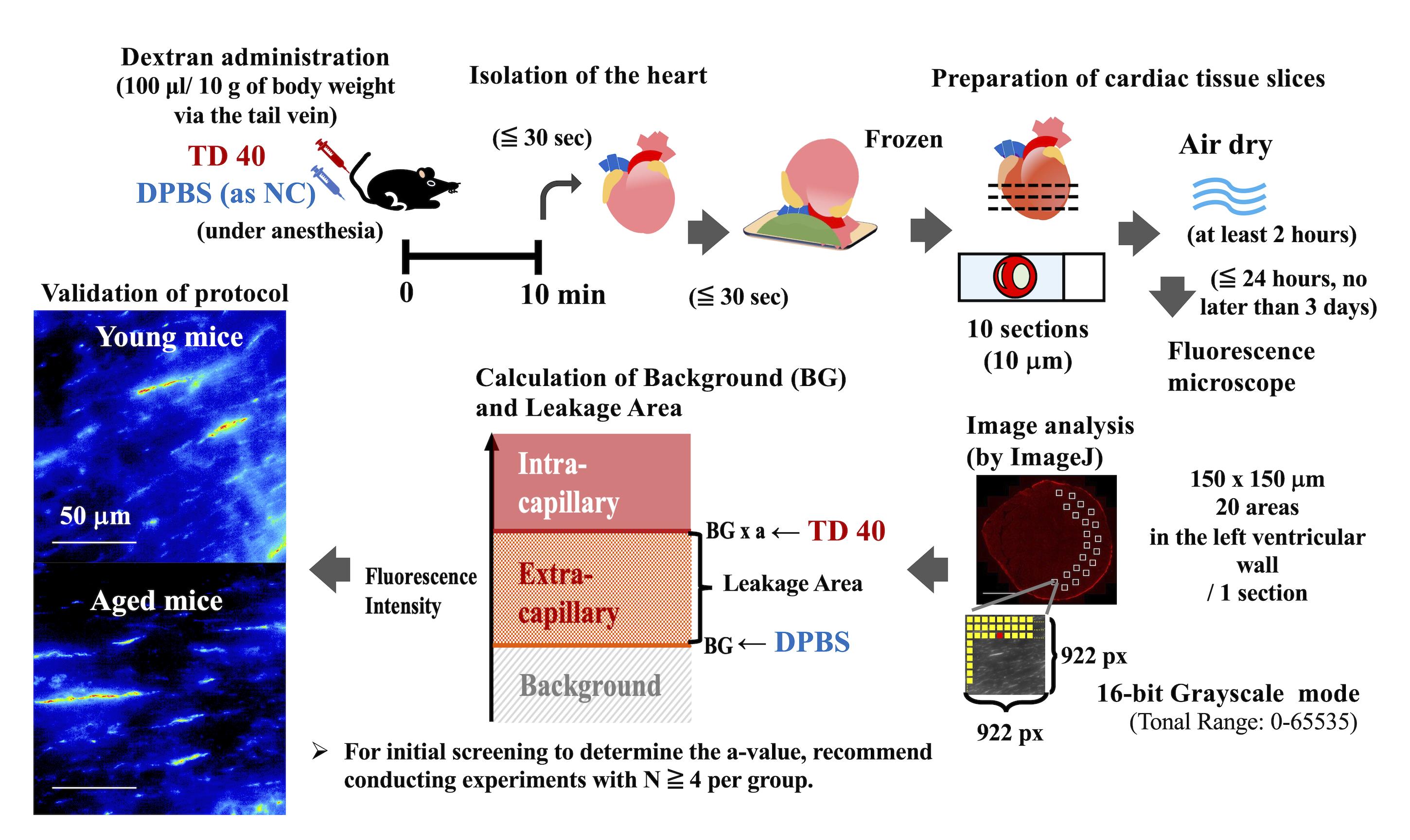
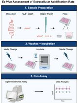
Assessment workflow for deep intracardiac capillary permeability. TD 40: TRITC-conjugated dextran 40, DPBS: Dulbecco's phosphate-buffered saline, NC: negative control, px: pixel(s), BG: background.
Background
Blood vessels extend throughout the body to circulate blood and supply cells in all tissues with sufficient oxygen and nutrients, as well as to collect and discharge cellular waste products. In particular, capillaries in peripheral tissues are considered sites of material exchange, where small molecular substances pass through the endothelial cell layer under colloid osmotic pressure (COP). Each tissue cannot function without nutrition and oxygen; normal physiological tissue function depends on the regulation of vascular permeability, an important function of capillaries [1].
Capillaries are composed of a single endothelial cell layer. Leakage from blood vessels into tissues is the source of interstitial fluid and reflects the underlying biological function of each organ. The homeostasis of organ-specific function lies in the diversity of vascular permeability. Hence, vascular permeability dysfunction is a major cause of disease. In recent years, abnormalities in microvascular function have been reported to be closely related to tissue function and disease development [2]. In fact, dysfunction of endothelial cells due to inflammation, aging, and viral and bacterial infections leads to organ damage. Endothelial dysfunction is critically involved in many chronic human diseases, such as diabetes, arthritis, and dementia [3–5]. A large cohort of human studies has shown that dysfunction of the tissue microvasculature, a nutritional supply channel, is associated with a wide range of subjective symptoms such as headache, palpitations, fatigue, and chest pain and increases the risk of more severe cardiovascular events [6,7]. Previous studies have noted that blood flow disturbance in cerebral blood vessels, especially in small blood vessels, caused by microplastic particles, could lead to neurological and cognitive disorders [8,9]. In addition, maintaining tissue microvascular function regulates individual lifespan in mice [10].
Permeability in microvascular vessels in superficial layers, such as tumors [11,12], skin, and retina [13–15], has been evaluated using fluorescent dextran or Evans blue. Several in vivo evaluations of cardiac permeability have also been reported [16–18]. These reports have evaluated capillaries visible under a microscope—such as those in transparent tissues at early developmental stages and on organ surfaces—and have demonstrated that capillary permeability increases in response to inflammation, cancer, and other diseases. Here, because most capillaries lie deep within tissues, our overall understanding remains incomplete. Therefore, substance exchange between capillaries and tissues is thought to occur via interstitial fluid, which permeates and circulates throughout the tissue, enveloping the cells. However, the mechanism underlying this process and its relationship to organ function remain unclear, and it is not yet known whether there are differences between healthy states and states of functional decline or disease.
In in vitro experiments, the selection and replacement of culture media are critical determinants of cellular status (such as proliferation, differentiation, and cell death) and of functional assessment. In contrast, in three-dimensional culture, cells (or cell clusters) have a high degree of freedom of movement, and the contact surface with the culture medium is constantly changing. Furthermore, in vivo, changes in the composition of cell populations alter intercellular interactions, and the interstitial fluid flowing between cells also changes daily in response to the environment. These changes in mechanical sensitivity affect cell dynamics [19,20]. Therefore, in drug discovery aimed at organ-specific efficacy, there is a need for novel validation methods that integrate imaging endpoints with functional measurements in translational toxicology and mechanobiology [21,22].
To date, no method is available to detect the exchange of substances in cardiac capillaries deep within the heart tissue or to evaluate what is detected. In this study, we develop a new method to detect and evaluate molecules leaking from cardiac capillaries. By administering fluorescent dextran to mice via the tail vein, followed by rapid processing of the heart tissue, we have detected leaking fluorescent material from intracardiac microvessels [23]. In addition, we calculated the diffusion area of the fluorescent substance and used it as an indicator of capillary permeability. Using this new method, we compared the diffusion area of molecules leaking from blood circulation into cardiac tissue in young and aged mice and found that it was significantly reduced in the latter [23]. In this way, we are now able to assess the presence of interstitial fluid that leaks from capillaries deep within tissues—a phenomenon that was previously impossible to observe. As a result, it has become clear that the flow of interstitial fluid within the heart changes with age, suggesting that this may be a contributing factor to age-related cardiac dysfunction and to the increased risk of disease.
Materials and reagents
Biological materials
1. Wild-type female mouse (Japan SLC, Inc., Shizuoka, Japan, C57BL/6NSlc)
Notes:
1. Mice were housed in separate cages, with a maximum of 5 mice per cage, in a specific-pathogen-free, temperature-controlled vivarium. They were maintained under a 12/12 h light/dark cycle with ad libitum access to food and water. The ambient room temperature was maintained at 22 ± 2 °C, and humidity was controlled at 55% ± 5%.
2. In this study, 6–7-month-old female mice (body weight: 20–25 g), whose body size had nearly reached maturity, were used as young mice, while 24-month-old female mice (body weight: 30–40 g) were used as aged mice.
3. C57BL/6NCrSlc strain (derived from NIH/Charles River, often referred to as B6N) was introduced to Japan SLC, Inc. from the Institute of Medical Science at the University of Tokyo in 1975.
4. All animal experiments were approved by the Institutional Animal Care and Use Committee at Tokyo Metropolitan Institute for Geriatrics and Gerontology (No. 20010 and 23016) and strictly adhered to the guidelines of Tokyo Metropolitan Institute for Geriatrics and Gerontology for animal experiments.
Reagents
1. Dulbecco’s phosphate-buffered saline (DPBS) (Fujifilm Wako Pure Chemical Co., catalog number: 045-29795)
2. Isoflurane (VIATRIS, catalog number: 901036504)
3. Domitor (Zenoaq, catalog number: AHE1)
4. Mitazolam (Sandoz K.K., catalog number: 614243022)
5. Vetorphale (Meiji Animal Health Co. Ltd., catalog number: VETLF5)
6. Tetramethylrhodamine (TRITC)-conjugated dextran 40 (average weight 40,000) (TdB Labs AB, catalog number: TD40)
7. Tragacanth gum, powder (FUJIFILM Wako Pure Chemical Co., catalog number: 200-2245)
8. Saline (Otsuka Pharmaceutical Factory Inc, catalog number: 3311401A2018)
9. Tissue-Tek® OCT compound (Sakura Finetek Japan Co., Ltd., catalog number: 4560133241825)
10. Isopentane (2-methylbutane) (FUJIFILM Wako Pure Chemical Co., Japan, catalog number: 166-00615)
Solutions
1. Three-drug anesthetic mixture (MMB) (see Recipes)
2. TRITC-conjugated dextran 40 solution (see Recipes)
3. Tragacanth gum solution (see Recipes)
Recipes
1. Three-drug anesthetic mixture (MMB)
| Reagent | Concentration | Storage | Final concentration | Quantity or volume |
|---|---|---|---|---|
| Domitor | 1 mg/mL | Lockable storage; room temperature | 75 μg/mL | 0.75 mL |
| Midazolam | 5 mg/mL | Lockable storage; room temperature | 0.4 mg/mL | 0.8 mL |
| Vetorphale | 5 mg/mL | Lockable storage; room temperature | 0.5 mg/mL | 1 mL |
| Saline | 7.45 mL | |||
| Total | 10 mL |
Note: Add 0.75 mL of Domitor, 0.8 mL of Midazolam, and 1 mL of Vetorphale to 7.45 mL of saline to make 10 mL of MMB. The administrative volume of MMB is 100 μL per 10 g of body weight.
2. TRITC-conjugated dextran 40 solution
| Reagent | Storage | Final concentration | Quantity or volume |
|---|---|---|---|
| TRITC-conjugated dextran 40 | Room temperature | n/a | 10 mg |
| DPBS (or saline) | Room temperature | n/a | 1 mL |
| Total | Divide the solution into 0.6 mL tubes, each containing 100–250 μL, and store at -20 °C | 10 mg/mL | 1 mL |
Note:
1. Store TRITC-conjugated dextran at -20 °C. Avoid repeated freeze–thaw cycles.
2. The administrative volume of the TRITC-conjugated dextran 40 solution is 100 µL per 10 g of body weight.
3. Tragacanth gum solution
| Reagent | Final concentration | Quantity or volume |
|---|---|---|
| Tragacanth gum, powder | n/a | 2 g |
| Saline | n/a | 5 mL |
| Total | n/a | 5 mL |
Note: Gradually add saline solution to the gum while mixing with a medicine spoon. Once it reaches a suitable softness, pack it into the syringe. Suitable softness means it should be firm enough to be squeezed out when the syringe is pressed.
Laboratory supplies
1. Microtube 1.5 mL (e.g., BM Equipment Co. Ltd., catalog number: NT-175)
2. Microtube 0.6 mL (e.g., Corning Inc., catalog number: MCT-060-C-S)
3. 1 mL disposable syringe (e.g., Terumo Co., catalog number: SS-01T10)
4. 5 mL disposable syringe (e.g., Terumo Co., catalog number: SS-05SZ)
5. 27 G (or 26 G) disposable needle (e.g., Terumo Co., catalog number: NN-2719S)
6. 30 G disposable needle (e.g., NIPRO Co., catalog number: 01-134)
7. Cork sheet, 3 mm (e.g., Green Stuff World SL, catalog number: 8436574509571ES)
8. White micro slide glass (Matsunami Glass Ind. Ltd., catalog number: FF-001)
9. Dry ice block (2 kg)
Equipment
1. Laboratory Animal Anesthesia System (Shinano Manufacturing Co., LTD., model: SN-487-OT Air)
2. Scissor (e.g., Kenis, Ltd., catalog number: 3-154-0262)
3. Tweezer (e.g., Kenis, Ltd., catalog number: 3-319-0281)
4. Ring tweezer (e.g., Kenis, Ltd., catalog number: 3-345-0555)
5. -80 °C freezer (e.g., Panasonic, catalog number: MDF-U33V-PJ)
6. Cryostat (e.g., Leica Microsystems, model: CM3050 S)
7. Upright fluorescence microscope (e.g., Leica Microsystems, model: DM6 B)
8. Microscope camera (e.g., Leica Microsystems, model: DFC9000 GT sCMOS camera)
Software and datasets
1. Leica Application Suite X (LAS X) (Leica, version 3.7.4.23463)
2. ImageJ (NIH, Version 1.54 g)
Note: To perform quantitative processing on multiple image files simultaneously, we create and execute the ImageJ macro shown in Figure S1.
3. GraphPad Prism (GraphPad Software, version 9.5.1)
Procedure
登录/注册后免费查看全文
文章信息
稿件历史记录
提交日期: Feb 26, 2026
接收日期: May 13, 2026
在线发布日期: Jun 1, 2026
出版日期: Jul 5, 2026
版权信息
© 2026 The Author(s); This is an open access article under the CC BY-NC license (https://creativecommons.org/licenses/by-nc/4.0/).
如何引用
Nakamura, M., Yoshida-Kikkawa, Y., Sugiura, K., Ito, Y. and Toyoda, M. (2026). Quantitative Assessment of Capillary Permeability in Deep Intracardiac Capillaries Using Fluorescent Dextran. Bio-protocol 16(13): e5723. DOI: 10.21769/BioProtoc.5723.

 Download Citation in RIS Format
分类
细胞生物学 > 组织分析 > 组织成像
医学 > 心血管疾病
细胞生物学 > 组织分析 > 生理学
您对这篇实验方案有问题吗?
在此处发布您的问题,我们将邀请本文作者来回答。同时,我们会将您的问题发布到Bio-protocol Exchange,以便寻求社区成员的帮助。
 发布问题
发布问题


