


Lodicule Isolation and Morphometric Analysis During Rice Floret Opening
水稻小花开放过程中浆片的分离与形态测量分析

(*contributed equally to this work) 发布: 2026年07月05日第16卷第13期 DOI: 10.21769/BioProtoc.5721 浏览次数: 417
评审: Shweta PanchalPragya BaruaAnonymous reviewer(s)
 PDF
PDF  Q&A
Q&A
参见作者原研究论文
The authors used this protocol in:
Feb 2026
Advertisement






Abstract
Rice lodicules are specialized floral organs located at the base of the ovary that undergo dynamic morphological changes during the flowering period. Water uptake–driven swelling and subsequent dehydration-induced shrinkage of the lodicules trigger floret opening and closure, respectively. Although lodicules play a central role in floret movement, standardized methods for quantitatively monitoring their temporal morphological changes remain limited. Here, we describe a detailed and reproducible workflow for lodicule sampling, dissection, imaging, and quantitative morphometric analysis. Florets are collected at predefined clock time points during the flowering period, and lodicules are carefully isolated under a stereomicroscope. High-resolution imaging is performed under consistent acquisition settings, followed by precise measurement of lodicule length, width, and thickness using image analysis software. This protocol emphasizes positional consistency in sampling, uniform imaging parameters, and standardized data analysis to enhance reproducibility. This method is suitable for evaluating the effects of genetic background or environmental conditions on lodicule morphology. By providing a standardized analytical framework, this protocol enables accurate and quantitative morphometric analysis of rice lodicules during floret opening.
Key features
• Standardized time-point sampling minimizes variability caused by diurnal fluctuations and handling during lodicule morphometric analysis.
• Enables reproducible isolation and imaging of rice lodicules while preserving native morphology and preventing dehydration-induced artifacts.
• Time-resolved workflow enables analysis of rapid morphological changes associated with floret opening and closure.
• Applicable for comparing genetic and environmental effects on lodicule morphology under controlled experimental conditions.
Keywords: Rice floret (水稻小花)
 Lodicule imaging (浆片成像)
Lodicule morphometry (浆片形态测量)
Time-course imaging (时间序列成像)
Morphometric analysis (形态测量分析)
Lodicule imaging (浆片成像)
Lodicule morphometry (浆片形态测量)
Time-course imaging (时间序列成像)
Morphometric analysis (形态测量分析)
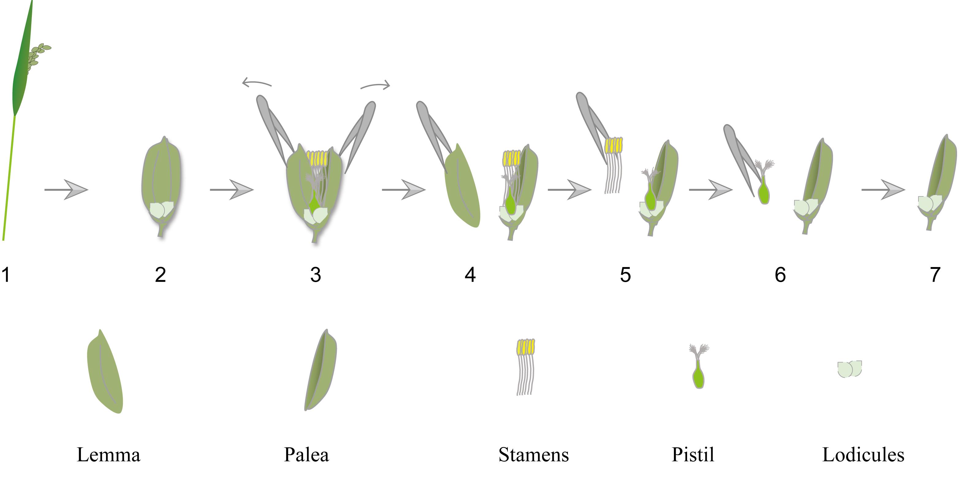
Graphical overview
Background
In rice (Oryza sativa), the floret consists of the lemma, palea, stamens, pistil, and two lodicules located at the base of the ovary. Lodicules function analogously to petals [1]. During the flowering period, lodicules absorb water and undergo rapid swelling. The resulting increase in volume generates a mechanical force that separates the lemma and palea, thereby enabling floret opening and pollination. After pollination is completed, lodicules lose water and shrink, which promotes the inward repositioning of the lemma and palea and leads to floret closure [2].
Accurate characterization of lodicule morphology is essential for understanding the mechanisms underlying floral movement, reproductive development, and genotype-dependent variation. Therefore, quantitative analysis of lodicule morphology at defined time points provides valuable phenotypic information. However, lodicule analysis is often influenced by technical variability. Inconsistent sampling intervals, variation of spikelet position in panicle, tissue dehydration during dissection, and differences in imaging parameters can introduce measurement bias. Previous studies have shown that lodicule morphology is highly sensitive to developmental stage, genetic background, and sampling conditions, often resulting in substantial variation in morphometric measurements across experiments [3–5]. In addition, the lack of standardized morphometric criteria limits comparability across studies.
To address these challenges, we describe a workflow for lodicule morphological analysis based on fixed clock-time sampling. This method avoids reliance on subjective developmental staging and instead emphasizes controlled sampling intervals, standardized dissection procedures, and calibrated imaging and measurement settings. By minimizing experimental variability, this protocol enables precise quantification of lodicule length, width, and thickness and facilitates reliable comparison across genotypes or experimental conditions.
Materials and reagents
Biological materials
1. Rice plants (Oryza sativa) grown under controlled conditions and sampled at the flowering stage (0–3 days post-heading)
Reagents
1. Ultrapure water
2. 70% (v/v) ethanol (for sterilization of tools)
3. Soil (Jiang Su Xing Nong Substrate&technology Co., Ltd., catalog number: 161102G0096N)
4. Vermicompost (Wuhan Jiyesheng Chemical Co., Ltd., catalog number: A00970)
Laboratory supplies
1. Fine forceps (RWD, catalog number: F12012-10)
2. Glass slide (MREDA, catalog number: M049985)
3. Dissection needles (MREDA, catalog number: M188991)
4. 1.5 mL microcentrifuge tubes
5. Parafilm (Sigma-Aldrich BS, catalog number: HS120667)
6. Ice and insulated foam box (for temporary storage of samples during transport and handling)
7. Filter paper (Tanon, catalog number: 586-1700)
Equipment
1. Fluorescence stereomicroscope (Leica, catalog number/model: M205 FA)
2. Growth chamber or greenhouse with controlled light and temperature conditions
Software and datasets
1. Leica Application Suite X (LAS X)
2. GraphPad Prism 10
Procedure
登录/注册后免费查看全文
文章信息
稿件历史记录
提交日期: Apr 3, 2026
接收日期: May 13, 2026
在线发布日期: May 27, 2026
出版日期: Jul 5, 2026
版权信息
© 2026 The Author(s); This is an open access article under the CC BY-NC license (https://creativecommons.org/licenses/by-nc/4.0/).
如何引用
Readers should cite both the Bio-protocol article and the original research article where this protocol was used:
- Hu, Y., Cui, S., Li, H., Hou, H., Xu, Z., Hao, B., Cai, L., Zhu, L., Wang, J., Chang, K., Li, W., Shao, W., Zhu, S., Li, C., Zhao, Z., Jiang, L., Tian, Y., Liu, X., Liu, S., Chen, L., Zhou, S. and Wan, J. (2026). Lodicule Isolation and Morphometric Analysis During Rice Floret Opening. Bio-protocol 16(13): e5721. DOI: 10.21769/BioProtoc.5721.
- Hu, Y., Li, H., Hou, H., Cui, S., Xu, Z., Hao, B., Cai, L., Zhu, L., Wang, J., Chang, K., et al. (2026). The multifunctional protein OsAIM1 regulates floret opening and closure timing via jasmonic acid-mediated lodicule dynamics in rice. Plant Physiol. https://doi.org/10.1093/plphys/kiaf690

 Download Citation in RIS Format
分类
植物科学 > 植物发育生物学 > 形态建成
植物科学 > 植物生理学 > 表型分析
植物科学 > 植物细胞生物学 > 组织分析
您对这篇实验方案有问题吗?
在此处发布您的问题,我们将邀请本文作者来回答。同时,我们会将您的问题发布到Bio-protocol Exchange,以便寻求社区成员的帮助。
 发布问题
发布问题


