


A Reliable Method for Thawing Primary AML and CMML Mononuclear Cells to Preserve Viability and Function
保持原代 AML 和 CMML 单个核细胞活性与功能的可靠复苏方法

(*Contributed equally to this work, §Technical contact: kelly.lim@adelaide.edu.au; verity.saunders@sahmri.org) 发布: 2026年06月05日第16卷第11期 DOI: 10.21769/BioProtoc.5720 浏览次数: 306
评审: Olga ZimmermannovaRakesh BamAnonymous reviewer(s)
 PDF
PDF  Q&A
Q&A
参见作者原研究论文
The authors used this protocol in:

Sep 2024
Advertisement







Abstract
Human mononuclear cells derived from peripheral blood and bone marrow are valuable resources for the study of hematological malignancies, including acute myeloid leukemia (AML) and chronic myelomonocytic leukemia (CMML). Cryopreservation enables long-term storage of patient samples for downstream assays; while thawing protocols have been described, subsequent recovery of viable cells after thawing can be challenging, particularly for fragile blast and monocyte populations. Here, we describe a reliable protocol for thawing cryopreserved AML and CMML mononuclear cells designed to preserve post-thaw viability, recovery, and functional integrity. The method incorporates controlled dilution of cells out of cryoprotectant with anticoagulant-supplemented thaw buffer, DNase I treatment, and gentle resuspension steps. Using this approach, post-thaw viability consistently exceeded 80% with a mean recovery of 55.6% across samples. Recovered cells retained functional capacity, as demonstrated by colony-forming assays, and maintained immunophenotypic characteristics by flow cytometry. This protocol provides a robust and reproducible method for the recovery of cryopreserved AML and CMML mononuclear cells and may be broadly applicable to other fragile or monocyte-rich patient-derived hematopoietic samples.
Key features
• Reliable thawing protocol that preserves viability and recovery of cryopreserved AML and CMML mononuclear cells.
• Controlled dilution with anticoagulant supplementation, DNase I treatment, and gentle resuspension minimizes cell aggregation of fragile blast and monocyte populations.
• Maintains cell functional capacity and immunophenotypic characteristics for downstream assays.
• Applicable to cryopreserved patient-derived mononuclear cells prone to aggregation, low recovery, or poor viability with conventional thawing.
Keywords: AML (AML) CMML (CMML)
Leukemia (白血病)
Monocyte (单核细胞)
Mononuclear cells (单个核细胞)
Cryopreserved (冻存)
Thaw (复苏)
Viability (细胞活性)
CMML (CMML)
Leukemia (白血病)
Monocyte (单核细胞)
Mononuclear cells (单个核细胞)
Cryopreserved (冻存)
Thaw (复苏)
Viability (细胞活性)
Graphical overview
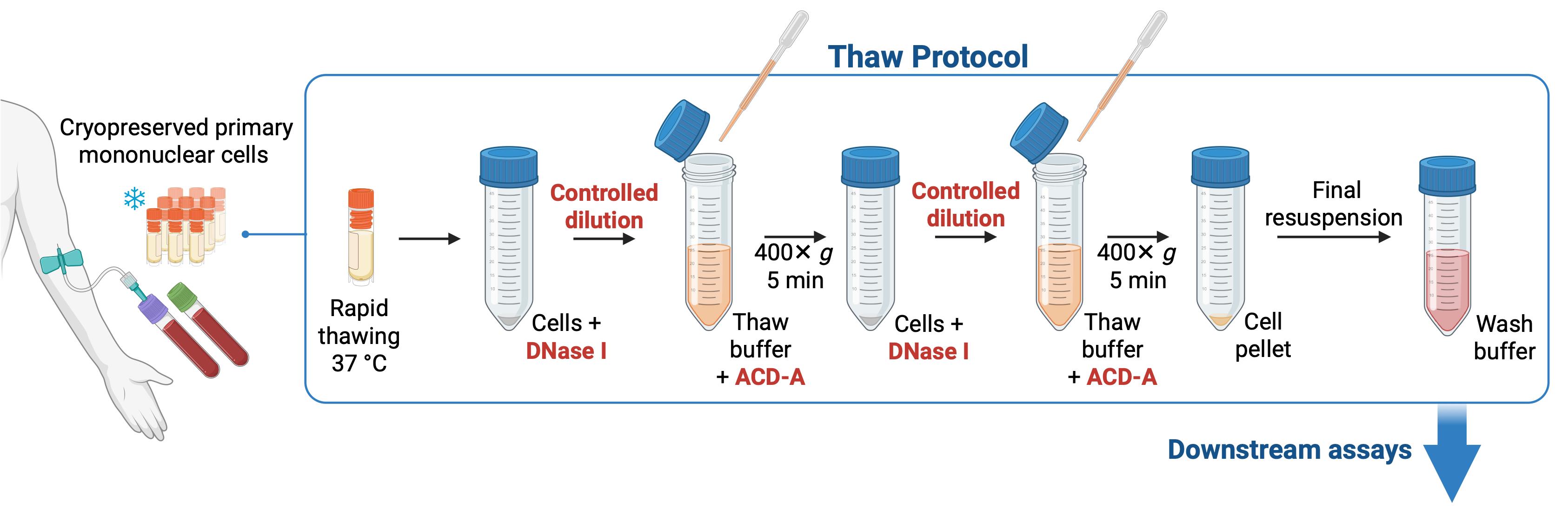
Overview of thaw protocol for cryopreserved primary acute myeloid leukemia (AML) and chronic myelomonocytic leukemia (CMML) mononuclear cells
Background
Human primary mononuclear cells isolated from peripheral blood and bone marrow are valuable in the study of cellular processes and disease mechanisms in hematological malignancies, including acute myeloid leukemia (AML) and chronic myelomonocytic leukemia (CMML). Because access to fresh patient material is limited and experiments cannot always be performed immediately following sample collection, cryopreservation is routinely used to enable long-term storage. However, recovery of viable cells can be challenging, since thawing exposes cells to several concurrent stressors, including dimethyl sulfoxide (DMSO) toxicity at room temperature, osmotic stress leading to membrane damage and extracellular DNA release, mechanical stress from vigorous pipetting, and metabolic stress associated with increased reactive oxygen species.
Certain molecular subtypes of AML, including mutant IDH1 and IDH2, and acute promyelocytic leukemia have been noted to be particularly difficult to recover after cryopreservation. Several protocols for thawing AML cells have been described, most commonly involving rapid thawing in a 37 °C water bath followed by washing steps in serum-containing media, but issues with low viability, poor recovery, and material loss persist [1–4].
To address these challenges, here we describe a reliable step-by-step protocol for thawing cryopreserved mononuclear cells that incorporates controlled dilution of cells out of cryoprotectant to reduce osmotic stress, DNase I treatment, and gentle resuspension in an anticoagulant-supplemented thaw buffer to limit cell aggregation during recovery. These steps are designed to preserve viability, facilitate recovery, reduce sample variability, and maintain the physiological integrity of AML and CMML cells for downstream applications. These include flow cytometry–based characterization of surface markers and cytokine profiling, as well as functional studies such as colony-forming assays, phagocytosis, macrophage differentiation, antigen processing and presentation, oxidative burst, and drug-response assays. We have found our method to be particularly well-suited for CMML samples, which are typically enriched for monocytes that are prone to aggregation during thawing and can be lost during washing steps. Using this method, we consistently achieved post-thaw viability exceeding 80% with a mean recovery of 55.6% across a range of total number of cells per ampoule of cryopreserved sample, with preserved colony-forming capacity and immunophenotypic characteristics.
We routinely apply this protocol for the recovery of cryopreserved AML and CMML mononuclear cells and anticipate it may be useful for other fragile or monocyte-rich primary hematopoietic patient-derived samples.
Materials and reagents
Biological materials
1. Cryopreserved AML and CMML mononuclear cells from peripheral blood or bone marrow, stored at -186 °C (vapor-phase liquid nitrogen tank) in cryoprotectant containing 90% fetal bovine serum and 10% dimethyl sulfoxide
Note: The average number of cells per cryovial used in this protocol was 2.62 × 10⁷ (range 3.0 × 10⁶–1 × 10⁸).
Reagents
1. Hanks’ balanced salt solution, modified with sodium bicarbonate, without calcium chloride and magnesium sulfate (Sigma-Aldrich, catalog number: H9394-500ML)
2. HEPES solution, 1 M (Sigma-Aldrich, catalog number: H0887-100ML)
3. Fetal bovine serum, triple sterile-filtered, pharmaceutical-grade, gamma-irradiated (CellSera, catalog number: AU-FBS/PG), heat-inactivated
4. FenwalTM anticoagulant citrate dextrose solution formula A (ACD-A) (Baxter, catalog number: AHB7898)
5. DNase I grade II 100 mg lyophilized (Roche, catalog number: 10104159001)
6. Trypan Blue stain, 0.4% (Gibco, catalog number: 15250-061)
7. Ethanol, 80% v/v (ChemSupply, catalog number: EL156-20L-P)
8. Glacial acetic acid (Sigma-Aldrich, catalog number: A6283-500ML)
9. Methyl violet (Sigma-Aldrich, catalog number: 69710-25G)
10. Dulbecco’s phosphate-buffered saline (PBS) (Sigma-Aldrich, catalog number: D8537-500ML)
Solutions
1. Wash buffer (see Recipes)
2. Thaw buffer (see Recipes)
3. DNase I (see Recipes)
4. White cell fluid stain (see Recipes)
Recipes
1. Wash buffer
| Reagent | Final concentration | Quantity or volume |
|---|---|---|
| Hanks’ buffered salt solution | 89% | 445 mL |
| Fetal bovine serum | 10% | 50 mL |
| 1 M HEPES | 10 mM | 5 mL |
| Total | 500 mL |
Store at 4 °C and use within 2 months.
2. Thaw buffer
| Reagent | Final concentration | Quantity or volume |
|---|---|---|
| Wash buffer | 97.5% | 39 mL |
| ACD-A | 2.5% | 1 mL |
| Total | 40 mL |
Prepare fresh immediately before using and warm to 37 °C.
3. DNase I
| Reagent | Final concentration | Quantity or volume |
|---|---|---|
| DNase I grade II 100 mg lyophilized | 2.4 mg/mL | 100 mg |
| Sterile distilled water | 99.8% | 41.7 mL |
| Total | 41.8 mL |
Store between -20 and -40 °C and thaw on ice.
4. White cell fluid stain
| Reagent | Final concentration | Quantity or volume |
|---|---|---|
| Glacial acetic acid | 2% | 2 mL |
| Distilled water | 98% | 98 mL |
| Methyl violet | 0.005% | 5 mg |
| Total | 100 mL |
Filter-sterilize and store at ambient temperature.
Laboratory supplies
1. 50 mL conical polypropylene centrifuge tubes (Nunc, catalog number: 339652)
2. Sterile 2.5 mL transfer pipette (Sarstedt, catalog number: 86.1172.001)
3. Mixing cannula (Fairmont Medical, catalog number: MIX1001)
4. Cell strainer, 70 μm (Greiner, catalog number: 542 070)
5. Serological pipette, 5 mL (Corning, Costar Stripette, catalog number: CLS4487-200EA)
6. Serological pipette, 10 mL (Corning Costar Stripette, catalog number: CLS4488-200EA)
7. Pipette tips, 200 μL (Axygen, catalog number: AXTT-200-Y)
Equipment
1. Class II Biosafety cabinet (Nuaire, Cellgard), with suction line for liquid waste removal
2. Benchtop centrifuge (Eppendorf, catalog number: 5810)
3. Light microscope (Olympus, model: BX45)
4. Water bath (Lauda AquaLine, model: AL25)
5. Hemocytometer counting chamber, Neubauer improved
6. 20–200 μL pipette (Gilson, model: Pipetman Classic P200)
7. 2–20 μL pipette (Gilson, model: Pipetman Classic P20)
Procedure
登录/注册后免费查看全文
文章信息
稿件历史记录
提交日期: Mar 17, 2026
接收日期: May 8, 2026
在线发布日期: May 21, 2026
出版日期: Jun 5, 2026
版权信息
© 2026 The Author(s); This is an open access article under the CC BY-NC license (https://creativecommons.org/licenses/by-nc/4.0/).
如何引用
Lim, K., Saunders, V., Kutyna, M., Robinson, N., Hiwase, D. and Thomas, D. (2026). A Reliable Method for Thawing Primary AML and CMML Mononuclear Cells to Preserve Viability and Function. Bio-protocol 16(11): e5720. DOI: 10.21769/BioProtoc.5720.

 Download Citation in RIS Format
分类
免疫学 > 免疫细胞功能
细胞生物学 > 细胞分离和培养 > 低温贮存
您对这篇实验方案有问题吗?
在此处发布您的问题,我们将邀请本文作者来回答。同时,我们会将您的问题发布到Bio-protocol Exchange,以便寻求社区成员的帮助。
 发布问题
发布问题


