


Protocol for In Vivo Two-Photon FCS to Measure Nanocarrier Number and Flow Velocity in Mouse Cerebral Microvasculature
利用活体双光子荧光相关光谱测量小鼠脑微血管中纳米载体数量与流动速度的实验方案


发布: 2026年07月05日第16卷第13期 DOI: 10.21769/BioProtoc.5717 浏览次数: 662
评审: Olga KopachJordi Boix-i-CollRupam Ghosh
 PDF
PDF  Q&A
Q&A
参见作者原研究论文
The authors used this protocol in:
Mar 2026
Advertisement






Abstract
Real-time measurement of blood flow and nanocarrier transport in the cerebral microvasculature is crucial for understanding neurovascular physiology and nanocarrier-based drug delivery. Existing techniques lack the ability to measure blood flow rates in individual vessels with high spatial and temporal resolution in real time. Two-photon fluorescence correlation spectroscopy (2P-FCS) provides a powerful approach for monitoring tracer molecules within a small confocal observation volume. This enables the simultaneous determination of particle number and flow dynamics in vivo. Here, we present a detailed protocol for in vivo 2P-FCS measurements in the mouse cerebral microvasculature. The protocol includes preparation of the cranial window, delivery of fluorescent dextran tracers for vascular visualization, and FCS measurements. It also includes two-photon imaging of the cerebrovascular network and acquisition and analysis of fluorescence correlation data. The protocol describes calibration of the confocal volume diameter and optimization of two-photon excitation parameters. This workflow enables real-time measurement of tracer concentration and flow velocity in individual cerebral microvessels with high spatial and temporal resolution. The method can be adapted to study blood flow dynamics, nanoparticle transport, and microvascular physiology in a variety of in vivo imaging systems equipped with multiphoton microscopy and FCS capabilities.
Key features
• Protocol for performing in vivo 2P-FCS for real-time measurements of nanocarrier flow and concentration in the mouse cerebrovasculature through an acute cranial window.
• Provides guidance on calibrating the confocal volume diameter and optimizing near-infrared (NIR) laser power and excitation wavelengths of fluorophores.
• Includes procedures for two-photon imaging of cerebral blood vessels.
• Applicable to studies of cerebral blood flow, nanoparticle transport, and microvascular dynamics using multiphoton microscopy systems equipped with FCS modules.
Keywords: Fluorescence correlation spectroscopy (FCS) (荧光相关光谱) Multiphoton microscopy (多光子显微镜)
Cerebral microvasculature (脑微血管)
In vivo Imaging (活体成像)
Nanocarrier transport (纳米载体运输)
Cerebral blood flow (脑血流)
Multiphoton microscopy (多光子显微镜)
Cerebral microvasculature (脑微血管)
In vivo Imaging (活体成像)
Nanocarrier transport (纳米载体运输)
Cerebral blood flow (脑血流)
Graphical overview
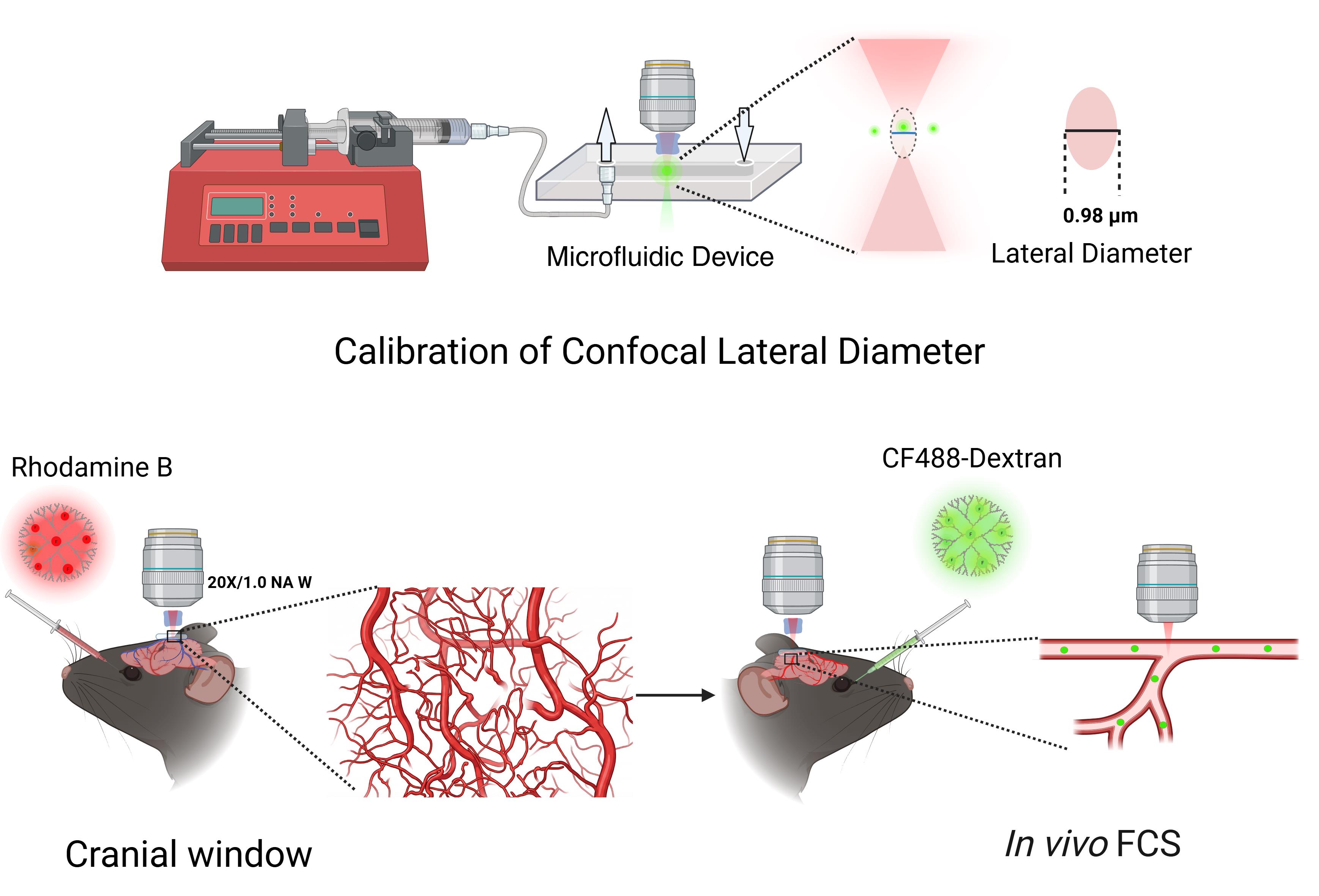
Workflow for in vivo two-photon fluorescence correlation spectroscopy (2P-FCS) measurements in mouse cerebrovasculature
Background
Cerebral blood vessels, such as veins, arteries, and capillaries, form the cerebrovascular network and are responsible for a variety of functions, including ion exchange regulation, molecular and oxygen transport, and waste removal [1]. Cerebral blood flow (CBF), particularly in capillaries, provides vital information about the central nervous system’s functionality. For instance, abnormalities in CBF patterns can indicate conditions such as ischemic stroke when blood flow slows down, brain tumors when flow patterns alter, or neurodegenerative diseases like Alzheimer's when CBF gradually declines [2,3]. Thus, CBF assessment can reveal how the brain’s autoregulation system maintains stable blood flow despite systemic changes in blood pressure. Various techniques, such as computed tomography (CT) [4], magnetic resonance imaging (MRI) [5], and fluorescence imaging [6,7] have been developed for measuring CBF. However, real-time measurement of blood flow rates in individual vessels using these existing technologies is challenging, especially in capillaries (<10 μm), due to their limited spatial resolution [8]. While CT and MRI provide whole-brain perfusion measurements, they lack the spatial resolution required to resolve capillary-scale flow dynamics [9,10]. Optical techniques such as laser speckle imaging and Doppler optical coherence tomography improve temporal resolution but do not achieve single-capillary sensitivity or single-particle detection [8,11]. Multiphoton laser-scanning microscopy (MPLSM) techniques can measure flow rates when a two-photon fluorescence dye provides contrast against red blood cells (RBCs). However, these approaches require line-scanning over vessel segments and are limited by scan speed, which prevents pixel-level flow readout and reduces temporal precision [12,13]. Furthermore, intensity-based imaging methods cannot directly quantify nanocarrier concentration or number in real time.
Fluorescence correlation spectroscopy (FCS) is a single-molecule-sensitive technique that measures and correlates the fluctuations of fluorescent molecules diffusing through a defined detection volume, often called the confocal volume. The correlation from fluctuations in fluorescent molecules provides information such as the molecules' diffusion time, surface binding, and the number of molecules within the confocal volume. The strength of FCS lies in its ability to distinguish the uncorrelated background from the correlated signal [14,15]. FCS has previously been widely applied for in vitro applications, including in microfluidic devices, but has now been expanded to study flow and diffusion dynamics in thick biological tissues using multiphoton excitation. By leveraging deeper penetration and reducing out-of-focus excitation artifacts of two-photon (2P) microscopy, in vivo FCS was used to measure pixel-by-pixel blood flow velocity in live mice. For example, Xu et al. employed FCS to measure CBF in live mice using optimized two-photon fluorophores with high spatial and temporal resolution at a depth of 300 μm within brain tissue [16]. Further developments in cerebral blood flow measurements were reported by Xiaojin et al., who used near-infrared-emitting DNA-stabilized silver nanoclusters (DNA-AgNCs) [17]. The liposomes-encapsulated near-NIR DNA-AgNCs showed a better spectral crosstalk with the blood vessel-lighting dye FITC. A recently published article by the same author shows that flow rate measurements were further improved by chronically labeling the cerebrovasculature with adeno-associated viral vectors (AAV) encoding albumin-mNeonGreen (Alb-mNG) expression [18]. A nanocarrier composed of loaded DNA-AgNCs, encapsulated in liposomes and loaded into cationic mesoporous silica nanoparticles, was used as an FCS probe for measuring cerebral blood flow. These studies established the feasibility of quantitative, real-time measurements of nanocarrier dynamics in the cerebrovasculature. Despite these published articles, guidance on the reproducibility of in vivo two-photon FCS remains challenging due to the complexity of the experimental procedures. Many experimental parameters, such as excitation and emission spectra for 2P dyes, confocal volume calibration, cranial window preparation, and correlation-based velocity extraction, among others, need to be optimized and are briefly described in the published articles. However, troubleshooting techniques to minimize motion artifacts are not discussed in detail in such articles. Hence, a step-by-step experimental procedure is needed to ensure the reproducibility of in vivo FCS measurements for the quantification of nanocarrier number in the cerebrovasculature.
The protocol described here provides detailed, reproducible experimental procedures for performing in vivo two-photon fluorescence correlation spectroscopy (2P-FCS) to measure the number of nanocarriers and their flow velocity in the mouse cerebral microvasculature in real time. This includes optimization of two-photon excitation, microfluidic calibration, cranial window preparation, and flow-diffusion autocorrelation modeling to determine absolute particle concentration and transport dynamics in real time. The benefit of this protocol is that it works well for a wide range of fluorescent nanocarriers, including dextran conjugates, liposomes, and polymeric nanoparticles. This technique can be used with various two-photon imaging systems that have photon-counting detectors. It is especially beneficial for researchers studying nanomedicine pharmacokinetics, cerebral blood flow, vascular transport dynamics, and in vivo nanoparticle clearance. Compared to earlier published in vivo FCS protocols, this one offers (i) a microfluidic calibration process to convert correlation decay times to absolute flow velocities, (ii) practical strategies to reduce motion artifacts in live animals, and (iii) a detailed data analysis framework for extracting both particle number and velocity from the same autocorrelation curve. Beyond cerebral blood flow measurement, this protocol can be adapted to investigate nanocarrier pharmacokinetics, blood–brain barrier transport, vascular permeability, tumor microvascular dynamics, and nanoparticle clearance in real time. By providing a standardized methodological framework, this protocol facilitates broader adoption of in vivo two-photon FCS for quantitative vascular and nanomedicine research.
Materials and reagents
Biological materials
1. Mice (C57BL/6), 5-6 months old, both male and female (The Jackson Laboratory, catalog number: 000664; stored in the institutional animal facility under standard housing conditions)
Reagents
1. FITC-dextran, 70 kDa (Millipore Sigma, CAS number: 60842-46-8); store at -20 °C and protect from light
2. CF488A-dextran, 250 kDa (Biotium, catalog number: 80117); store at -20 °C and protect from light
3. Rhodamine B-dextran, 70 kDa (Thermo Fisher Scientific, catalog number: D1841); store at -20 °C and protect from light
4. Phosphate-buffered saline (PBS), 20×, pH 7.4 (Thermo Fisher Scientific, catalog number: 28348); store at room temperature
5. Isoflurane (size: 250 mL) (Piramal Healthcare), store at 15–30 °C
6. Meloxicam 5 mg/mL (Entirely Pets Pharmacy, catalog number: MWI119887); store at 20 °C
7. Dexamethasone sodium phosphate (Sigma-Aldrich, catalog number: D2915); store at 4 °C
8. Bupivacaine hydrochloride, 0.25% (Meitheal, NDC: 71288-723-52)
9. Sterile 0.9% saline solution (Baxter, catalog number: 2B1324); store at room temperature
10. Deionized water (18.2 MΩ·cm) (Millipore system)
11. Gelfoam absorbable gelatin sponge (Pfizer, catalog number: 00009031501)
12. Chlorhexidine solution 0.2% (Heartland Vet Supply & Pharmacy, catalog number: 71142)
13. 70% ethanol wipes (Uline, catalog number: S-18560)
14. GenTeal tears lubricant eye gel (Alcon, NDC: 0065-8064-01)
Solutions
1. 1× PBS, pH 7.4 (see Recipes)
2. Rhodamine B-dextran (70 kDa) working injection solution (see Recipes)
3. CF488-dextran (250 kDa) working injection solution (see Recipes)
Recipes
1. 1× PBS, pH 7.4
| Reagent | Final concentration | Quantity or volume |
|---|---|---|
| 10× PBS | 1/10 | 10 mL |
| MilliQ H2O | n/a | 90 mL |
| Total | 1× | 100 mL |
2. Rhodamine B-dextran (70 kDa) working injection solution
| Reagent | Final concentration | Quantity or volume |
|---|---|---|
| Rhodamine B-dextran | 10 mg/mL | 100 μL |
| 1× PBS | n/a | 100 μL |
| Total | 5 mg/mL | 200 μL |
3. CF488-dextran (250 kDa) working injection solution
| Reagent | Final concentration | Quantity or volume |
|---|---|---|
| CF488-dextran | 10 mg/mL | 1 μL |
| 1× PBS | n/a | 99 μL |
| Total | 0.1 mg/mL | 100 μL |
Laboratory supplies
1. Round glass coverslips, 5 mm diameter (Fisher Scientific, catalog number: 50-949-439)
2. Scalpel blades, No. 10 (Integra Miltex, catalog number: 4-110)
3. Surgical scissors (Fine Science Tools, catalog number: 14001-12)
4. Fine forceps (Fine Science Tools, catalog number: 11251-10)
5. 1 mL syringes (BD, catalog number: 309659)
6. 27 G insulin syringes (BD, catalog number: 328468)
7. 0.22 μm syringe filters (Millipore, catalog number: SLGP033RS)
8. Dental cement (Parkell C&B Metabond, catalog number: S380)
9. Cyanoacrylate adhesive (Loctite 401)
10. 96-well glass bottom plates (Cellvis, catalog number: P96-1.5H-N)
11. Elbow Luer connector male (ibidi, catalog number:10802)
12. Luer connector female (ibidi, catalog number:10825)
13. Biocompatible silicone tubing (ibidi, catalog number: 10840)
14. MAT professional-grade wide electrical tape white (Amazon, 1.5 inch× 66 ft.)
15. 25 × 25 mm No. 1.5 coverslip (VWR, catalog number: 48366-249)
16. Greiner Petri dish (Sigma-Aldrich, catalog number: 664161)
17. O-Ring Kit (Great Western Seal and Gasket, model: O-KIT-568-N70-436-36)
18. Cotton tip applicators (Medline, catalog number: MIIMDS202105)
Equipment
1. Two-photon laser scanning microscope (Zeiss LSM 880 NLO, Carl Zeiss Microscopy, model: LSM 880)
2. Tunable femtosecond pulsed laser (Spectra Physics X3 or equivalent, 140 fs, 80 MHz repetition rate)
3. Water-immersion objective lens (Zeiss 20×/1.0 NA W Plan-Apochromat)
4. Photon-counting detector (Becker & Hickl SPC module or equivalent TCSPC system)
5. Hardware correlator (ALV-7004 or equivalent)
6. Isoflurane vaporizer system (VetEquip, model: 911103)
7. Heating pad with temperature controller (Harvard Apparatus, catalog number: 50-7059)
8. Dental drill with micro-burr (Foredom, model: K.1070)
9. Flex Station 3 microplate reader (Molecular device, model: FLEX3)
10. Digital handheld optical power and energy meter consoles (THOR LABS, model: PM100D2)
11. Sticky-Slide I0.2 Luer (ibidi, catalog number: 80176)
12. Single-channel syringe pump (United States Plastic Corp., catalog number: 98242)
13. Optical power sensor (Thorlabs, model: PM100D or equivalent)
14. Stereotaxic frame/head holder (Kent Scientific or equivalent)
15. Laser safety curtain with smart table (Newport, model: OTS-LSC-512)
16. Mouse Surgical Kit (a complete 7-piece surgical set for mice) (Kent Scientific, model: INSMOUSEKIT)
17. 525/50 nm bandpass filter (Semrock, custom order)
18. 590/100 nm bandpass filter (Semrock, custom order)
19. Dichroic mirror (Carl Zeiss, model: BS MP 760)
Software and datasets
1. Zen Black 2.3 (Carl Zeiss Microscopy, Germany), released in 2018; requires a commercial license provided with the Zeiss LSM 880 microscope system.
2. Microsoft Excel 365 (Microsoft, USA), released in 2023; a commercial license is required.
Procedure
登录/注册后免费查看全文
文章信息
稿件历史记录
提交日期: Mar 13, 2026
接收日期: May 6, 2026
在线发布日期: May 27, 2026
出版日期: Jul 5, 2026
版权信息
© 2026 The Author(s); This is an open access article under the CC BY-NC license (https://creativecommons.org/licenses/by-nc/4.0/).
如何引用
Pande, S., Wang, X., Fu, X., Prathapasinghe, H. and Richards, C. I. (2026). Protocol for In Vivo Two-Photon FCS to Measure Nanocarrier Number and Flow Velocity in Mouse Cerebral Microvasculature. Bio-protocol 16(13): e5717. DOI: 10.21769/BioProtoc.5717.

 Download Citation in RIS Format
分类
神经科学 > 神经解剖学和神经环路 > 荧光成像
生物物理学 > 显微技术 > 双光子激光扫描显微镜
生物工程 > 生物医学工程 > 药物递送
您对这篇实验方案有问题吗?
在此处发布您的问题,我们将邀请本文作者来回答。同时,我们会将您的问题发布到Bio-protocol Exchange,以便寻求社区成员的帮助。
 发布问题
发布问题


