


Hemispherectomy-Based Optical Window for In Vivo Visualization of Trigeminal Ganglion Neurons in Mice
基于大脑半球切除术建立光学窗口,实现小鼠三叉神经节神经元的活体成像

发布: 2026年07月05日第16卷第13期 DOI: 10.21769/BioProtoc.5716 浏览次数: 530
评审: Olga KopachRaniki KumariAnonymous reviewer(s)
 PDF
PDF  Q&A
Q&A
参见作者原研究论文
The authors used this protocol in:

Jul 2025
Advertisement






Abstract
Functional imaging of neural structures at the base of the cranium, including the trigeminal ganglion (TG), is technically challenging due to limited optical access. The TG—the largest sensory ganglion in the head—houses primary afferent neurons that relay information from the teeth, oral cavity, and face, yet investigation of somatosensory processing at the population level has remained limited. Here, we present a surgical procedure for an optical-window preparation that enables direct optical access to the TG. The ganglion is exposed by a large temporal craniotomy with removal of overlying tissue, and a glass cuboid is then placed in direct contact with the TG to suppress motion while maintaining the cranial cavity as a closed compartment without continuous perfusion. This preparation allows reliable visualization and recording of individual TG neurons during controlled stimulation of diverse facial and intraoral sites. Our approach provides a practical platform to map peripheral sensory representations within the TG and to investigate mechanisms underlying dental sensation, orofacial pain, and trigeminal circuit function.
Key features
• Establishes stable optical access to the mouse trigeminal ganglion using a hemispherectomy-based glass cuboid cranial window preparation.
• Reduces motion artifacts through direct cuboid-TG contact, enabling robust single-neuron calcium imaging.
• Provides a large field of view of the whole TG with fluorescence microscopy hardware and supports imaging in vivo.
Keywords: Trigeminal ganglion (三叉神经节) In vivo calcium imaging (活体钙成像)
Epifluorescence microscopy (宽场荧光显微镜)
Cranial window (颅窗)
Hemispherectomy (大脑半球切除术)
In vivo calcium imaging (活体钙成像)
Epifluorescence microscopy (宽场荧光显微镜)
Cranial window (颅窗)
Hemispherectomy (大脑半球切除术)
Graphical overview

Schematic overview of the trigeminal ganglion (TG) window implantation. The head plate is horizontally fixed to the skull, followed by placement of a glass cuboid onto the exposed TG after hemispherectomy. This enables stable optical access and visualization of the TG through the glass window. V1–V3 denote the three branches of the trigeminal nerve: V1, ophthalmic; V2, maxillary; and V3, mandibular. A, anterior; P, posterior; M, medial; L, lateral.
Background
The trigeminal system provides the principal gateway by which mechanical, thermal, and nociceptive signals from the face and oral cavity reach the brain [1–5]. These inputs shape everyday behaviors—feeding, speech-related movements, grooming—and are also central to common clinical conditions such as dental hypersensitivity, temporomandibular disorders, migraine, and trigeminal neuralgia. Trigeminal ganglion (TG) neurons are the first-order sensory afferents for these modalities, projecting to brainstem nuclei and higher-order somatosensory regions where structured representations of the periphery emerge [2,6–15]. Analogous to the dorsal root ganglia, which convey somatosensory information from the body, the TG contains, together with satellite glial cells, pseudounipolar sensory neurons of diverse sizes and functions. TG neurons are highly heterogeneous, with relatively fewer proprioceptive neurons and a larger proportion of cold-sensitive neurons, in addition to tactile-responsive neurons [16–18]. Despite extensive work on downstream somatosensory processing in the brainstem and cortex, how ensembles of TG neurons collectively encode distinct orofacial structures and stimulus features in vivo remains poorly resolved.
A key limitation has been methodological: the TG is deep and mechanically coupled to surrounding tissues, making it difficult to achieve optical access with sufficient stability for single-neuron imaging. Traditional extracellular recordings [19,20] and anatomical tracing [21] have provided important insights, but they sample limited numbers of neurons at a time and do not readily capture the coordinated population dynamics that may underlie sensory coding and plasticity. To address this knowledge gap, we developed a surgical preparation for in vivo calcium imaging [22–29] that combines direct exposure of the TG [30–33] via hemispherectomy with placement of a rigid glass cuboid window that mechanically stabilizes the ganglion. This protocol enables wide-field monitoring of large TG neuronal populations in genetically encoded calcium indicator mice (Figure 1) [5] and can be combined with optogenetic and chemogenetic manipulation, facilitating functional mapping of facial and intraoral representations and offering a versatile entry point for studying trigeminal sensory processing and disease-relevant mechanisms [5].
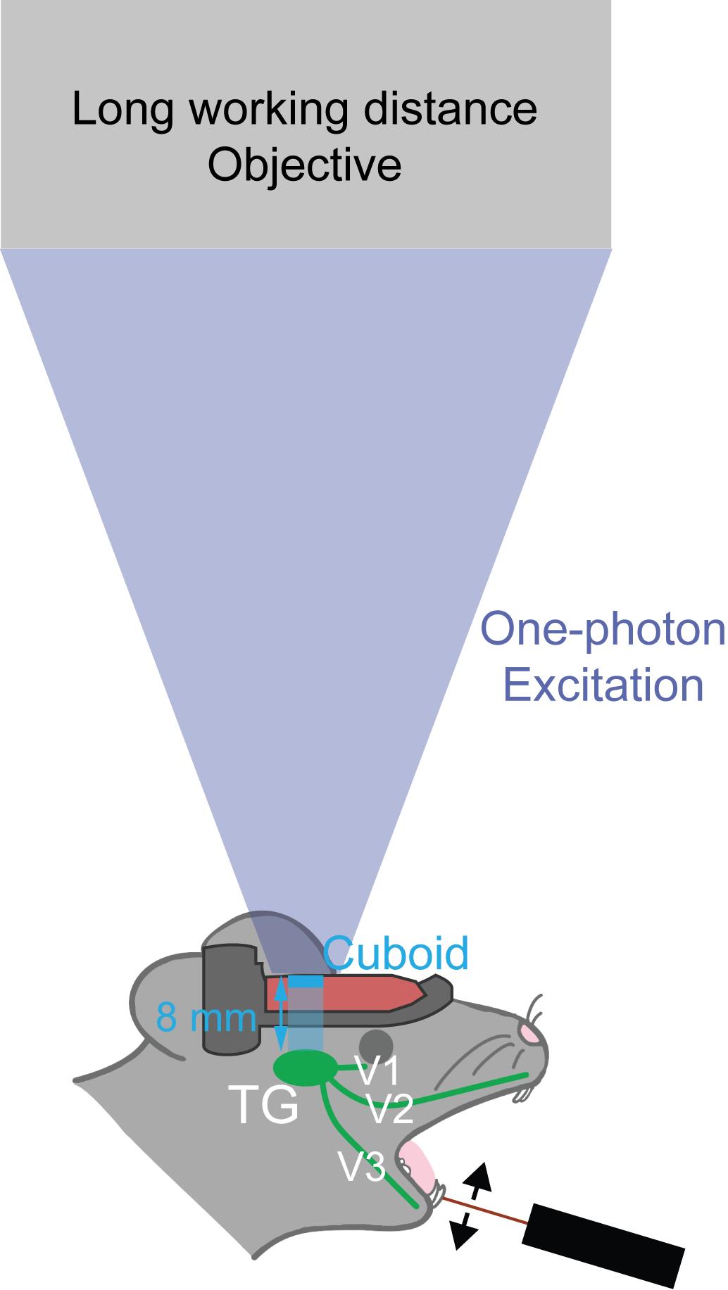
Figure 1. Schematic illustration of trigeminal ganglion (TG) innervation of the face, including the oral cavity.The glass cuboid enables in vivo calcium imaging of the entire TG. In our previous work [5], sensory stimulation was applied to the left mandibular incisor using a piezoelectric bender. V1–V3 denote the three branches of the trigeminal nerve: V1, ophthalmic; V2, maxillary; and V3, mandibular. Working distance of the objective lens needs to be longer than 8 mm (100 mm for MV PLAPO 1×).
Materials and reagents
Biological materials
1. Thy1-GCaMP6f mice (aged >8 weeks) (The Jackson Laboratory, stock number: 025393)
Reagents
1. Carprofen (Rimadyl, catalog number: VetRx MW 026357)
2. Lidocaine hydrochloride jelly, 2% (IMS, catalog number: 76329-3015-5)
3. Cyanoacrylate adhesive (Aron Alpha, TOAGOSEI, catalog number: 04613)
4. Dental cement (Jet Denture Repair Package, LANG, catalog number: 1223CLR)
5. Phosphate-buffered saline (PBS), 10× (Gibco, catalog number: 70011-044)
6. Hair remover cream (Veet, catalog number: 3299655)
7. Antibiotic ointment (Medi-First, catalog number: 22373)
8. Povidone-iodine, 10% (Betadine, catalog number: 67618-150-01)
9. Isoflurane (Piramal Critical Care, catalog number: 66794-017-25)
10. Ketamine 100 mg/mL (Patterson Veterinary, catalog number: 07-894-8462)
11. Xylazine 100 mg/mL (AKORN animal health, catalog number: 59399-111-50)
12. Bupivacaine 5 mg/mL (Cook-Waite, catalog number: 99184)
Laboratory supplies
1. Glass cuboid, 3 × 3 × 8 mm (UQG Optics; glass cuboid, 3 mm × 3 mm × 8 mm)
2. Head post/head plate (316L stainless steel, Craftcloud)
3. Forceps (FST, catalog number: 11252-00)
4. Micro-scissors (FST, catalog number: 15000-04)
5. Scissors (FST, catalog number: 14060-09)
6. Surgifoam (Ethicon, catalog number: 1972)
7. Needles: 20G (BD, catalog number: 305176), 18G (Air-Tite, catalog number: 14-817-220), 16G (Air-Tite, catalog number: 14-817-104)
8. Cotton swabs (Puritan, catalog number: 826-WC)
9. Kimwipes (Kimberly-Clark Professional, catalog number: 34155)
10. 3 mL transfer pipettes (Falcon, catalog number: 357575)
11. Tissue culture dish (Fisherbrand, catalog number: FB012920)
12. Insulin syringe with fixed needle (Sol-Vet, catalog number: V12905)
13. Sandpaper, 80 grit
14. Toothpicks
15. Erlenmeyer flask, 500 mL (KIMAX, catalog number: 26500)
16. Hose couplers (ROTH, catalog number: E806.1-E809.1)
17. PFA tubing (ROTH, catalog number: 1NAY.1-1NAT.1)
18. Two-hole rubber stopper (Fisherbrand, catalog number: 14-140S)
19. Sandpaper (Johnson abrasives, catalog number: 10110-15)
20. Toothpicks (KingSeal, catalog number: 77150)
Equipment
1. Anesthetic vaporizer (E-Z Systems, model: EZ-108SA)
2. Oxygen concentrator (VARON, model: Y-105)
3. Stereotaxic alignment system (KOPF, model: 1900)
4. Auxiliary ear bars (Narishige, model: EB-5N)
5. Animal temperature controller (WPI, model: ATC 1000)
6. Micro drill (Harvard Apparatus, model: 75-1874)
7. LED illumination (Amscope, model: LED-50WY)
8. Germinator (Braintree Scientific, model: NC9956482)
9. Vacuum regulator (Ohio Medical, model: PISA)
10. Epifluorescence microscope (Olympus, model: MVX-10)
11. Blue LED (Thorlabs, model: SOLIS-470)
12. 1× objective lens (MV PLAPO 1×/0.25 NA)
13. Filter cube (U-MF/XL, Olympus), containing an excitation bandpass filter of 470 nm, a dichroic filter of 495 nm, and an emission bandpass filter of 525/50 nm
14. Monochrome CMOS camera (Thorlabs, model: CS135MU)
15. Piezoelectric bender (Thorlabs, model: PB4NB2S)
16. Wixey digital angle gauge (Wixey, model: WR300 Type 2)
Software and datasets
1. Tinkercad (Autodesk, https://www.tinkercad.com/)
Procedure
登录/注册后免费查看全文
文章信息
稿件历史记录
提交日期: Feb 11, 2026
接收日期: May 10, 2026
在线发布日期: Jun 1, 2026
出版日期: Jul 5, 2026
版权信息
© 2026 The Author(s); This is an open access article under the CC BY-NC license (https://creativecommons.org/licenses/by-nc/4.0/).
如何引用
Iwamoto, R., Matunis, A., Stacy, E., Abe, K., Tamura, S., Kambe, Y., Itokazu, T., Hikida, T., Sato, T. K. and Sato, T. R. (2026). Hemispherectomy-Based Optical Window for In Vivo Visualization of Trigeminal Ganglion Neurons in Mice. Bio-protocol 16(13): e5716. DOI: 10.21769/BioProtoc.5716.

 Download Citation in RIS Format
分类
神经科学 > 神经解剖学和神经环路 > 荧光成像
神经科学 > 感觉和运动系统 > 动物模型
您对这篇实验方案有问题吗?
在此处发布您的问题,我们将邀请本文作者来回答。同时,我们会将您的问题发布到Bio-protocol Exchange,以便寻求社区成员的帮助。
 发布问题
发布问题


