


Quantification of Spatial Patterns of Microtubule Transport by Kinesin-1 Head and Tail
驱动蛋白-1头部与尾部介导的微管运输空间模式定量分析

发布: 2026年05月20日第16卷第10期 DOI: 10.21769/BioProtoc.5697 浏览次数: 547
评审: Sushma KalmodiaAnonymous reviewer(s)
 PDF
PDF  Q&A
Q&A
参见作者原研究论文
The authors used this protocol in:
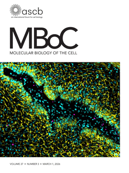
Mar 2026
Advertisement








Abstract
The conventional kinesin-1 is a plus-end-directed microtubule-dependent motor protein with distinct motor head, stalk, and tail domains. Along with the motor head, which binds and walks along microtubules in an adenosine 5’-triphosphate (ATP) dependent manner, kinesin also contains a C-terminal microtubule binding tail. Motor-driven collective motility is well characterized using in vitro gliding assays, which show uninterrupted, smooth trajectories of transport. However, gliding assays driven by the full-length Drosophila kinesin-1 with both head and tail resulted in the emergence of spontaneous spatial microtubule patterns and stop-and-go motion. This was reproduced by an equimolar ratio of the active head and passive tail. Here, we describe the detailed protocol to reconstitute these microtubule gliding assays using multiple motor types: the full-length kinesin-1, the motor head or tail, mixtures of both head and tail, and a rigor mutant of the kinesin. We provide details of the approach taken to acquire the image time-series, to then quantify the spatial patterns that result from these motor combinations. Our approach provides a framework to systematically characterize the spatiotemporal effects of molecular motor-driven collective microtubule transport.
Key features
• This protocol highlights the cloning and expression of two major Drosophila kinesin-1 constructs: the microtubule binding tail and isoleucine-alanine-lysine (IAK)-deleted kinesin-1 full length.
• This protocol describes a typical gliding assay setup for the kinesin motor domain, alone as well as in combination with the kinesin tail.
• We present a systematic framework for typical gliding assays, including experimental acquisition as well as quantitative analysis of microtubule collective transport.
• This protocol gives a quantitative metric for microtubule spatial patterns, which enables systematic analysis of microtubule curvature, both in vitro and in vivo.
Keywords: Microtubule (微管) Kinesin-1 (驱动蛋白-1)
Gliding assay (滑行实验)
Microscopy (显微成像)
Image analysis (图像分析)
Gibson assembly (Gibson 组装)
Kinesin-1 (驱动蛋白-1)
Gliding assay (滑行实验)
Microscopy (显微成像)
Image analysis (图像分析)
Gibson assembly (Gibson 组装)
Graphical overview
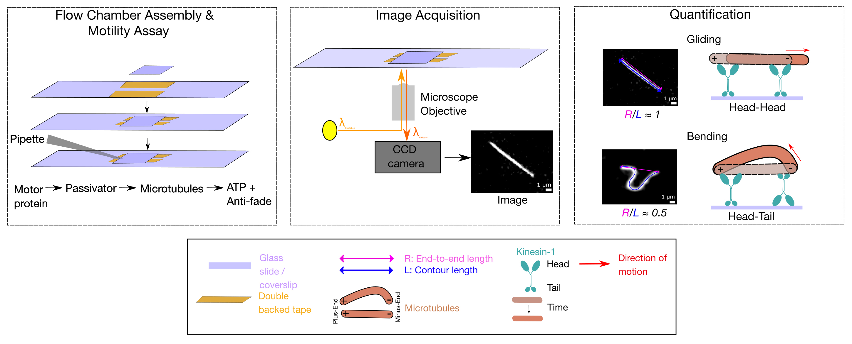
Background
The plus-end-directed kinesin-1 is an essential microtubule-associated motor protein regulating intracellular transport. Kinesin-1 motor walks along microtubule filaments in an ATP-dependent manner, which is studied in vitro using gliding assays [1]. A classical gliding assay setup involves motor proteins immobilized on the surface, allowing microtubules to bind to motors. Force generated by the motor walk causes microtubules to glide in a direction opposite to the direction of motor walk. Microtubule motility driven by both kinesin [2] and dynein [3] can then be observed using fluorescence microscopy. Conventionally, motor-driven collective microtubule transport properties are identified using truncated motor domain constructs of kinesin-1 [4]. Interestingly, kinesin-1 full-length also consists of a C-terminal cargo binding tail [5], which can bind microtubules in an electrostatic manner [6] in an ATP-independent manner [7]. While motor-driven transport is well characterized, the role of the tail in gliding assays is less well known. Recently, we have shown that full-length Drosophila kinesin-1-driven gliding assays result in the emergence of spatiotemporal patterns such as bending, looping, and oscillations along with stop-and-go motion of microtubules [8]. These can be reproduced by an equimolar mixture of the active head and passive tail.
Similar spatial patterns have been reported both in cells [9] and modified gliding assays with artificial constraints for microtubules, resulting in patterns such as buckling [10], flagellar oscillation [11], and loops or spirals [12]. Thus, a systematic analysis of filament curvature, independent of the types of motion, helps us better understand microtubule mechanical properties, such as bending rigidity and persistence length. In previous work, the spontaneous bending patterns of microtubules in a thermal bath resulted in a measure of ~1 mm persistence length [13]. Motor forces exceeding 4–6 pN generated by kinesin in a gliding assay with one end of the filament clamped resulted in highly bent microtubules [10]. This has also been used to explain the highly curved microtubules inside cells as a result of the intracellular forces [14]. We have also observed such patterns in kinesin-1 gliding assays arising out of buckling instabilities due to the same filament being occasionally bound by an immobilizing tail and multiple active heads [8]. These patterns allow us to examine the effect of collective mechanics of both motors and microtubules in a simplified in vitro setup.
Here, we describe a gliding assay setup in order to explore spatial patterns in microtubules, driven by the activity of kinesin heads and tails. To this end, we have made a Drosophila kinesin-1 tail domain construct. In combination with the motor domain of kinesin-1, we have experimentally reconstituted gliding assays that result in bending patterns and quantified the spatial and temporal properties of microtubules. This approach of using gliding assays and quantitative analysis may be generally applicable to measure motor-driven cytoskeletal mechanics in vitro.
Materials and reagents
Biological materials
1. Escherichia coli DH5α competent cells (The Coli Genetic Stock Center, Cheshire, CT, USA, Strain No: CGSC#: 14231)
2. Escherichia coli Bl21(DE3) competent cells (Thermo Fisher Scientific, USA, catalog number: EC00114)
3. K401, purified as previously described [15]
4. K980, purified as previously described [16]
5. Anti-GFP nanobody, purified as described [17]
Reagents
1. D(+)-Biotin (Sigma-Aldrich, catalog number: 8512090001)
2. 5(6)-Carboxytetramethylrhodamine N-succinimidyl ester (Sigma-Aldrich, catalog number: 21955)
3. Piperazine 1,4-bis (2-ethanesulphonic acid) (PIPES) (Sigma-Aldrich, catalog number: P6757-500G)
4. Magnesium chloride (Sigma-Aldrich, catalog number: 208337)
5. Ethylene glycol-bis(2-aminoethylether)-N,N,N′,N′-tetraacetic acid (EGTA) (Sigma-Aldrich, catalog number: E3889-100G)
6. GTP-disodium salt (Sigma-Aldrich, catalog number: 36051-31-7)
7. Adenosine 5′-triphosphate (ATP) disodium salt hydrate (Sigma-Aldrich, catalog number: A2383-10G)
8. Adenosine 5′-triphosphate (ATP) magnesium salt (Sigma-Aldrich, catalog number: A9187-1G)
9. 2-(N-Morpholino)ethanesulfonic acid (MES) hydrate (Sigma-Aldrich, catalog number: M8250- 250G)
10. Calcium chloride (Sigma-Aldrich, catalog number: C4901)
11. Paclitaxel (lyophilized powder) (Cytoskeleton Inc, catalog number: TXD01)
12. Casein sodium salt from bovine milk (Sigma-Aldrich, catalog number: C-8654)
13. Streptavidin, Streptomyces avidinii (Sigma-Aldrich, catalog number: 85878)
14. Sodium dihydrogen phosphate (Sigma-Aldrich, catalog number: 1.06370)
15. di-sodium hydrogen phosphate dihydrate (Sigma-Aldrich, catalog number: 1.06580)
16. β-Mercaptoethanol (Sigma-Aldrich, catalog number: 444203)
17. Phenyl methane sulfonyl fluoride (PMSF) (Sigma-Aldrich, catalog number: P7626)
18. Isopropyl-β-D-Thiogalactopyranoside (IPTG) [SRL Biochem, catalog number: 67208 (094866)]
19. Glucose oxidase (SRL Biochem, catalog number: 9001-37-0)
20. Catalase (SRL Biochem, catalog number: 9001-05-2)
21. D-(+)-Glucose (Sigma-Aldrich, catalog number: G5400-1KG)
22. Acetone (SRL Biochem, catalog number: 66951)
23. Potassium hydroxide (KOH) (Sigma-Aldrich, catalog number: 221473)
24. Bovine serum albumin (Sigma-Aldrich, catalog number: A7906-50G)
25. Glycerol (Sigma-Aldrich, catalog number: G901-2)
26. Luria Bertani (Lysogeny) broth, Miller (HiMedia, catalog number: G1245)
27. Tryptone (HiMedia Laboratories, catalog number: RM014-500G)
28. Yeast extract (HiMedia Laboratories, catalog number: RM027-500G)
29. Potassium phosphate dibasic (HiMedia Laboratories, catalog number: MB044-500G)
30. Potassium phosphate monobasic (HiMedia Laboratories, catalog number: MB050-500G)
31. NEBuilder® HiFi DNA Assembly master mix (New England Biology, catalog number: E2621S)
32. Q5® high-fidelity DNA polymerase (New England Biology, catalog number: M0491S)
33. Deoxynucleotide (dNTP) solution mix (New England Biology, catalog number: N0447L)
34. T4 polynucleotide kinase (New England Biology, catalog number: M0201S)
35. T4 DNA ligase (Promega, catalog number: M1801)
36. DpnI (New England Biology, catalog number: R0176S)
37. XbaI (New England Biolabs, catalog number: R0145)
38. Imidazole (Sigma-Aldrich, catalog number: 5710-OP)
39. HisPurTM cobalt resin (Thermo Fisher Scientific, catalog number: 89965)
40. Ampicillin (Sigma-Aldrich, catalog number: A9518)
42. Rabbit anti-tetramethyl rhodamine antibody (Thermo Fisher Scientific, catalog number: A-6397)
43. QIAprep Spin Miniprep Kit (Qiagen, catalog number: 27104)
44. Pluronic F127 (Sigma-Aldrich, catalog number: P2443)
45. Primers
a. Forward primer for K910-980 (vector), ATGAACTATATAAAGGTACTAAACAGAAGATTTCCTTC (Sigma-Aldrich)
b. Reverse primer for K910-980 (vector), AGTTCTTCTCCTTTGCTCATATGTATATCTCC (Sigma-Aldrich)
c. Forward primer for K910-980 (insert), TACATATGAGCAAAGGAGAAGAACTTTTCA (Sigma-Aldrich)
d. Reverse primer for K910-980 (insert), TGTTTAGTACCTTTATATAGTTCATCCATGCCAT (Sigma-Aldrich)
e. Forward primer for K936, AACTCGCTTGTTCCGCGT (Sigma-Aldrich)
f. Reverse primer for K936, ACGTCTGCCCAGATGCTTC (Sigma-Aldrich)
46. Recombinant DNA plasmids
a. Drosophila Khc(1–980)-6xHis (K980), Addgene (unpublished), Addgene plasmid no. 129762, https://www.addgene.org/129762/
b. Drosophila Khc(1–401)-BCCP-6XHis (K401), Addgene [15], Addgene plasmid no. 15960, https://www.addgene.org/15960/
c. Human KIF5B(1–560)-GFP-6xHis (K560), Addgene [23], Addgene plasmid no. 15219, https://www.addgene.org/15219/
d. pGEX6P1-GFP-nanobody, Addgene [17], Addgene plasmid no. 61838, https://www.addgene.org/61838/
Solutions
1. 5× PIPES-EGTA-MgCl2 (PEM) buffer (see Recipes)
2. K401 purification buffer (see Recipes)
3. K980/K560/KRigor purification buffer (see Recipes)
4. Taxol buffer (see Recipes)
5. Antifade mix (see Recipes)
6. Motility buffer (see Recipes)
7. PCR mix (see Recipes)
8. Gibson assembly mix (see Recipes)
9. Digestion–phosphorylation–ligation (DPL) buffer (see Recipes)
10. Digestion buffer (see Recipes)
11. High-molarity PIPES buffer (HMPB) (pH 6.8) (see Recipes)
12. Depolymerization buffer (pH 6.6) (see Recipes)
13. High pH cushion buffer (pH 8.6) (see Recipes)
14. Low pH cushion buffer (pH 8.6) (see Recipes)
15. Labeling buffer (pH 8.6) (see Recipes)
16. Quenching buffer (pH 7) (see Recipes)
Recipes
1. 5× PEM buffer (pH 6.8)
| Reagent | Final concentration | Quantity or volume |
| PIPES | 400 mM | 6.048 g |
| EGTA | 5 mM | 0.951 g |
| MgCl2 | 25 mM | 0.508 g |
| Double-distilled H2O | Make up to 50 mL |
2. K401 purification buffer
| Reagent | Final concentration | Quantity or volume |
| 200 mM Na-Phosphate (pH 7.2) | 20 mM | 10 mL |
| 1 M β-mercaptoethanol | 10 mM | 1 mL |
| 1 M MgCl2 | 4 mM | 0.4 mL |
| 100 mM ATP | 50 μM | 0.05 mL |
| 50 mM PMSF | 0.1 mM | 0.2 mL |
| Double-distilled H2O | Make up to 100 mL |
3. K980/K560/KRigor purification buffer
| Reagent | Final concentration | Quantity or volume |
| 200 mM Na-Phosphate (pH 8) | 50 mM | 25 mL |
| 5 M NaCl | 300 mM | 6 mL |
| 100 mM β-mercaptoethanol | 5 mM | 0.5 mL |
| 1 M MgCl2 | 1 mM | 0.1 mL |
| 100 mM ATP | 100 μM | 0.1 mL |
| 50 mM PMSF | 0.1 mM | 0.2 mL |
| Double-distilled H2O | Make the volume up to 100 mL |
4. Taxol buffer
| Reagent | Final concentration | Quantity or volume |
| 2 mM Paclitaxel | 20 μM | 1 μL |
| 5× PEM buffer | 1× | 20 μL |
| Double-distilled H2O | Make the volume up to 100 μL |
5. 10× antifade mix
| Reagent | Final concentration | Quantity or volume |
| 125 μM glucose oxidase | 10 μM | 8 μL |
| 40 μM catalase | 15 μM | 37.5 μL |
| 1 M glucose | 500 mM | 50 μL |
| 5× PEM buffer | 1× | 20 μL |
| Deionized H2O | Make the volume up to 100 μL |
6. Motility buffer
| Reagent | Final concentration | Quantity or volume |
| 100 mM MgCl2 | 5 mM | 2 μL |
| 10× antifade mix | 1× | 5 μL |
| 1,400 M β-mercaptoethanol | 140 mM | 25 μL |
| 2 mg/mL casein | 0.5 mg/mL | 12.5 μL |
| 100 mM MgATP | 1 mM | 2 μL |
| 5× PEM buffer | 1× | 10 μL |
| Double-distilled H2O | Make up to 50 μL |
7. PCR mix
| Reagent | Final concentration | Volume for 1 reaction |
| 5× Q5 reaction buffer | 1× | 10 μL |
| 10 mM DNTP mix | 200 μM | 1 μL |
| 10 μM forward primer | 0.5 μM | 2.5 μL |
| 10 μM reverse primer | 0.5 μM | 2.5 μL |
| Q5 high-fidelity DNA polymerase | 0.02 U/μL | 0.5 μL |
| Template DNA | 1 μg | 1 μL |
| Autoclaved water | Make the volume up to 50 μL |
8. Gibson assembly mix
| Reagent | Final concentration | Volume for 1 reaction |
| 0.05 pM vector | 0.0125 pM | 2.5 μL |
| 0.5 pM insert | 0.1 pM | 2 μL |
| 2× NEBuilder HiFi DNA Assembly master mix | 1× | 5 μL |
| Autoclaved double-distilled water | Make the volume up to 10 μL |
9. DPL buffer
| Reagent | Final concentration | Volume for 1 reaction |
| DNA | ≤1 μg | 40 μL |
| DpnI | 0.02 U/μL | 0.5 μL |
| T4 kinase | 0.2 U/μL | 1 μL |
| T4 ligase | 0.01 U/μL | 1 μL |
| 10× T4 ligase buffer | 1× | 5 μL |
| Autoclaved double-distilled water | Make up to 50 μL |
10. Digestion buffer
| Reagent | Final concentration | Volume for 1 reaction |
| DNA | 1 μg | 2 μL |
| XbaI | 0.4 U/μL | 0.2 μL |
| 10× CutSmart buffer | 1× | 1 μL |
| Autoclaved double-distilled water | Make the volume up to 10 μL |
11. HMPB (pH 6.9)
| Reagent | Final concentration | Quantity or volume |
| PIPES | 1 M | 151.2 g |
| EGTA | 10 mM | 1.016 g |
| MgCl2 | 20 mM | 3.804 g |
| Double-distilled H2O | Make the volume up to 500 mL |
12. Depolymerization buffer (pH 6.6)
| Reagent | Final concentration | Quantity or volume |
| MES | 50 mM | 4.85 g |
| CaCl2 | 1 mM | 0.073 g |
| Double-distilled H2O | Make the volume up to 500 mL |
13. High pH cushion buffer (pH 8.6)
| Reagent | Final concentration | Quantity or volume |
| HEPES | 0.1 M | 1.1915 g |
| EGTA | 1 mM | 19.02 g |
| MgCl2 | 1 mM | 4.75 mg |
| Glycerol | 60% | 30 mL |
| Double-distilled H2O | Make the volume up to 50 mL |
14. Low pH cushion buffer (pH 8.6)
| Reagent | Final concentration | Quantity or volume |
| Glycerol | 60% | 6 mL |
| 5× PEM | 1× | 2 mL |
| Double-distilled H2O | Make the volume up to 10 mL |
15. Labeling buffer (pH 8.6)
| Reagent | Final concentration | Quantity or volume |
| HEPES | 0.1 M | 1.1915 g |
| EGTA | 1 mM | 19.02 g |
| MgCl2 | 1 mM | 4.75 mg |
| Glycerol | 40% | 20 mL |
| Double-distilled H2O | Make the volume up to 50 mL |
16. Quenching buffer (pH 7)
| Reagent | Final concentration | Quantity or volume |
| K-glutamate | 50 mM | 0.0736 g |
| 100 mM MgCl2 | 0.5 mM | 1 mL |
| 5× PEM buffer | 1× | 2 mL |
| Double-distilled H2O | Make the volume up to 10 mL |
Laboratory supplies
1. Pipette tips: 2–20 μL (Tarsons, catalog number: 521000A), 10–100 μL (Tarsons, catalog number: 521010A), 100–1,000 μL (Tarsons, catalog number: 521020A)
2. Microtubes (microcentrifuge tubes): 500 μL (Tarsons, catalog number: 524060), 1,500 μL (Tarsons, catalog number: 524070A)
3. Ultracentrifuge polypropylene tube for TLA 100.3 ultracentrifuge rotor: 1.5 mL polypropylene tube (Beckmann Coulter, catalog number: 343169)
4. Ultracentrifuge polypropylene tube for TLA 120.2 ultracentrifuge rotor: 1 mL open-top thick-walled polypropylene tube (Beckmann Coulter, catalog number: 357656)
5. Ultracentrifuge tube for Ti 70 ultracentrifuge rotor: 26.3 mL polycarbonate bottle with cap assembly (dimensions: 25 × 89 mm) (Beckmann Coulter, catalog number: 337922)
6. Micro cover glass 22 mm × 22 mm No. 1.5 (VWR, catalog number: 48366-227)
7. Glass slides 22 mm × 60 mm (HiMedia Laboratories, catalog number: BG003-5 × 50NO)
8. Double-sided Kapton® polyimide tape (Ted Pella Inc, catalog number: 16087-12)
Equipment
1. Pipettes: 1–10 μL, 2–20 μL, 20–200 μL, and 200–1,000 μL (PIPETMAN, Gilson)
2. Optima MAX-XP ultracentrifuge (Beckmann Coultier, model: 393315)
3. Optima XE-100 floor model ultracentrifuge (Beckmann Coultier, model: A94516)
4. Eppendorf high-speed centrifuge 5810R (Eppendorf, model: 5810R)
5. Benchtop ultracentrifuge rotor (Beckman Coulter, model: TLA 100.3)
6. Benchtop ultracentrifuge rotor (Beckman Coulter, model: TLA 120.2)
7. Floor model ultracentrifuge rotor (Beckman Coulter, model: Ti 70)
8. Eppendorf BioSpectrometerR basic (Eppendorf, catalog number: 6135HQ004308)
9. Thermomixer comfort dry heating block (Eppendorf)
10. Vibra Cell Ultrasonics (Sonics & Materials, Inc., model: VCX130)
11. Inverted epifluorescence microscope (Nikon Corp., model: Nikon TiE)
12. Lenses: CFI Plan Apochromat VC 60XA WI N.A. 1.20, W.D. 0.29 mm, CFI Plan Apochromat VC 100XH N.A.1.40, W.D. 0.13mm (Nikon Corp.)
13. Camera
14. Andor Clara2 CCD camera (Andor, Oxford Instruments)
15. Temperature-controlled chamber (Oko Labs)
16. Filters: TRITC filter: fC-FL Epi-Fl Filter Block TRITC, excitation: 385–425 nm; emission: 550–660 nm (Nikon Corp.); FITC filter: EGFP (FITC/Cy2) fluorescence filter, excitation: centered at 470 nm with a 40 nm bandwidth; emission: centered at 525 nm with a 45 nm bandwidth (Chroma Technology Corp)
Software and datasets
1. Fiji version 1.54p [18]
2. Python (version: v3.9.12) with packages SciPy (v1.7.3) [7] and NumPy (v1.21.5) [2]
3. MATLAB (version: R2022b)
4. FIESTA [1]
5. Inkscape 1.2
Procedure
登录/注册后免费查看全文
文章信息
稿件历史记录
提交日期: Feb 11, 2026
接收日期: Apr 14, 2026
在线发布日期: May 12, 2026
出版日期: May 20, 2026
版权信息
© 2026 The Author(s); This is an open access article under the CC BY-NC license (https://creativecommons.org/licenses/by-nc/4.0/).
如何引用
Readers should cite both the Bio-protocol article and the original research article where this protocol was used:
- Basu, J., Singh, K., Jannasch, A. and Athale, C. A. (2026). Quantification of Spatial Patterns of Microtubule Transport by Kinesin-1 Head and Tail. Bio-protocol 16(10): e5697. DOI: 10.21769/BioProtoc.5697.
- Basu, J., Singh, K., Jannasch, A. and Athale, C. A. (2026). Spatial coin-tossing by kinesin-1 head and tail binding collectively drives microtubule patterns. Mol Biol Cell. 37(3): ee25–09–0427. https://doi.org/10.1091/mbc.e25-09-0427

 Download Citation in RIS Format
分类
细胞生物学 > 细胞结构 > 微管
生物化学 > 蛋白质 > 定量
生物化学 > 蛋白质 > 活性
您对这篇实验方案有问题吗?
在此处发布您的问题,我们将邀请本文作者来回答。同时,我们会将您的问题发布到Bio-protocol Exchange,以便寻求社区成员的帮助。
 发布问题
发布问题


