


Chemoenzymatic Labeling Method for Detection of O-GlcNAcylated α-Synuclein Proteins by Western Blot
利用化学酶法标记和蛋白质印迹检测 O-GlcNAc 修饰的 α-突触核蛋白

发布: 2026年05月20日第16卷第10期 DOI: 10.21769/BioProtoc.5692 浏览次数: 360
评审: Elena A. OstrakhovitchWeidong AnAnonymous reviewer(s)
 PDF
PDF  Q&A
Q&A
参见作者原研究论文
The authors used this protocol in:
Oct 2025
Advertisement








Abstract
α-Synuclein (α-syn) aggregation has emerged as a key pathogenetic feature in several neurodegenerative disorders. The α-syn protein has various conformational strains, each with unique structural features that influence their cytotoxicity, propagation, and neuroinflammation. A post-translational modification known as O-GlcNAcylation has been found to influence the toxicity of α-syn and its propensity to aggregate. Difficulties in detecting and quantifying this modification are a major challenge to understanding its roles among the conformational forms of α-syn. We now describe a protocol for detecting O-GlcNAcylated α-syn that combines a click chemistry labeling approach and western blotting. This chemoenzymatic method involves the transfer of azido-modified galactose (GalNAz) from UDP-GalNAz to O-GlcNAcylated proteins, enabling their further functionalization with alkyne-containing polyethylene glycol of defined molecular weight. This protocol facilitates the determination of the glycosylation status of varying conformations of α-syn and their stoichiometric ratios.
Key features
• The method outlines a chemoenzymatic mass-tagging approach for the identification and quantification of O-GlcNAcylated α-syn.
• The process involves using GalT 289L for GalNAz transfer, followed by PEG mass tag conjugation, and then subjected to western blotting.
• Visualizable modified α-syn with a noticeable molecular weight shift, enabling estimation of its relative stoichiometry.
• This protocol provides a rapid and simple strategy that can be completed within two or three days.
Keywords: Post-translational modifications (翻译后修饰) O-GlcNAc modification (O-GlcNAc 修饰)
α-Synuclein (α-突触核蛋白)
β-1,4-galactosyltransferase 1 (β-1,4-半乳糖基转移酶 1)
Western blot (蛋白质印迹法)
O-GlcNAc modification (O-GlcNAc 修饰)
α-Synuclein (α-突触核蛋白)
β-1,4-galactosyltransferase 1 (β-1,4-半乳糖基转移酶 1)
Western blot (蛋白质印迹法)
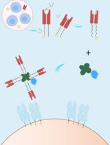
Graphical overview
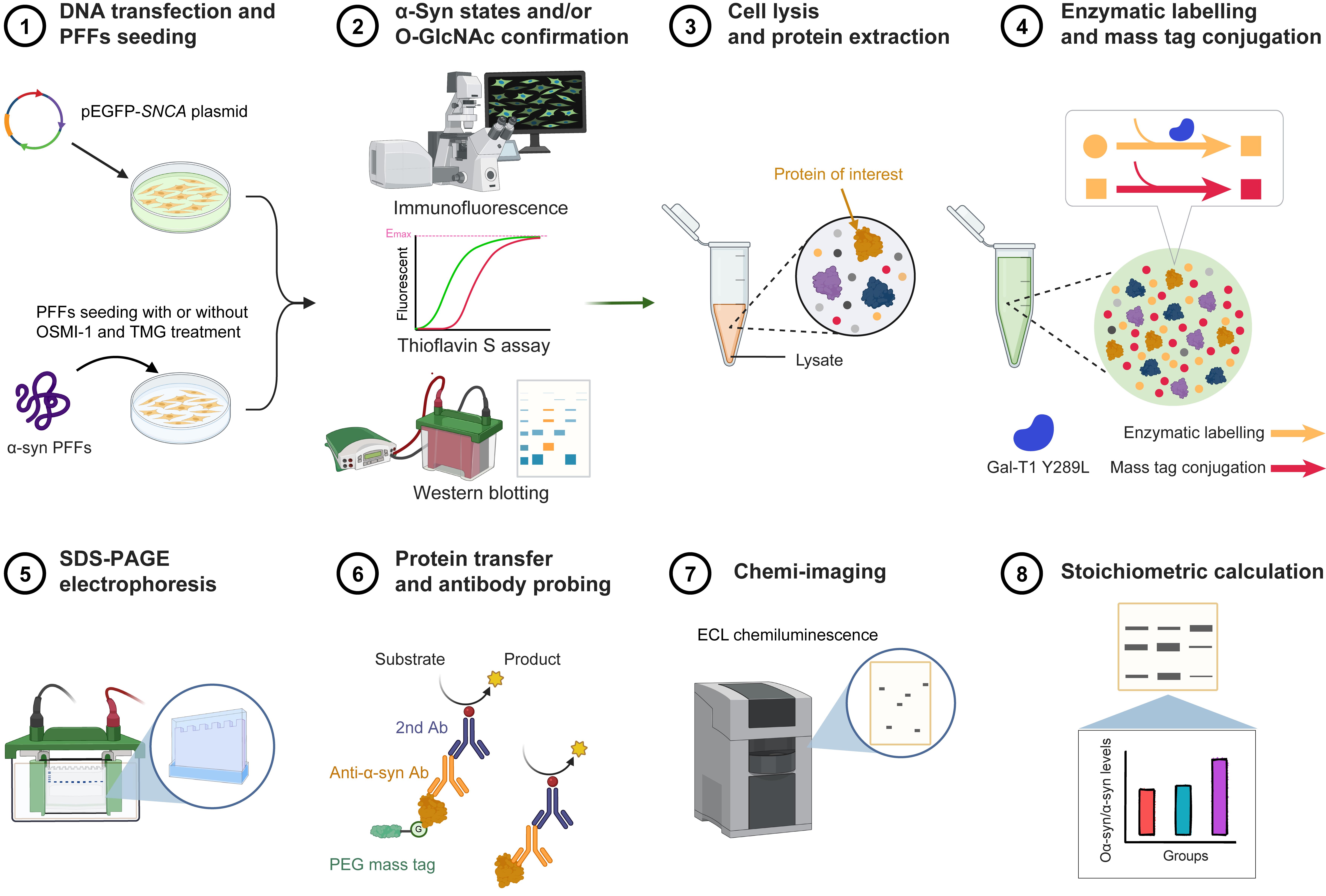
Chemoenzymatic labeling procedure for detecting O-GlcNAcylated α-Synuclein (α-syn) proteins by western blot
Background
Pathological aggregation of α-Synuclein (α-syn) is a defining characteristic of various neurodegenerative disorders, including Parkinson's disease and multiple system atrophy [1]. α-syn, a 140 amino acid protein, exhibits intrinsic disorder and impressive conformational flexibility, allowing it to take on a wide range of structural forms, including oligomers, protofibrils, and mature fibrils [2]. These heterogeneous strains have different conformational structures that are closely related to their diverse cytotoxicity, selective neuronal vulnerability, non-cell-autonomous propagation, and neuroinflammatory effects [3–5]. Emerging evidence implicates post-translational modifications (PTMs) in the modulation of α-syn structure and function, with dysregulated PTMs contributing to its propensity for misfolding and aggregation [6,7].
O-GlcNAcylation is a ubiquitous intracellular process entailing the enzymatic attachment of a single monosaccharide, N-acetylglucosamine, to cytoplasmic, nuclear, and organelle proteins [8,9]. O-GlcNAc transferase (OGT) catalyzes the addition of GlcNAc from uridine diphosphate N-acetylglucosamine (UDP-GlcNAc) to the hydroxyl moiety of serine or threonine residues in proteins, while O-GlcNAcase (OGA) removes it [10]. Regulating the O-GlcNAc modification process can affect the pathology of α-syn by altering the structure of its aggregates, which reduces their cytotoxicity, cell-to-cell propagation, and neuroinflammation [11–13] (Figure 1). A deeper understanding of the interplay of roles of O-GlcNAcylation in α-syn pathology in disease pathways could lead to targeted therapies. However, investigation of the functions of O-GlcNAcylation is challenging due to complexities associated with the detection and quantification of this modification [14,15]. Development of reliable detection methods is crucial for investigating O-GlcNAcylated α-syn and to understand its pivotal roles in regulating α-syn proteins, associated cellular processes, and disease pathology [11].
An effective method for observing O-GlcNAcylated proteins involves a chemoenzymatic labeling approach that utilizes recombinant bovine β-1,4-galactosyltransferase 1 (GalT Y289L) to tag O-GlcNAcylated proteins with an N-azidoacetylgalactosamine (GalNAz) group [16]. This method enables accurate, unbiased, and comprehensive labeling of O-GlcNAcylated proteins by combining them with a peptide. A versatile set of tags can be fused and combined with the proteins using copper-catalyzed or strain-promoted azide-alkyne cycloaddition, allowing them to be visualized [17,18]. By adding polyethylene glycol (PEG) of defined molecular weight, the modification status of two O-GlcNAcylated proteins, nucleoporin 62 (Nup62) and cyclic AMP-response element binding protein (CREB), was determined in protein lysate based on observed elevated molecular weight bands. This approach facilitates the investigation of the O-GlcNAcylation status of various strains of amyloid α-syn with different conformational states.
In this protocol, we describe a chemoenzymatic method to rapidly and sensitively assess O-GlcNAcylated α-syn. By labeling O-GlcNAcylated proteins with GalNAz using the GalT Y289L enzyme, followed by conjugation with defined DBCO-PEG mass tags via alkyne functional groups, the modification of α-syn can be identified and quantified by use of a specific antibody. This method, employed in models of α-syn ectopic expression and preformed fibril seeding, rapidly, sensitively, and quantitatively detected changes in O-GlcNAc modification of α-syn following inhibition of OGA and OGT.
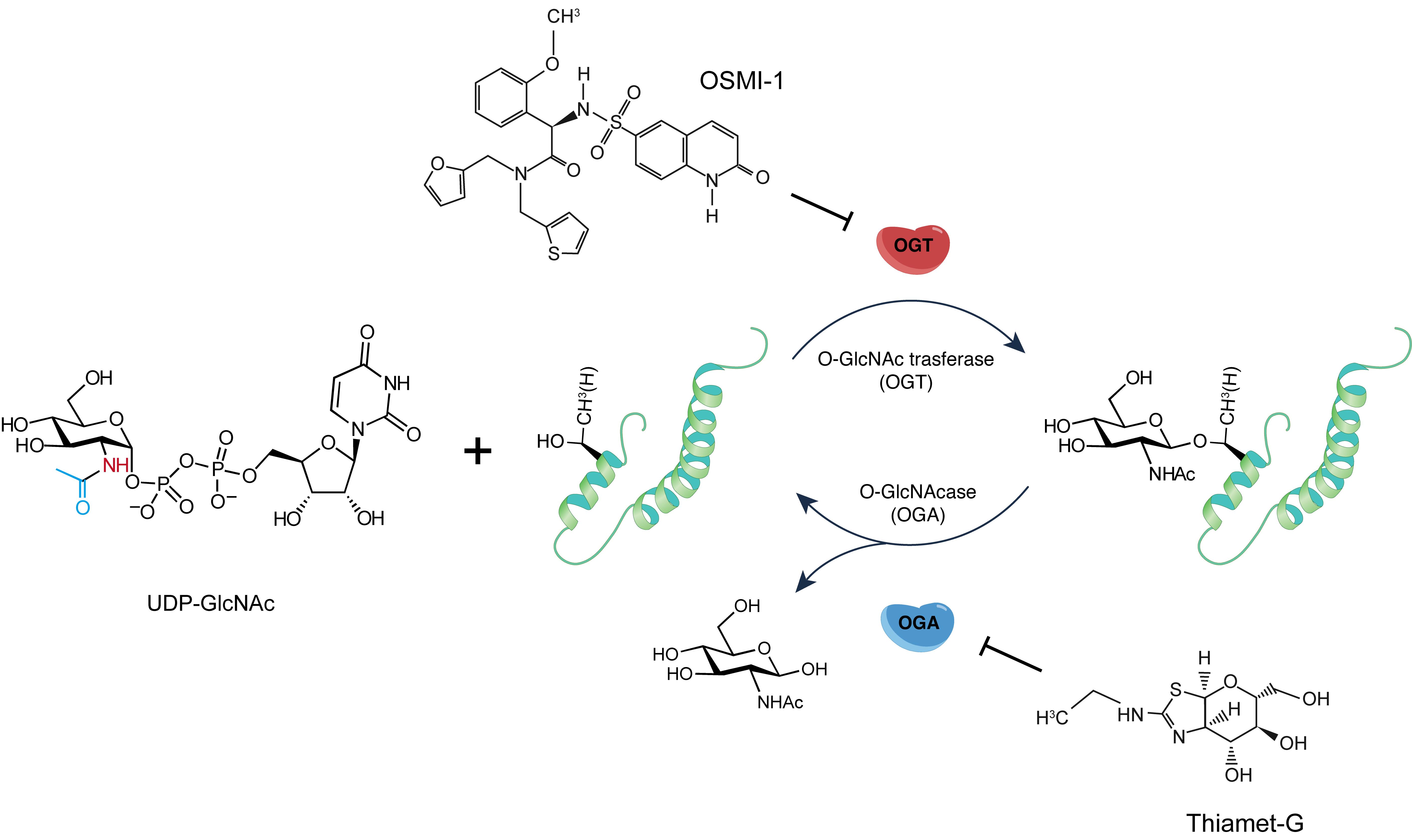
Figure 1. Schematic of protein O-GlcNAcylation cycling. O-GlcNAc modification installs a monosaccharide onto proteins catalyzed by O-GlcNAc transferase (OGT). Removal is catalyzed by O-GlcNAcase (OGA) [19]. Thiamet-G (TMG), a potent chemical inhibitor of OGA, increases O-GlcNAcylation. OSMI-1, a cell-permeable inhibitor of OGT, reduces O-GlcNAcylation without affecting other glycosyltransferases.
Materials and reagents
Biological materials
1. SH-SY5Y cells (YaJi Biological, catalog number: YS267C)
2. pEGFP-SNCA plasmids (Addgene ID: 40822)
3. Recombinant human α-Synuclein proteins
Note: The plasmid was transformed into BL21(DE3) cells, and proteins were expressed and purified using Capto Q ion-exchange chromatography resin [13].
4. Recombinant Gal-T1 Y289L proteins
Note: The construction of the Gal-T1 Y289L mutant plasmid and the purification of the recombinant protein were performed as described in the previous report [20].
5. PVDF membrane (Millipore, catalog number: IPVHO0010)
6. Thiamet G (TargetMol, catalog number: T6056)
7. OSMI-1 (TargetMol, catalog number: T16409)
Reagents
1. DMEM complete medium (Corning, catalog number: 10-013-CVRC)
2. Fetal bovine serum (Gibco, catalog number: 10099141C)
3. Penicillin-streptomycin solution (Gibco, catalog number: 15140122)
4. NaCl (Yonghua Chemical Co., Ltd., catalog number: S105802)
5. KCl (Sigma-Aldrich, catalog number: 7447-40-7)
6. Na2HPO4 (Aladdin, catalog number: D743055)
7. KH2PO4 (Aladdin, catalog number: P434887)
8. 1M Tris-HCl pH8.0 (Servicebio, catalog number: T1150)
9. 1M Tris-HCl pH7.5 (Servicebio, catalog number: T1140)
10. NP-40 (Macklin, catalog number: N885725)
11. Sodium deoxycholate (Beyotime, catalog number: ST2049)
12. jetPRIME DNA transfection reagent (Polyplus, catalog number: 101000046)
13. 50 mM iodoacetamide (IAA) (Accela, catalog number: SY009900)
14. Methanol (Yonghua Chemical Co., Ltd., catalog number: M104903)
15. Chloroform (Sigma, catalog number: 102442)
16. Thioflavin S (Sigma, catalog number: T1892-25G)
17. MOPS (Beyotime, catalog number: ST302)
18. SDS (Servicebio, catalog number: GC204005)
19. 100 mM MnCl2 (Merck, catalog number: 20-309)
20. 0.5 mM UDP-GalNAz (NewCan BioTech, catalog number: NC13303) in 10 mM HEPES, pH 7.9
21. DBCO-PEG 5 kDa (TargetMol, catalog number: T17753)
22. 500 U/μL PNGase F (UA BIOSCIENCE, catalog number: UA070014) in H2O
23. 4% paraformaldehyde (PFA) (Fdbio Science, catalog number: FD9679)
24. Dithiothreitol (DTT) (Fdbio Science, catalog number: FD7927)
25. Skim milk (Fdbio Science, catalog number: FD0080)
26. Tween-20 (Fdbio Science, catalog number: FD0020)
27. Triton X-100 (Solar bio, catalog number: T8200)
28. PUGNAc (TargetMol, catalog number: T38722)
29. Triethanolamine (Macklin, catalog number: 102-71-6)
30. Glycine (Aladdin, catalog number: A110749)
31. Bovine serum albumin (BSA) (Aladdin, catalog number: A104912)
32. Rabbit anti-GFP (Abcam, catalog number: ab209)
33. Mouse anti-α-syn LB509 (Abcam, catalog number: ab27766)
34. Rabbit anti-α-syn MJFR1 (Abcam, catalog number: ab138501)
35. Rabbit anti-α-syn pS129 antibody (Abcam, catalog number: ab51253)
36. Mouse anti-O-GlcNAc RL2 antibody (Invitrogen, catalog number: MA1-072)
37. Fluoromount-G slide mount agent (SouthernBiotech, catalog number: 0100-20)
38. Mouse anti-GAPDH (Fdbio Science, catalog number: FD0063)
39. Goat anti-mouse HRP (Fdbio Science, catalog number: FDM007)
40. Goat anti-rabbit HRP (Fdbio Science, catalog number: FDR007)
41. Goat anti-rabbit IgG (H+L) cross-adsorbed secondary antibody, FITC (Invitrogen, catalog number: F2765)
42. F(ab')2-goat anti-mouse IgG (H+L) cross-adsorbed, Alexa FluorTM 594 (Invitrogen, catalog number: A-11020)
43. Donkey anti-mouse IgG H&L (Alexa Fluor® 647) (Abcam, catalog number: ab150107)
44. BCA protein assay kit (Fdbio Science, catalog number: FD2001)
45. Fdbio-Femto ECL kit (Fdbio Science, catalog number: FD8030)
46. Pre-stained protein ladder marker (Vazyme, catalog number: MP201-02)
Solutions
1. Cell culture medium (see Recipes)
2. Phosphate-buffered saline (PBS) pH 7.4 (see Recipes)
3. Lysis buffer (see Recipes)
4. Homogenization buffer (see Recipes)
5. Mass-labeling buffer (see Recipes)
6. 2.5× labeling buffer pH 7.9 (see Recipes)
7. Resuspension solution A (see Recipes)
8. Resuspension solution B (see Recipes)
9. 1× PBST wash buffer (see Recipes)
10. Permeabilization solution (see Recipes)
11. Blocking buffer (see Recipes)
12. 4× SDS loading buffer (see Recipes)
13. TBST pH 7.6 (see Recipes)
14. Transfer buffer (see Recipes)
15. 10× running buffer for SDS-PAGE electrophoresis (see Recipes)
16. Blocking buffer (see Recipes)
17. Primary antibody dilution (see Recipes)
18. Primary antibody dilution (see Recipes)
Recipes
1. Cell culture medium
| Reagent | Final concentration | Amount |
|---|---|---|
| DMEM | 1× | 500 mL |
| Fetal bovine serum | 10% | 50 mL |
| 100× Penicillin-streptomycin | 1× | 5.5 mL |
| Total | n/a | 555.5 mL |
2. PBS pH 7.4
| Reagent | Final concentration | Amount |
|---|---|---|
| NaCl | 137 mM | 8 g |
| KCl | 2.7 mM | 0.2 g |
| Na2HPO4 | 10 mM | 1.44 g |
| KH2PO4 | 2 mM | 0.24 g |
| Milli-Q H2O | n/a | 800 mL |
| Total | n/a | 1,000 mL |
3. Lysis buffer
| Reagent | Final concentration | Amount |
|---|---|---|
| NaCl (5 M) | 150 mM | 3 mL |
| 1 M Tris-HCl (pH 8.0) | 25 mM | 2 mL |
| NP-40 | 1% | 1 mL |
| Sodium deoxycholate | 1% | 1 mL |
| SDS | 0.1% | 0.1 mL |
| Milli-Q H2O | n/a | up to 100 mL |
| Total | n/a | 100 mL |
Note: Add protease and phosphatase inhibitors before use.
4. Homogenization buffer
| Reagent | Final concentration | Amount |
|---|---|---|
| 1 M Tris-HCl (pH 7.5) | 25 mM | 2 mL |
| NaCl (5 M) | 150 mM | 3 mL |
| Triton X-100 | 1% | 1 mL |
| PUGNAc | 20 μM | 0.707 mg |
| Milli-Q H2O | n/a | up to 100 mL |
| Total | 100 mL |
Note: Add protease and phosphatase inhibitors before use.
5. Mass-labeling buffer
| Reagent | Final concentration | Amount |
|---|---|---|
| Triethanolamine (TEA) (pH 7.4) | 10 mM | 1.49 mg |
| NaCl | 150 mM | 8.77 mg |
| SDS | 1% | 10 mg |
| DBCO-PEG | 1 mM | 5 mg |
| Milli-Q H2O | n/a | up to 1 mL |
| Total | 1 mL |
6. 2.5× labeling buffer pH 7.9
| Reagent | Final concentration | Amount |
|---|---|---|
| HEPES | 50 mM | 11.9 mg |
| NaCl | 125 mM | 73 mg |
| NP-40 | 5% | 0.5 mL |
| Milli-Q H2O | n/a | up to 10 mL |
| Total | n/a | 10 mL |
7. Resuspension solution A
| Reagent | Final concentration | Amount |
|---|---|---|
| HEPES | 20 mM | 47.7 mg |
| SDS | 1% | 100 mg |
| Milli-Q H2O | n/a | up to 10 mL |
| Total | n/a | 10 mL |
8. Resuspension solution B
| Reagent | Final concentration | Amount |
|---|---|---|
| Triethanolamine (TEA) (pH 7.4) | 10 mM | 14.9 mg |
| NaCl | 150 mM | 87.7 mg |
| SDS | 1% | 100 mg |
| Milli-Q H2O | n/a | up to 10 mL |
| Total | n/a | 10 mL |
9. 1× PBST wash buffer
0.05% (v/v) Tween 20 detergent
1× PBS
10. Permeabilization solution
0.1% (v/v) Triton X-100 detergent
1× PBS
11. Blocking buffer
1% BSA (w/v)
1× PBS
12. 4× SDS loading buffer
250 mM Tris-HCl (pH 6.8)
8% (w/v) sodium dodecyl sulfate (SDS)
50% (v/v) glycerol
0.2% (w/v) bromophenol blue
20% (v/v) β-mercaptoethanol (β-ME)
13. TBST pH 7.6
20 mM Tris-HCl
137 mM NaCl
0.1% (v/v) Tween-20
14. Transfer buffer
25 mM Tris
192 mM glycine
20% (v/v) methanol
15. 10× running buffer for SDS-PAGE electrophoresis
250 mM Tris
1.91 M glycine
1% (w/v) SDS
16. Blocking buffer
5% (w/v) skim milk
10 mL of TBST
17. Primary antibody dilution
Dilute each primary antibody in TBST
18. Primary antibody dilution
Dilute each HRP-conjugated secondary antibody in TBST
Equipment
1. Cell culture incubator (Thermo Scientific, model: 905-ULTS)
2. High-speed centrifuge (Thermo Scientific, model: Sorvall LegendTM Micro 17R)
3. Ultrasonic cell crusher (Shanghai Jingxin Industrial Development Co., Ltd, model: XM-150T)
4. Thermostatic shaker (Shanghai Jingxin Industrial Development Co., Ltd, model: JXH-200)
5. Vortex mixer (M&S Instruments, model: VORTEX-GENIE 2 Mixer)
6. Mini LabRollerTM rotator (Sigma-Aldrich BS, model: Z674591-1EA)
7. Nunc Lab-Tek II CC2 Chamber slide system (Thermo Scientific, model: 154917)
8. Ultrasonic cell crusher (Shanghai Jingxin Industrial Development Co., Ltd, model: XM-150T)
9. Mini vertical gel electrophoresis cell (Tanon, model: VE-180)
10. Mini transfer cell (Tanon, model: VE-586)
11. Power supply (Tanon, model: EPS-600)
12. Digital systems for imaging western blotting (Bio-Rad, model: ChemiDoc MP Imaging System)
13. Microplate reader (TECAN, model: INFINITE 200 PRO)
14. Confocal laser scanning microscope (ZEISS, model: LSM900)
Software and datasets
1. Fiji/ImageJ, https://imagej.nih.gov/, https://imagej.net/software/fiji/
2. Microsoft Excel, https://www.microsoft.com/en-in/microsoft-365/excel
3. GraphPad Prism, https://www.graphpad.com/scientific-software/prism/
Procedure
登录/注册后免费查看全文
文章信息
稿件历史记录
提交日期: Jan 26, 2026
接收日期: Apr 3, 2026
在线发布日期: Apr 29, 2026
出版日期: May 20, 2026
版权信息
© 2026 The Author(s); This is an open access article under the CC BY-NC license (https://creativecommons.org/licenses/by-nc/4.0/).
如何引用
Meng, H., Ma, Z., Miao, Y. and Liu, C. (2026). Chemoenzymatic Labeling Method for Detection of O-GlcNAcylated α-Synuclein Proteins by Western Blot. Bio-protocol 16(10): e5692. DOI: 10.21769/BioProtoc.5692.

 Download Citation in RIS Format
分类
神经科学 > 神经系统疾病 > 神经退行性病变
生物化学 > 蛋白质 > 翻译后修饰
生物化学 > 蛋白质 > 标记
您对这篇实验方案有问题吗?
在此处发布您的问题,我们将邀请本文作者来回答。同时,我们会将您的问题发布到Bio-protocol Exchange,以便寻求社区成员的帮助。
 发布问题
发布问题


