


Analysis of Cauline Leaf Development in Arabidopsis thaliana Using Time-Lapse Confocal Microscopy
利用延时共聚焦显微成像分析拟南芥茎生叶发育过程

发布: 2026年05月20日第16卷第10期 DOI: 10.21769/BioProtoc.5691 浏览次数: 738
评审: Pawan KumarAnonymous reviewer(s)
 PDF
PDF  Q&A
Q&A
参见作者原研究论文
The authors used this protocol in:
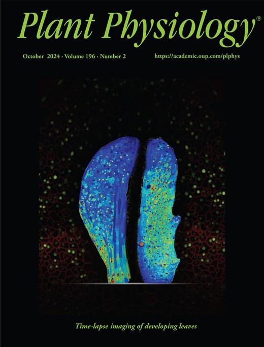
Aug 2024
Advertisement







Abstract
Understanding cellular growth dynamics in plants requires precise, long-term imaging of developing tissues. Cauline leaves are produced during the transition from vegetative to reproductive development and provide a useful system for studying how laminar organs diversify in form and function. While other laminar organs, such as rosette leaves and sepals, have been extensively studied, early cauline leaf development remains technically challenging to capture due to their concealed position, curved morphology, and the presence of dense trichomes. Here, we provide a complete pipeline for the dissection, confocal imaging, 2.5D segmentation, and image analysis of initiating cauline leaves in Arabidopsis thaliana. This method enables reproducible, high-resolution imaging of cauline leaves, supporting robust quantitative analysis of growth across developmental stages at cellular scale resolution.
Key features
• Fine dissection method for exposing initiating cauline leaves in Arabidopsis thaliana.
• Long-term confocal live imaging of cauline leaf development at cellular resolution.
• Optimized imaging parameters for high-fidelity 2.5D segmentation and growth analysis in MorphoGraphX.
Keywords: Confocal microscopy (共聚焦显微镜) Live imaging (活体成像)
Cauline leaf (茎生叶)
Growth dynamics (生长动态)
Image analysis (图像分析)
Organ development (器官发育)
Arabidopsis thaliana (拟南芥)
Live imaging (活体成像)
Cauline leaf (茎生叶)
Growth dynamics (生长动态)
Image analysis (图像分析)
Organ development (器官发育)
Arabidopsis thaliana (拟南芥)
Graphical overview
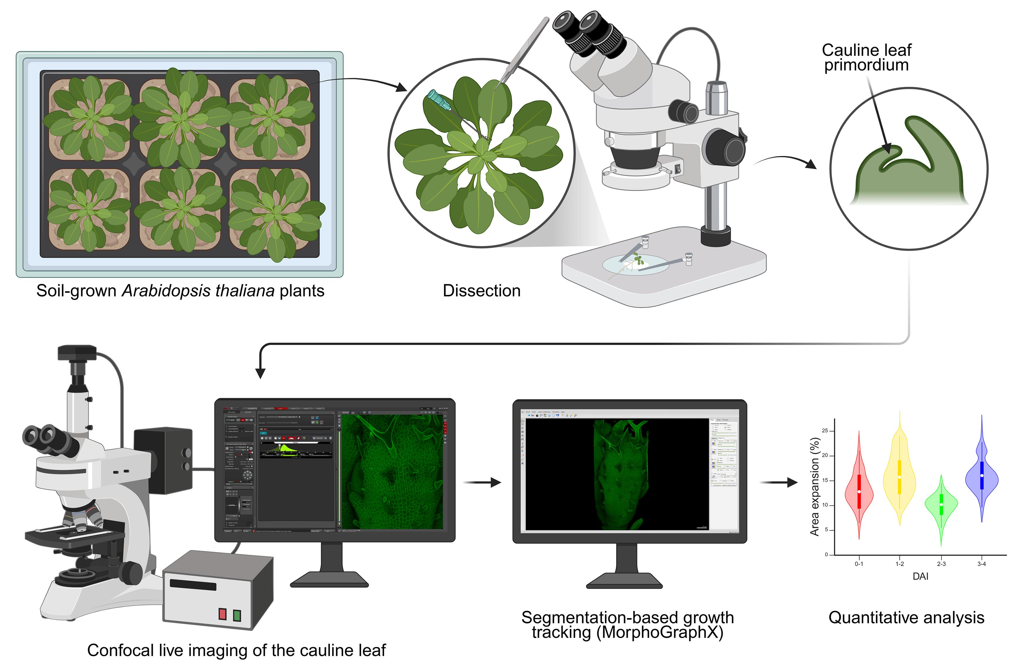
Workflow for long-term confocal imaging and growth tracking of cauline leaves. In the quantitative analysis step, area expansion refers to the increase in area of a cell lineage between successive time points; when a mother cell divides, the areas of the daughter cells are summed so that growth is quantified independently of cell division. Created in BioRender. Lab, M. (2026).
Background
Plant lateral organs, such as leaves and flowers, originate as simple protrusions at the shoot apical meristem but diversify greatly in size and shape to fulfill specific roles at maturity. While the developmental dynamics of rosette leaves and floral organs have been well characterized through live imaging and growth analysis, those of cauline leaves remain poorly understood [1–3].
Cauline leaves are initiated during the transition from vegetative to reproductive growth and combine characteristics of both rosette leaves and floral organs. They therefore provide a useful system for studying how developmental transitions reshape laminar organ identity, growth, and function. However, their early development is difficult to observe, as they are the last leaves to emerge from the meristem—prior to bolting and flower initiation—and are partially concealed by surrounding tissues. In addition, their pronounced curvature and dense trichome coverage on the abaxial surface make them challenging to image using existing approaches optimized for more accessible structures. Previous studies employing scanning electron microscopy or leaf removal have provided insights into morphology and developmental timing, but these techniques lack the spatial and temporal resolution required to capture dynamic growth processes [4–5].
In this paper, we present a protocol for confocal live imaging of initiating cauline leaves in Arabidopsis thaliana. We detail procedures for plant preparation, dissection, and long-term imaging of the same sample with minimal disruption to its development. The protocol is optimized for cellular-resolution imaging and subsequent quantitative analysis, including segmentation-based growth tracking. This method expands the live imaging toolkit to include a previously elusive organ type, enabling investigations into the cellular basis of heteroblasty and the developmental transitions underlying the diversification of laminar organs.
Materials and reagents
Note: The original workflow used in Le Gloanec et al. [6] was developed by the author at the University of Montreal, Canada. For the present protocol paper, the workflow was independently re-implemented at the National University of Singapore, Singapore, in order to validate reproducibility and to generate the illustrative material presented here. The illustrative images shown in this protocol were generated from the Singapore re-implementation, whereas the time-lapse datasets and macroscopic measurements used for validation are from Le Gloanec et al. [6]. This manuscript therefore presents the protocol in a stand-alone format, with expanded methodological detail, updated figures, and implementation notes from both locations. Where materials and reagents differed between the two locations, both versions are listed in the following sections, separated by “or.”
Biological materials
1. Arabidopsis thaliana Columbia-0 (Col-0) carrying the pUBQ10::myr:YFP construct, which encodes a plasma membrane-localized fluorescent marker, was used for time-lapse experiments [7]; other plasma membrane marker lines can also be used, like pUBQ10::PM-tdTomato [8] or p35S::LTI6b-GFP [9]
Reagents
1. ½ MS medium
a. Murashige and Skoog (MS) basal salt mixture (Sigma, catalog number: M552450L)
b. Murashige and Skoog (MS) vitamin solution (Sigma, catalog number: M3900-50ML)
c. Sucrose (Fisher, catalog number: S5-3)
d. Agar (Fisher, catalog number: BP1423-2)
or
a. Murashige and Skoog (MS) medium, including vitamins (Duchefa Biochemie, catalog number: M0222.0100)
b. MES monohydrate (Duchefa Biochemie, catalog number: M1503.1000)
c. Sucrose crystallized (Duchefa Biochemie, catalog number: S0809.5000)
d. Plant agar biotechnology grade (1st base, catalog number: 4020)
2. Plant preservative mixture (PPM) (Plant Cell Technology, catalog number: 71806-1)
3. 95% denaturated alcohol (Fisher, catalog number: HC-1100-1GL) or ethanol approx. 96% grade AR (QRëC, catalog number: E7045-1-2500)
Solutions
1. ½ MS medium (see Recipes)
2. 0.1% PPM solution (see Recipes)
3. 70% ethanol solution (see Recipes)
Recipes
1. ½ MS medium
| Recipe version 1 (Montreal) | Recipe version 2 (Singapore) |
|---|---|
| 2.15 g of MS basal salt mixture | 2.2 g of MS medium including vitamins |
| 10 g of sucrose | 10 g of sucrose crystallized |
| 1 mL of MS vitamin solution | 0.5 g of MES monohydrate |
| Add deionized H2O to 1 L | Add deionized H2O to 1 L |
| Adjust pH to 5.8 | Adjust pH to 5.8 |
| 15 g of agar | 15 g of plant agar |
| After autoclaving | |
| 1 mL of PPM | 1 mL of PPM |
2. 0.1% PPM immersion solution
1 L deionized H2O
1 mL of PPM
3. 70% ethanol solution
737 mL of 95% denatured alcohol
263 mL of sterile deionized H2O
Laboratory supplies
1. Plastic pots, trays, and lids for potting: thermoformed square pots with drainage 2.63” × 2.63” × 2.25”, no-hole trays 21” × 11” × 2.5”, and compatible plastic dome 21.50” × 11” × 2.10” (Teris) or thermoformed pots with drainage 95 × 78 × 75 mm, no-hole trays 54 × 28 × 6.5 cm, and compatible plastic domes 54 × 28 × 9 cm (Linyi Jiaxin Plastic Products Co., Ltd.)
2. All-purpose growing soil mixture (ASB Greenworld Grower Mix) or Jiffy FloraFleur potting soil M.70L/T002 (FarEastFlora, catalog number: 0000785)
3. 100 μL, 200 μL, and 1 mL pipettes with corresponding tips
4. 60 × 15 mm Petri dish (SARSTEDT, catalog number: 82.1194.500 or Corning®, catalog number: 430196)
5. Laboratory film (ParafilmTM)
6. Low lint tissue wipe (KimwipesTM, Kimberly-Clark)
7. Deionized H2O
8. Precision tweezers with fine point (Dumont No. 5)
9. Syringe needles, 18G × 1 1/2 and 27G × 1 1/2 (BD®, catalog number: 305196 and 301629)
10. Scalpel blades (FEATHER, #10)
11. Surgical tape (MicroporeTM tape 3M)
Equipment
1. Growth chamber, Conviron GEN1000 or Percival AR-66L3 with custom LED lighting (Grow Light C75 NS12, Valoya)
2. Autoclave
3. Laminar flow cabinet
4. Dissection stereomicroscope (Stemi 35, Zeiss or S8APO, Leica, equipped with KL 300 LED, Schott)
5. Upright confocal microscope equipped with long-working-distance water-dipping lenses (LSM800, Zeiss, with W Plan-Apochromat 40×/1.0 DIC M27 FWD = 2.5 mm or Stellaris 8 DM6 CFS, Leica, with HCX APO L 20×/1.0 W)
Software and datasets
1. Microscope software, Zeiss Zen 2.6 blue edition (Carl Zeiss Microscopy GmbH), or LAS X STELLARIS (Leica Microsystems)
2. MorphoGraphX 2.0.1 (https://morphographx.org/software/) (access date, 2026-02-26) [10–11]
3. All codes have been deposited to OSF: https://osf.io/uth78/ (access date, 2026-02-26)
Procedure
登录/注册后免费查看全文
文章信息
稿件历史记录
提交日期: Feb 27, 2026
接收日期: Apr 10, 2026
在线发布日期: Apr 27, 2026
出版日期: May 20, 2026
版权信息
© 2026 The Author(s); This is an open access article under the CC BY-NC license (https://creativecommons.org/licenses/by-nc/4.0/).
如何引用
Readers should cite both the Bio-protocol article and the original research article where this protocol was used:
- Le Gloanec, C. (2026). Analysis of Cauline Leaf Development in Arabidopsis thaliana Using Time-Lapse Confocal Microscopy. Bio-protocol 16(10): e5691. DOI: 10.21769/BioProtoc.5691.
- Le Gloanec, C., Gómez-Felipe, A., Alimchandani, V., Branchini, E., Bauer, A., Routier-Kierzkowska, A. L. and Kierzkowski, D. (2024). Modulation of cell differentiation and growth underlies the shift from bud protection to light capture in cauline leaves. Plant Physiol. 196(2): 1214–1230. https://doi.org/10.1093/plphys/kiae408

 Download Citation in RIS Format
分类
植物科学 > 植物发育生物学 > 形态建成
细胞生物学 > 细胞成像 > 共聚焦显微镜
植物科学 > 植物细胞生物学 > 细胞成像
您对这篇实验方案有问题吗?
在此处发布您的问题,我们将邀请本文作者来回答。同时,我们会将您的问题发布到Bio-protocol Exchange,以便寻求社区成员的帮助。
 发布问题
发布问题


