


Lipid Analysis in Live Caenorhabditis elegans Using Solution-State NMR Spectroscopy
利用溶液态 NMR 光谱分析活体秀丽隐杆线虫中的脂质

发布: 2026年05月05日第16卷第9期 DOI: 10.21769/BioProtoc.5686 浏览次数: 484
评审: Pablo HoijembergAlba BlesaAnnmary Paul Erinjeri
 PDF
PDF  Q&A
Q&A
参见作者原研究论文
The authors used this protocol in:

Sep 2024
Advertisement







Abstract
Unsaturated fatty acids (UFAs) play key roles in essential cellular functions such as membrane dynamics, metabolism, and animal development. Disruptions in UFA metabolism are linked to metabolic, cardiovascular, and neurodegenerative disorders. Cellular UFAs composition and quantification are normally determined using methods such as gas chromatography and/or mass spectrometry, which require extraction procedures and prevent analysis of live specimens. Here, we describe a protocol that employs uniform 13C isotope labeling and high-resolution 2D solution-state nuclear magnetic resonance (NMR) spectroscopy to analyze lipid composition and fatty acid unsaturation directly in the model organism Caenorhabditis elegans. The approach enables in vivo assessment of lipid storage compositions with sufficient resolution and sensitivity to distinguish wild-type animals from those with altered fatty acid desaturation. Complementary analysis of total lipid extracts provides information regarding lipid molecules that are not detected in vivo, such as phospholipid molecules organized in biological membranes. Overall, this non-destructive NMR-based method offers a powerful tool for investigating lipid metabolism in C. elegans and other small model systems that can be isotopically enriched.
Key features
• Solution-state NMR spectroscopy is not destructive and can be used on live cells and multicellular organisms.
• 13C isotopic enrichment is required for high-resolution NMR analysis of lipids in live C. elegans.
• Lipid signals from live worms arise from the mobile lipid phase in lipid droplets.
• NMR provides readouts of lipid compositions in live animals at a highly sensitive rate, enabling precise interpretation of the whole cell lipid metabolism.
Keywords: In vivo NMR spectroscopy (体内 NMR 光谱) Unsaturated fatty acids (UFAs) (不饱和脂肪酸(UFAs))
Lipids (脂质)
Caenorhabditis elegans (秀丽隐杆线虫)
13C isotope labeling (13C 同位素标记)
Unsaturated fatty acids (UFAs) (不饱和脂肪酸(UFAs))
Lipids (脂质)
Caenorhabditis elegans (秀丽隐杆线虫)
13C isotope labeling (13C 同位素标记)
Graphical overview
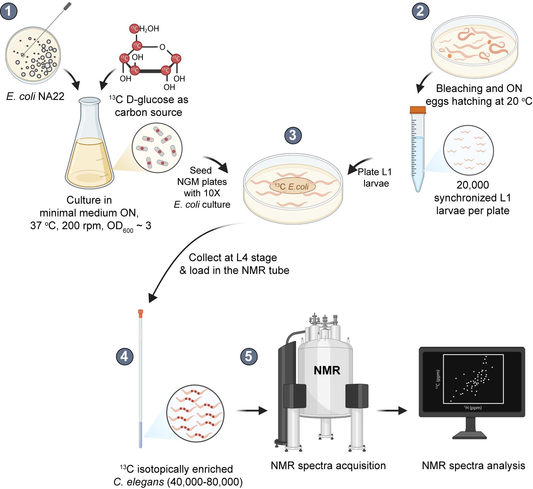
High-resolution solution-state nuclear magnetic resonance (NMR) of live C. elegans. Schematic representation of the experimental setup for analyzing lipids in live C. elegans using NMR. (1) E. coli cells are grown overnight (ON) at 37 °C in M9 minimal medium containing 13C-D-glucose as the sole carbon source, resulting in uniformly 13C-enriched bacteria. (2) Worms are synchronized by bleaching gravid adults and allowing eggs to hatch overnight at 20 °C. (3) Approximately 20,000 L1 larvae are transferred to each of four 9 cm NGM plates seeded with 1 mL of 10× 13C-enriched bacteria concentrated from the ON culture, which typically reaches an OD600 of ~ 3. The larvae are incubated at 20 °C for 48 h until they reach the L4 stage. During this period, worms feed exclusively on the labeled bacteria and incorporate 13C into their biomolecules. (4) L4 worms are collected from each plate in 5 mL of 1× M9 buffer and washed at least two times by sequential resuspension and centrifugation steps to remove residual bacteria. The worm pellet is transferred to microconical tubes and resuspended in approximately 600 μL of 1× M9 buffer. An aliquot of 60 μL of 99.9% deuterated water (D2O) is added, and the slurry is transferred to a Shigemi tube using an automatic pipette and a cut pipette tip. The tube is gently shaken to decant the slurry to the bottom and load it into the NMR spinner without applying the plunge. (5) The tube is loaded in the NMR spectrometer, the probe is tuned to the appropriate 1H and 13C frequencies (standard automatic tuning and matching), magnetic field inhomogeneities introduced by the sample are corrected (standard automatic shimming), the hard and shaped pulses in the pulse sequences are calibrated, and the NMR acquisitions are started. After NMR acquisitions, the slurry is removed, and worms are recovered for viability analysis and/or further experiments. These may include the preparation of total lipid extracts to quantify lipid species at low concentrations or membrane phospholipids, which are not detected in the in vivo experiments due to their restricted motions.
Background
Lipids are essential cellular biomolecules [1], and disruptions in lipid metabolism have been directly linked to disease onset at different degrees in multicellular organisms, including humans [2–4]. Over the years, research in Caenorhabditis elegans has revealed many of the genetic and biochemical pathways that regulate lipid metabolism and fat storage [5,6]. Understanding how lipid metabolism is regulated requires experimental approaches that can directly capture lipid dynamics in living organisms. Caenorhabditis elegans is a powerful model for investigating the physiological roles of specific fatty acids in growth, development, and nervous system function. Unlike mammals, C. elegans does not require dietary essential fatty acids, as it can synthesize polyunsaturated fatty acids (PUFAs) de novo using saturated and monounsaturated fatty acids as precursors [7]. This metabolic flexibility relies on the presence of plant-like Δ12 desaturases together with elongase activities characteristic of animals. Moreover, C. elegans lacks specialized adipocytes and stores neutral lipids in lipid droplets, enabling the direct study of lipid storage dynamics [7]. C. elegans is genetically and cytologically tractable, with a rapid life cycle and extensive conservation of genes and biochemical pathways with humans (≈60%–80%), making it a highly predictive system for studying lipid metabolism in vivo [7].
Traditional approaches to studying lipid composition usually rely on solvent extraction followed by chromatographic or mass spectrometric analysis [8]. While highly sensitive, these methods disrupt cells and tissues, which means that valuable information about how lipids behave and interact inside living systems is lost. Staining methods such as Nile Red, Oil Red, or BODIPY provide spatial localization of lipid particles, but they cannot reveal detailed chemical information about their compositions [9]. Advanced imaging techniques like coherent Raman spectroscopy (CARS) can map lipid-rich regions in live worms, but identifying specific lipid groups with high resolution is not possible [10]. As a result, analyzing lipid composition with highly precise chemical details while maintaining the native context of live cells and multicellular organisms is not possible with the existing methodologies.
Nuclear magnetic resonance (NMR) spectroscopy has been extensively used to analyze the lipid composition of complex samples such as oils, fats, tissue extracts, and body fluids [11–13]. Compared to other analytical techniques, especially mass-spectrometry approaches, NMR sensitivity is low and permits the detection of molecules that are present at high concentration, typically above 1 μM [14], while it fails to detect scarce species. Notwithstanding this limitation, NMR offers a unique, non-destructive way to study biomolecules with atomic resolution in the native environments of cells and multicellular organisms without the need for tags or dyes, and provides both qualitative and quantitative information [15–22].
In this work, we describe the details and experimental setup of a method that combines uniform 13C isotope labeling with bidimensional, solution-state NMR spectroscopy to analyze the lipid composition of live C. elegans. Traditional 1H-NMR strategies, typically used in the in vitro analysis of biological samples, are not suitable for in vivo studies because macromolecular crowding, viscosity, and magnetic field inhomogeneities increase NMR line widths and prevent accurate spectral analysis [15,17]. Using 13C and two-dimensional NMR techniques helps overcome these issues, producing spectra with much higher resolution. Because natural levels of 13C are very low, uniform isotopic labeling is necessary to obtain high-quality spectra within reasonable experimental times.
NMR analysis of live worms and complementary total lipid extracts showed that the lipid signals detected in vivo originate from lipids stored within lipid droplets [15,23]. This corresponds to the mobile lipid phase, and it has also been observed in other systems such as cultured mammalian cells [24,25]. Comparison of the spectral features of N2 worms and fat-3 mutants with defective fatty acid desaturation confirmed the absence of lipid products generated by the FAT-3 enzyme [15].
Thus, with this approach, we can monitor the degree of fatty acid unsaturation and identify different lipid classes within lipid droplets directly in living worms. In addition, it enables us to differentiate the lipid content of wild-type and mutant animals with impaired fatty acid desaturation. The high spectral quality of solution-state NMR of 13C-enriched worms indicates that this type of analysis can be used not only for lipids but also for other abundant metabolites such as sugars, amino acids, and other small molecules [20]. We anticipate that this methodology, in combination with genetic, environmental, or pharmacological approaches, will be useful for studying lipid metabolism under physiological or pathological conditions.
Materials and reagents
Biological materials
1. E. coli OP50 (CGC, University of Minnesota, name: OP50-1, genotype [pyre-, Strr, uracil auxotroph, streptomycin-resistant)]
2. E. coli NA22 (CGC, University of Minnesota, name: NA22, genotype: prototroph)
3. E. coli HT115 (CGC, University of Minnesota, name: HT115(DE3), genotype: [F-, mcrA, mcrB, IN(rrnD-rrnE)1, rnc14::Tn10(DE3 lysogen: lacUV5 promoter -T7 polymerase])
4. C. elegans N2 (CGC, University of Minnesota, name: CGC1, genotype: wild isolate, var Bristol)
5. C. elegans fat-3 (CGC, University of Minnesota, name: VC788, genotype: ok1126)
6. C. elegans NL5901 (CGC, University of Minnesota, name: NL5901, genotype: pkIs2386[unc-54p::α-synuclein::YFP + unc-119(+)]
Reagents
1. KH2PO4 (Supelco, catalog number: 1.04873)
2. K2HPO4 (Supelco, catalog number: 1.05104)
3. Na2HPO4 (Supelco, catalog number: 1.06580)
4. NaOH (Sigma-Aldrich, catalog number: 484024)
5. KOH (Sigma-Aldrich, catalog number: 06103)
6. NaCl (Biopack Productos Químicos, catalog number: 2000164600)
7. CaCl2 (Sigma-Aldrich, CAS: 10043-52-4)
8. MgSO4 (Supelco, CAS: 7487-88-9)
9. NH4Cl (Cicarelli, CAS: 7783-20-2)
10. D-Glucose-6-13C (Cortecnet, catalog number: CC860P10)
11. Bacteriological peptone (Thermo Scientific, Oxoid, catalog number: LP0037B)
12. Agar (Difco, catalog number: 214530)
13. Cholesterol (Sigma-Aldrich, catalog number: C8667)
14. Ethanol (Supelco, catalog number: 1.00983)
15. LB broth (Difco, catalog number: 244620)
16. Streptomycin sulphate salt (Sigma-Aldrich, catalog number: S6501)
17. Ampicillin sodium salt (Millipore, catalog number: 171254)
18. Tetracycline (Sigma-Aldrich, catalog number: 87128)
19. Isopropyl β-D-thiogalactoside (IPTG) (Promega, catalog number: V3951)
20. Methanol (Supelco, catalog number: 1.06018)
21. Chloroform (Supelco, catalog number: 1.02445)
22. Deuterium oxide (D2O) (Sigma-Aldrich, catalog number: 151882, 99.9 atom% D)
23. Deuterated chloroform (CDCl3, 99.8%) supplemented with 0.03% v/v tetramethyl-silane (TMS) for spectral referencing (Sigma-Aldrich, catalog number: 225789)
24. Butylated hydroxytoluene (BHT) (Sigma-Aldrich, catalog number: PHR1117)
25. 3-trimethylsilyl-1-propanesulfonic acid sodium salt (DSS) (CIL, catalog number DLM-32-1)
26. KCl (Sigma-Aldrich, catalog number: P3911)
27. H3PO4 (Biopack, catalog number: 9742.08)
Solutions
1. E. coli minimal medium 10× (see Recipes)
2. E. coli minimal medium working solution (see Recipes)
3. Nematode growth medium (NGM) (see Recipes)
4. Complete NGM (see Recipes)
5. 1 M phosphate buffer (pH 6) (see Recipes)
6. M9 10× (see Recipes)
7. Miller Luria-Bertani broth (LB) (see Recipes)
8. Bleaching solution (see Recipes)
Recipes
1. E. coli minimal medium 10×
| Reagent | Final concentration (M) | Quantity for 250 mL |
|---|---|---|
| KH2PO4 | 1.1 | 37.5 g |
| NaH2PO4 | 5.3 | 160.0 g |
| NaCl | 0.43 | 6.25 g |
| H2Od | - | 250 mL |
Dissolve all reagents in distilled water (dH2O) to a final volume of 250 mL. Adjust the pH to 7.3 using NaOH. Sterilize by autoclaving (121 °C, 20 min). Store at room temperature. The solution can be stored indefinitely. If precipitation or visible contamination occurs, discard and prepare fresh. Dilute 1:10 in sterile dH2O before use as instructed in Recipe 2. This concentrated solution is enough for approximately 30 experiments.
2. E. coli minimal medium working solution
| Reagent | Final concentration (mM) | Quantity for 100 mL |
|---|---|---|
| E. coli minimal medium 10× (Recipe 1) | - | 10 mL |
| 0.1 M CaCl2 | 0.1 | 100 μL |
| 1 M MgSO4 | 2.0 | 200 μL |
| NH4Cl | 18.7 | 100 mg |
| D-Glucose-6-13C | 11.0 | 200 mg |
| Sterile dH2O | - | 90 mL |
Prepare just before use. Dissolve all reagents in dH2O to a final volume of 100 mL. Ensure that all solutions used are sterile. After adding the solid reagents, allow them to fully dissolve. Sterilize the final solution by filtering through a 0.22 μm membrane filter.
3. Nematode growth medium (NGM)
| Reagent | Final concentration (% w/v) | Quantity for 1 L |
|---|---|---|
| Peptone | 0.25 | 2.5 g |
| NaCl | 0.3 | 3 g |
| Agar | 1.7 | 4× 4.25 g (17 g) |
| dH2O | - | 1 L |
Dissolve all reagents in dH2O (except for agar) to a final volume of 1 L. Weigh 4.25 g of agar into each of four 500 mL glass bottles. Distribute the NaCl/peptone solution equally among the four bottles (250 mL each). Sterilize by autoclaving (121 °C, 20 min). Store at room temperature.
4. Complete NGM medium
| Reagent | Final concentration | Quantity for 250 mL |
|---|---|---|
| 1 M CaCl2 | 1 mM | 250 μL |
| 1 M MgSO4 | 1 mM | 250 μL |
| 1 M phosphate buffer (pH 6) (Recipe 5) | 25 mM | 6.25 mL |
| Cholesterol (5 mg/mL in EtOH) | 1.25 mg/mL | 250 μL |
Add all the supplements listed above to the melted NGM primary solution (250 mL) under sterile conditions before use. Wait until the medium has cooled to 55–60 °C before adding the supplements. Mix gently to ensure even distribution. Avoid re-melting the solution, as supplements may deteriorate and/or precipitate.
5. 1 M phosphate buffer (pH 6)
| Reagent | Final concentration (M) | Quantity for 100 mL |
|---|---|---|
| KH2PO4 | 0.8 | 10.83 g |
| K2HPO4 | 0.2 | 3.56 g |
| dH2O | - | 100 mL |
Dissolve all reagents in dH2O to a final volume of 100 mL. Adjust the pH to 6.0 with 2 M KOH. Add dropwise while stirring and monitor with a calibrated pH meter. Sterilize by autoclaving (121 °C, 20 min). Store at room temperature.
6. M9 10×
| Reagent | Final concentration (M) | Quantity for 250 mL |
|---|---|---|
| KH2PO4 | 0.22 | 7.5 g |
| Na2HPO4 | 0.42 | 15 g |
| NaCl | 0.17 | 2.5 g |
| 1 M MgSO4 | 0.01 | 2.5 mL |
| dH2O | - | 250 mL |
Dissolve all reagents in dH2O (except for MgSO4) to a final volume of 250 mL. Sterilize by autoclaving (121 °C, 20 min) and then add 2.5 mL of 1 M MgSO4. Store at room temperature. Dilute 1:10 in sterile dH2O before use (M9 1×).
7. LB
| Reagent | Final concentration (% w/v) | Quantity for 1 L |
|---|---|---|
| LB broth | 2.5 | 25 g |
| dH2O | - | 1 L |
Dissolve LB broth in dH2O to a final volume of 1 L. Divide the solution into 100 mL aliquots in 250 mL glass bottles. Sterilize by autoclaving (121 °C, 20 min). Store at room temperature.
8. Bleaching solution
| Reagent | Final concentration | Quantity for 50 mL (mL) |
|---|---|---|
| 6% bleach (60 g chlorine/L) | 1.2% (12 g chlorine/L) | 10 |
| 1 M NaOH | 0.5 M | 25 |
| dH2O | - | 15 |
Prepare preferably on the same day of use. Dissolve all reagents in dH2O and store for up to one week at room temperature. Protect from light.
Laboratory supplies
1. 15 mL conical tubes (Tarson, catalog number: 546121-RK)
2. 50 mL conical tubes (Tarson, catalog number: 546041-RK)
3. 10 μL pipette tips (ExtraGene, catalog number: TIP-10-C)
4. 200 μL pipette tips (Tarson, catalog number: 521010Y)
5. 1,000 μL pipette tips (Tarson, catalog number: 521016B)
6. 1.5 mL microconical (Eppendorf type) tubes (Tarson, catalog number: 500020N)
7. Petri dishes 6 cm diameter (Φ60 × 15mm) (Tarson, catalog number: 460061)
8. Petri dishes 9 cm diameter (Φ90 × 15mm) (Tarson, catalog number: 460095)
9. Glass Pasteur pipettes (Deltalab, catalog number: 702)
10. Glass tubes (Thermo Fisher Scientific, catalog number: 99449-16)
11. 125 mL glass bottles (CEP, catalog number: Tanja125CC)
12. 250 mL glass bottles (Boeco, catalog number: BOE50806365)
13. 500 mL glass bottles (Boeco, catalog number: BOE5080445)
14. 500 mL Erlenmeyer flask (Fisher catalog number: FB500500)
15. 5 mm water-compatible NMR tubes (Shigemi, corp Japan, catalog number: BMS-005)
16. 5 mm standard NMR tubes (Wilmad, catalog number: WG-1000-7)
17. Glass beads 3 mm diameter (Merck, catalog number: Z143928)
18. Membrane filter (Millex-GV Filter, 0.22 μm) (Millipore, catalog number: SLGV004SL)
19. 100 mL graduated cylinder (Glassco, catalog number: 137.204.05)
Equipment
1. Shaker (Thermo Scientific, model: MaxQ 6000, catalog number: 11754948)
2. Centrifuge (4,000× g) (Thermo Scientific, model: Sorvall ST 16R, catalog number: 75004380)
3. Sterile hood (BIOBASE Vertical Laminar Flow Cabinet BBS-V800)
4. Centrifuge for 15 mL conical tubes (500–2,000× g) (Rolco, catalog number: CM 2036.4, swinging buckets)
5. Stereomicroscope (ZEISS, model: Stemi 305)
6. Refrigerated incubator (20 °C) (Numak, model: HPI-250L)
7. Centrifuge (500–2,000× g) (Eppendorf, model: 5418 R, catalog number: 5401000013)
8. Hand centrifuge (1,298× g) (Hettich, catalog number: 10210722)
9. NMR spectrometer (Bruker, model: 700 MHz Avance III) equipped with a room temperature, triple resonance inverse NMR probe (5 mm 1H/D-13C/15N TXI)
10. Sonicator (Diagenode Bioruptor Sonicaton System, model: UCD-200TM-X, catalog number: 302902)
11. Ultrasonic water bath (0 °C) (Faithful FSF-020S Ultrasonic Cleaner, catalog number: 33F68020S)
12. Water-trap vacuum pump [Faithful, model: SHZ-DIII(P)]
13. Nitrogen evaporator (Organomation, model: 12 Position N-EVAP)
14. Refrigerator (2–8 °C) (Gafa, model: HGF388AFP 374L)
15. Ultra-freezer (-80 °C) (Thermo Scientific, model: Forma 900 Series, catalog number: 904TS)
16. NMR tube spinner (Bruker Biospin, catalog number: Z42516)
17. NMR tube depth gauge (Bruker Biospin, catalog number: Z10627)
18. Micropipettes (Gilson, P20, catalog number: F144056M; P200, catalog number: F144058M; P1000, catalog number: F144059M)
Software and datasets
1. Topspin (Bruker Biospin, version 3.5)
2. The Human Metabolome Database (https://www.hmdb.ca/) [26]
3. The Biological Magnetic Resonance Bank (BMRB) (https://bmrb.io/) [27]
4. The American Oil Chemists’ Society (AOCS) Resource Lipid Library (https://www.aocs.org/publications-resources/resource-library/)
Procedure
登录/注册后免费查看全文
文章信息
稿件历史记录
提交日期: Dec 30, 2025
接收日期: Mar 18, 2026
在线发布日期: Apr 17, 2026
出版日期: May 5, 2026
版权信息
© 2026 The Author(s); This is an open access article under the CC BY-NC license (https://creativecommons.org/licenses/by-nc/4.0/).
如何引用
Guastaferri, F. V., Delprato, C. B., Cravero, B. H., Prez, G., de Mendoza, D. and Binolfi, A. (2026). Lipid Analysis in Live Caenorhabditis elegans Using Solution-State NMR Spectroscopy. Bio-protocol 16(9): e5686. DOI: 10.21769/BioProtoc.5686.

 Download Citation in RIS Format
分类
生物物理学 > 核磁共振波谱法 > 体内NMR光谱法
生物化学 > 脂质 > 脂质测定
系统生物学 > 代谢组学 > 脂类组学
您对这篇实验方案有问题吗?
在此处发布您的问题,我们将邀请本文作者来回答。同时,我们会将您的问题发布到Bio-protocol Exchange,以便寻求社区成员的帮助。
 发布问题
发布问题


