


Extraction and Isolation of Extracellular Vesicles From Piper betle Leaves Using the Apoplastic Fluid Washing and Size Exclusion Chromatography Method
基于质外体液洗脱与尺寸排阻色谱法提取和分离蒌叶来源的细胞外囊泡


发布: 2026年05月05日第16卷第9期 DOI: 10.21769/BioProtoc.5685 浏览次数: 988
评审: Pawan KumarMarisa ConteAnonymous reviewer(s)
 PDF
PDF  Q&A
Q&A
参见作者原研究论文
The authors used this protocol in:

Nov 2024
Advertisement








Abstract
Plant-derived extracellular vesicles (PDEVs) have emerged as important mediators of intercellular communication and hold growing potential in therapeutic applications. However, standardized methods for their isolation, particularly from Piper betle leaves (PBL), remain unexplored. Existing apoplastic fluid washing (AFW) extraction techniques typically rely on manual syringe infiltration, which often leads to inconsistent pressure control, variable yields, and increased risk of tissue damage. This protocol describes a vacuum-assisted AFW extraction method optimized for the recovery of intact extracellular vesicles (EVs) from PBL. The workflow features controlled negative pressure using a vacuum pump and chamber to achieve more efficient leaf infiltration compared to infiltration using the syringe method and reproducible apoplastic fluid (AF) collection with subsequent low-speed centrifugation steps, to ensure minimal contamination and preservation of vesicle integrity. Piper betle–derived extracellular vesicle (PBdEV) isolation and purification steps are performed using size exclusion chromatography (SEC). The size and concentration of PBdEVs were confirmed using nanoparticle tracking analysis (NTA), whereas the cup-shaped and lipid bilayer morphology of the EVs were confirmed using transmission electron microscopy (TEM). The method is scalable and adaptable to various leaf morphologies and physiological states, making it suitable for both exploratory and high-throughput studies. Overall, this protocol provides a more consistent, efficient, and tissue-preserving alternative to traditional syringe-based AF extraction methods, offering higher-quality EV preparations for plant EV research.
Key features
• This protocol focuses on the extraction of Piper betle–derived extracellular vesicles (PBdEVs) using a gentle approach to maintain the vesicles’ morphology.
• This protocol is suitable for large-scale experiments with multiple biological replicates or different leaf samples of similar morphology.
Keywords: Apoplastic fluid washing (质外体液洗脱) Piper betle leaves (蒌叶)
Extracellular vesicles (细胞外囊泡)
Piper betle–derived extracellular vesicles (蒌叶来源细胞外囊泡)
Piper betle leaves (蒌叶)
Extracellular vesicles (细胞外囊泡)
Piper betle–derived extracellular vesicles (蒌叶来源细胞外囊泡)
Graphical overview
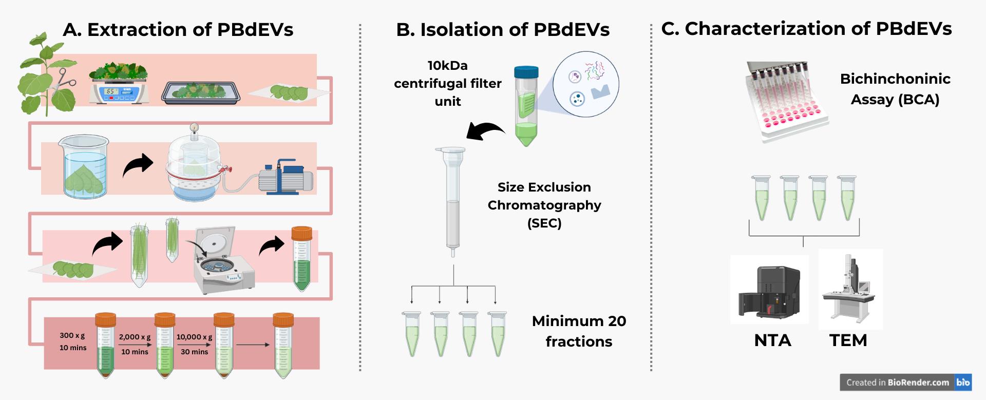
Overview of the workflow to extract and isolate Piper betle–derived extracellular vesicles (PBdEVs). The complete workflow of extracting, isolating, and characterizing PBdEVs involves (A) extracting the PBdEVs using the apoplastic fluid washing (AFW) method and (B) applying size exclusion chromatography (SEC) to gently isolate PBdEVs. After that, (C) bicinchoninic assay (BCA) is performed to determine the protein-to-particle ratio, before identification of the isolated particles using nanoparticle tracking analysis (NTA) and transmission electron microscopy (TEM). The identification methods are not included in this protocol.
Background
Extracellular vesicles (EVs) are nanoscale, membrane-bound particles released by both animal and plant cells [1]. In plants, EVs mediate intercellular communication, participate in defense responses, and transport bioactive cargoes such as proteins, lipids, nucleic acids, and small metabolites [2] across cell walls into the apoplast [3]. Plant-derived EVs are gaining popularity due to their roles in interspecies communication [4], potential application in nanoscale drug delivery, and disrupting oncogenic communication [5–8]. Several methods have been developed to isolate plant EVs, including differential ultracentrifugation, density gradient separation, and precipitation-based approaches [9–11]. While these methods have enabled biochemical characterization and functional studies, they often face challenges such as low yield, co-isolation of contaminants, the requirement of specialized equipment, or limited applicability to different plant tissues [12]. Apoplastic fluid washing (AFW) is a proposed method for harvesting plant extracellular contents with minimal intracellular contamination [13–15], making it well-suited for the isolation of EVs for biochemical characterization and functional testing. Due to their richness in secondary metabolites exhibiting antibacterial and anti-inflammatory activities [16,17] and their abundance in Malaysia [18], Piper betle leaves represent a promising source of plant-derived EVs for further exploration of their therapeutic potential [19–21].
Materials and reagents
Biological materials
1. Piper betle leaves from the family Piperaceae; young to mature leaves, aged 20-30 days old
Note: Young to mature Piper betle leaves have light to deep green–colored leaves (refer to Figure 1A, B), besides being soft to the touch instead of feathery, and can be bent without breaking. The reason why age matters in choosing the leaves is because of the integrity level of the cell wall. In young to mature leaves, cell walls are less developed, thus better in permitting the infiltration of buffer with gentle pressure.
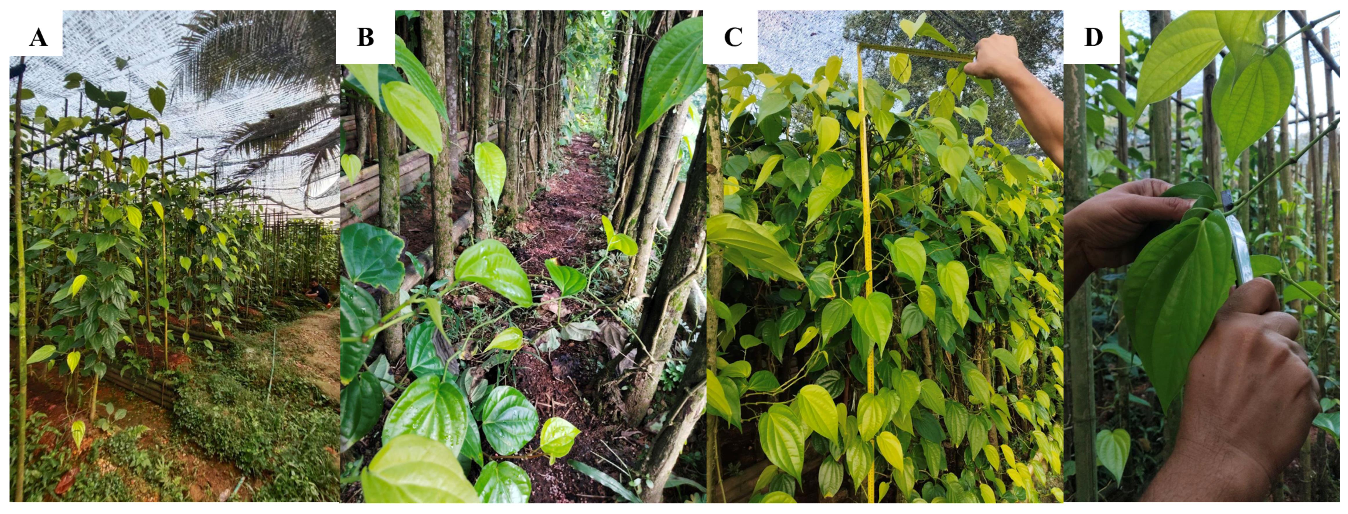
Figure 1. Harvesting Piper betle leaves. (A) Propagation and cultivation conditions of Piper betle. The plant is propagated vegetatively using stem cuttings containing 2–3 nodes, planted on a raised bed at 60–90 cm spacing with 2 m of supporting pole for climbing, and grown under partial shade of the black net. (B) The Piper betle is supplied with NPK fertilizer to support healthy leaf growth. (C) The plant can reach up to 2 m high after 6 months, when it is ready to be harvested. (D) The farmer is instructed to cut the desired leaves at the petiole with a clean knife.
Reagents
1. Phosphate-buffered saline (PBS), pH 7.4 (Biomedia, catalog number: 820301-1); store at 4 °C
2. 70% ethanol (EtOH) (Chemiz, CAS number: 64-17-5); store at room temperature (RT) in a flammable liquid storage cupboard
Caution: This chemical is flammable.
3. SepharoseTM CL-2B (Cytiva Sweden AB, catalog number: 10346215); store at RT away from direct sunlight and heat
Caution: This chemical is flammable.
4. Milli-Q (Merck Millipore); keep at 4 °C for the purpose of this protocol; store at RT if not used
Laboratory supplies
1. Glass chamber A (height: 220 mm; diameter: 190 mm; thickness: 5 mm) (PLT Scientific Sdn Bhd)
Note: This glass chamber was ordered and modified based on our needs. You may need a bigger or smaller chamber depending on the size of your leaves, vacuum chamber, vacuum pump, and experiment scale.
2. Glass chamber B (height: 200 mm; diameter: 150 mm; thickness: 5 mm) (PLT Scientific Sdn Bhd)
Note: This glass chamber was ordered and modified based on our needs. You may need a bigger or smaller chamber depending on the size of your leaves, vacuum chamber, vacuum pump, and experiment scale.
3. Parafilm M All-Purpose Laboratory Film (American Can, catalog number: LMA-PM996)
4. 50 mL conical centrifuge tubes (Biomedia, catalog number: 940204)
5. 20 mL needleless syringe (Terumo, catalog number: 250203B)
6. Pipette tips, 200 and 1,000 μL
7. Econo-Pac® Chromatography Columns kit (Bio-Rad, catalog number: 7321010)
8. Serological pipette, 10 and 25 mL (Bio-Rad, catalog numbers: 031024CM09, 051922CM08)
9. Blunt forceps (LLG Labware, catalog number: 4008476)
10. 500 mL glass beaker (waste container)
11. Cling wrap (Analisa Resources, catalog number: 39201025)
12. Amicon® Ultra Centrifugal Filter, 10 kDa MWCO (Millipore, catalog number: UFC9010)
13. Eppendorf Safe-Lock® tubes biobased 1.5 mL (Eppendorf, catalog number: 0030123093)
Equipment
1. Vacuum chamber and pump (HEXO, model: RS-1.5)
Note: The pump oil may not be provided and needs to be bought separately, if not provided by the manufacturer.
2. Refrigerated centrifuge (Centrifuge 5804 R) (Eppendorf, catalog number: 5805FM568504) (fixed-angle centrifuge)
3. Tabletop centrifuge (Trabota, model: J74276-M000) (fixed-angle centrifuge)
4. Micropipette 20–200 μL (Joanlab, model: 01235556)
5. Micropipette 100–1,000 μL (Joanlab, model: 01242536)
6. Analytical laboratory scale (OHAUS, model: PA214)
7. Freezer (-80 °C)
8. Retort stand or box with holes
9. Electronic pipette with stand (Joanlab, model: EP100Pro)
10. Vegetable washing basket (KECH, catalog number: 1079134)
Procedure
登录/注册后免费查看全文
文章信息
稿件历史记录
提交日期: Dec 26, 2025
接收日期: Mar 17, 2026
在线发布日期: Apr 16, 2026
出版日期: May 5, 2026
版权信息
© 2026 The Author(s); This is an open access article under the CC BY-NC license (https://creativecommons.org/licenses/by-nc/4.0/).
如何引用
Sanddy, I., Adawiyah, W. R., Ooi, D. J., Ekanayake, G., Fazeli, A., Rahman, N. I. A. and Khan, N. L. A. (2026). Extraction and Isolation of Extracellular Vesicles From Piper betle Leaves Using the Apoplastic Fluid Washing and Size Exclusion Chromatography Method. Bio-protocol 16(9): e5685. DOI: 10.21769/BioProtoc.5685.

 Download Citation in RIS Format
分类
植物科学 > 植物细胞生物学 > 细胞器分离
细胞生物学 > 细胞器分离 > 胞外囊泡
您对这篇实验方案有问题吗?
在此处发布您的问题,我们将邀请本文作者来回答。同时,我们会将您的问题发布到Bio-protocol Exchange,以便寻求社区成员的帮助。
 发布问题
发布问题


