


A Suspension-Trapping Protocol for Bottom-Up Proteomics Sample Preparation
基于悬浮捕获法的自下而上蛋白质组学样品制备方案

发布: 2026年05月05日第16卷第9期 DOI: 10.21769/BioProtoc.5681 浏览次数: 587
评审: Joyce ChiuARPITA DAVETingting Chen
 PDF
PDF  Q&A
Q&A
参见作者原研究论文
The authors used this protocol in:
Oct 2025
Advertisement










Abstract
Bottom-up proteomics workflows encompass several key stages, including sample preparation, data acquisition, and data analysis. Of these, sample preparation is the initial and critical stage, as it significantly influences the depth, reproducibility, and reliability of subsequent mass spectrometry–based analyses. While several main digestion strategies exist, including in-gel, in-solution, and filter-aided methods, each presents distinct trade-offs in terms of throughput, contamination removal, and applicability to complex biological matrices. The Suspension Trapping (S-Trap) method offers a compelling alternative by efficiently capturing and digesting proteins while removing interferents like sodium dodecyl sulfate (SDS), which can compromise downstream LC–MS/MS performance. This protocol details a S-Trap workflow optimized for biofluid proteomics, specifically plasma, serum, and cerebrospinal fluid (CSF). We describe two complementary formats: a manual tube-based procedure for individual or small-batch samples and a 96-well-plate-based system enabling high-throughput processing. The protocol integrates optional high-abundance protein depletion to enhance coverage of low-abundance analytes and includes steps for reduction, alkylation, digestion, and peptide elution for low total protein content samples, such as plasma, serum, and cerebrospinal fluid. By providing a detailed protocol, this work aims to improve the consistency and accessibility of S-Trap-based sample preparation, facilitating robust and reproducible discoveries in bottom-up proteomics.
Key features
• Plasma/serum/cerebrospinal fluid sample preparation for bottom-up proteomics.
• Lab Suspension Trapping (S-Trap)-based digestion for efficient detergent removal and high peptide recovery.
• Optimized for challenging samples (e.g., CSF, plasma) with low protein concentration or high lipid content.
• Includes both single-tube and high-throughput 96-well plate formats for flexible experimental design.
Keywords: Mass spectrometry (质谱) LC-MS (液相色谱-质谱联用)
Proteomics (蛋白质组学)
Sample preparation (样品制备)
S-trap (S-trap)
LC-MS (液相色谱-质谱联用)
Proteomics (蛋白质组学)
Sample preparation (样品制备)
S-trap (S-trap)
Graphical overview
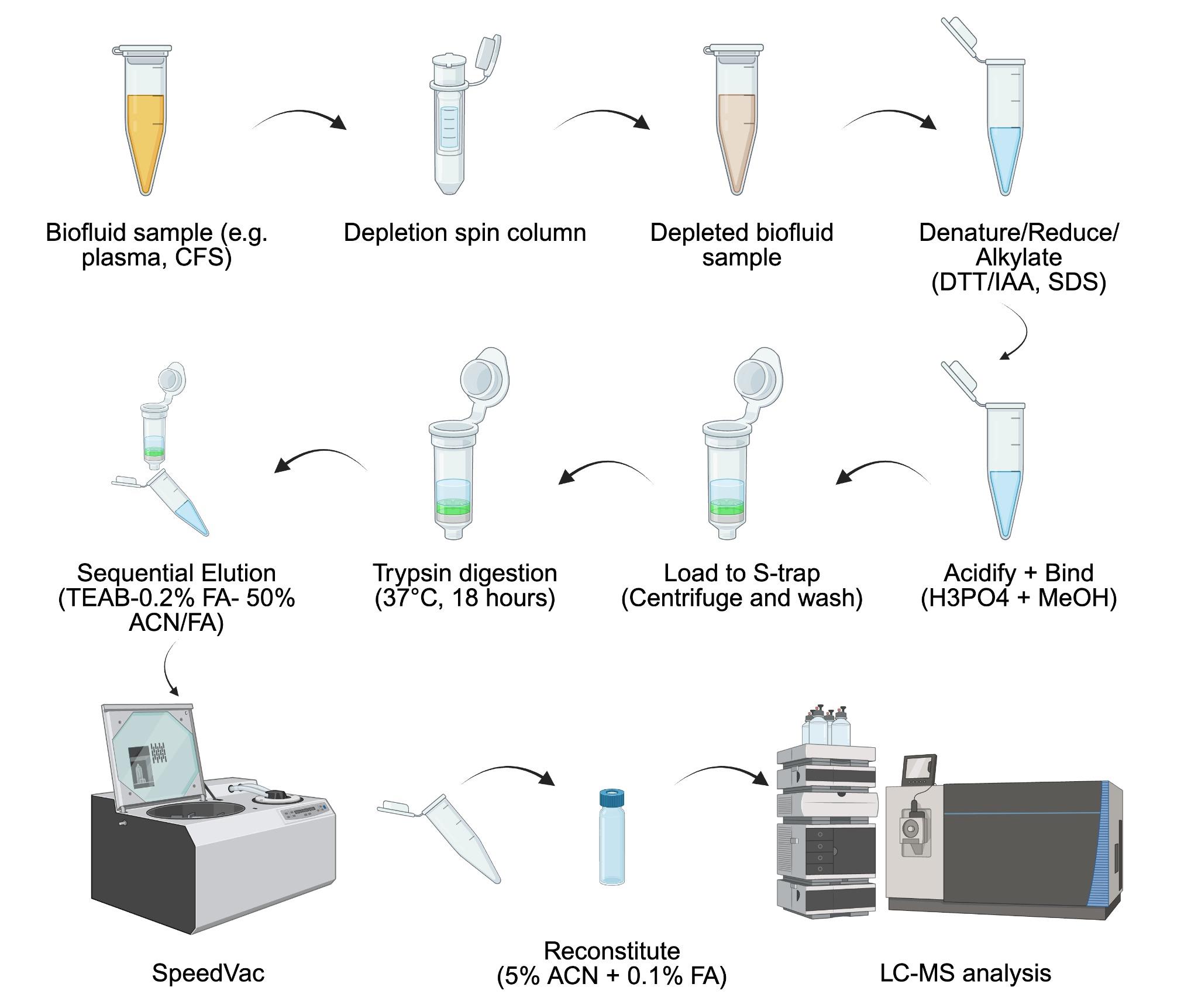
Biofluid sample Suspension Trapping (S-Trap) protein extraction workflow for bottom-up proteomics analysis
Background
In the field of bottom-up proteomics, protein digestion strategies are primarily categorized into in-gel, in-solution, and on-filter digestion, each offering unique strengths and limitations already summarized [1,2]. Additionally, comparative studies that aimed to address the advantages and disadvantages of different approaches have also been reported [2]. In-solution digestion is one of the most commonly used methods for tissue and cell samples [3]. It involves denaturing, reducing, alkylating, and digesting proteins in the liquid phase, offering simplicity and operational efficiency [3]. Compared to in-gel digestion, this method minimizes sample loss and is less labor-intensive, making it particularly suitable for routine proteomic workflows [2]. However, challenges such as protein aggregation and incomplete solubilization can contribute to sample loss. In-gel digestion, by contrast, separates proteins based on size via SDS-PAGE before digestion [4]. This approach effectively reduces contaminants and allows for the fractionation of complex samples, enhancing mass spectrometry analysis for high-complexity proteomes [4]. Proteins are excised from the gel, destained, and digested directly within the gel pieces. While robust and reproducible, in-gel digestion is time-consuming and requires significant manual effort, which can limit throughput [2,4]. On-filter digestion, exemplified by filter-aided sample preparation (FASP), processes proteins retained on molecular-weight cutoff membranes, removing contaminants and recovering peptides through centrifugation [2,5]. FASP is widely used due to its robustness against detergents and gel-free nature [6]. However, the reliance on repetitive centrifugation, requiring over 10 cycles for a complete workflow, significantly increases processing time [2]. Recent adaptations, such as performing cell lysis, reduction, and alkylation outside the filter, have attempted to address this bottleneck, though limitations inherent to membrane-based approaches persist.
The Suspension Trapping (S-Trap) method, introduced by Zougman et al. in 2014, has gained significant attention in proteomics due to its efficiency, simplicity, and high protein recovery [7]. Its key features include a quartz fiber filter stack and a unique protein solubilization protocol using 5% SDS, 12% phosphoric acid, and 90% methanol [7]. This process creates protein particulates that are trapped on the filter, followed by in-filter digestion and peptide elution. The availability of commercial S-Trap filters and 96-well plates further streamlines workflows, improving reproducibility and reducing manual variability.
Sample preparation is a crucial yet challenging step in bottom-up proteomics, often hindered by time constraints, user variability, and labor-intensive processes [8]. Even within the same category of digestion protocols—whether in-gel, in-solution, or on-filter—variations in methods, chemicals, and reagents across proteomics facilities and research groups can significantly impact results [8]. Recognizing the importance of standardization, we previously established a robust chloroform/methanol protein extraction and in-solution trypsin digestion protocol [9]. Building on this foundation, we now expand our workflow to include the S-Trap method, providing a detailed protocol to support consistency and reproducibility. The peptides generated in this protocol are compatible with most liquid chromatography systems and data acquisition methods, including data-dependent acquisition (DDA) and data-independent acquisition (DIA). Moreover, this protocol includes both a tube format and a 96-well plate format, optimizing it for high-throughput proteomics analysis.
Materials and reagents
Biological materials
1. Frozen rat cerebrospinal fluid sample
Note: In our work, 31 samples were used. This protocol has also been successfully conducted using human cerebrospinal fluid and plasma samples.
Reagents
1. Trizma base (Tris) (Sigma-Aldrich, catalog number: T1503-1KG)
2. Phosphoric acid (Honeywell, catalog number: 79606-500 mL)
3. PierceTM BCA Protein Assay kit with Dilution-FreeTM BSA protein standards (Thermo Fisher Scientific, catalog number: A55865).
4. High-SelectTM Top14 abundant protein depletion resin (Thermo Fisher Scientific, catalog number: A36366)
5. S-TrapTM micro column (≤100 μg; PROTIFI, CO2-micro-10, CO2-micro-40, or CO2-micro-40, depending on numbers of samples processed)
6. Dithioreitol (DTT) (Sigma-Aldrich, catalog number: D9779-1G)
7. Iodoacetamide (IAA) (Sigma-Aldrich, catalog number: I1149-5G)
8. Trypsin w/ CaCl2 (TPCK-treated, 500 μg), 10 pack (SCIEX, catalog number: 4445250)
9. Milli-Q water
10. Formic acid (Sigma-Aldrich, catalog number: 695076-100ML)
11. Acetonitrile (ACN) CHROMASOLV LC-MS (Honeywell, catalog number: 34967-4X4L)
12. Sodium deoxycholate (DOC) (Sigma-Aldrich, catalog number: 30970-25G)
13. Ammonium bicarbonate (ABC) (Sigma-Aldrich, catalog number: A6141-25G)
14. Triethylammonium bicarbonate buffer (TEAB) (Sigma-Aldrich, catalog number: T7408-100 mL)
15. Dithiothreitol (DTT) solution, 1 M (ThermoFisher Scientific, catalog number: P2325)
16. PBS solution, pH 7.4 (1×, 10 mM) (GibcoTM, ThermoFisher Scientific, catalog number: 10010023)
Note: Trypsin platinum, mass spectrometry grade (100 μg, Promega, catalog number: VA900) is recommended for samples such as cerebrospinal fluid. For tissue or cell lysate samples, suitable alternatives include trypsin platinum, mass spectrometry grade (100 μg, Promega, catalog number: V5280) or sequencing-grade modified trypsin (lyophilized, 5 × 20 μg, Promega, catalog number: V5111)
Solutions
1. Lysis buffer (see Recipes)
2. 50 mM ammonium bicarbonate (pH ~8) containing 3% (w/v) DOC (see Recipes)
3. Aqueous buffer (see Recipes)
4. S-trap binding buffer, pH 7.1 (see Recipes)
5. Buffer A (see Recipes)
6. Buffer B (see Recipes)
Recipes
1. Lysis buffer
| Reagent | Final concentration |
|---|---|
| SDS | 2% |
| Tris (pH 8.5) | 100 mM |
Note: The composition of the lysis buffer may vary in SDS concentration, typically 2% or 10%. SDS functions primarily to denature proteins, prevent aggregation, and maintain protein solubility. Higher concentrations (e.g., 10%) can be helpful for challenging samples such as skin tissue or membrane protein complexes; however, they also increase the complexity of downstream cleanup. In this protocol, we used 2% SDS, which was effective for cerebrospinal fluid (CSF) samples. A detailed recipe for high-concentration SDS and other buffer formulations is available upon request.
2. 50 mM ammonium bicarbonate (pH ~8) containing 3% (w/v) DOC
| Reagent | Final concentration |
|---|---|
| Ammonium bicarbonate | 50 mM |
| DOC | 3% |
3. Aqueous buffer
| Reagent | Final concentration |
|---|---|
| Acetonitrile | 47.5% |
| Water | 47.5% |
| Formic acid | 5% |
4. S-trap binding buffer, pH 7.1
| Reagent | Final concentration |
|---|---|
| Methanol | 90% |
| TEAB | 1 M |
Note: Rinse glassware with 100% methanol before preparing S-Trap binding buffer.
5. Buffer A
| Reagent | Final concentration |
|---|---|
| Water (LC-MS grade) | 99.4% |
| Acetonitrile | 0.5% |
| Formic acid | 0.1% |
6. Buffer B
| Reagent | Final concentration |
|---|---|
| Acetonitrile | 99.9% |
| Formic acid | 0.1% |
Laboratory supplies
1. Pipette tips (TipOne 10, 200, 1,000 μL) (USA Scientific, catalog numbers: 1111-3800, 1110-1800, 1111-2821)
2. Pipettes (ErgoOne single-channel pipette 2.5, 10, 200, 1,000 μL) (USA Scientific, catalog numbers: 7100-0125, 7100-0510, 7100-2200, 7110-1000)
3. Pour boat weigh dish 2-1/4”l ×1-3/4”w × 5/16”d, 20 mL cap (Wilkem Scientific, catalog number: 10177901)
4. Pipette tip gel loading 0.57 mm O.D. 200 μL round non-sterile (Wilkem Scientific, catalog number: LABB13790)
5. Microcentrifuge tube 0.5 mL non-sterile (Cell Treat, Wilkem Scientific, catalog number: 72316004)
6. Microcentrifuge tube 1.5 mL non-sterile (Cell Treat, Wilkem Scientific, catalog number: 229441)
7. Microcentrifuge tube 1.7 mL non-sterile (Cell Treat, Wilkem Scientific, catalog number: 229441)
8. S-trapTM 96-well mini plate (PROTIFI, catalog number: P002-96MINI)
9. 96-well sample collection plate (Waters, catalog number: 186002842)
10. Sealing mat for 96-well plate, polypropylene, round well (Waters, catalog number: 186002483)
11. 96-well filter plate, 0.2 µm, 1 mL (AcroPrep Advance, Cytiva, catalog number: 8686)
Note: Low protein-binding microcentrifuge tubes (e.g., SARSTEDT, catalog number: 72.706.600) are recommended to minimize peptide adsorption and improve recovery, especially for protein-limited samples such as cerebrospinal fluid.
Equipment
1. Eppendorf Vacufuge plus 5305 (Eppendorf AG, catalog number: 5305FQ525373)
2. Savant SpeedVac SPD210 vacuum concentrator (Thermo Scientific, catalog number: SPD210-P1)
3. Nanodrop One (ThermoFisher, catalog number: ND-ONE-W)
4. Eppendorf Thermomixer F1.5 (Eppendorf AG, model: 5384)
5. Grant-bio PMS-1000i (Grant Instrument Ltd., model: V.2GW.001)
6. Vortex MAXI MIX 1 (Thermo Scientific, Model: M16715)
7. Barnstead Thermolyne LABQUAKE Shaker Rotisserie (catalog number: 3625485)
8. Tecan (Resolvex) A200 Positive Pressure Manifold System
9. Thermo Fisher Orbitrap Ascend Tribrid Mass Spectrometer
Software and datasets
1. Spectronaut® (Version 18.0, Biognosys AG)
3. Rat UniProt FASTA database (UP000002494_10116_Rat_Reference_20240602.fasta) and Universal Contaminant Protein FASTA (created 06.01.22, used 06.03.25) were used for protein identification and to generate the library for DirectDIA analysis
Procedure
登录/注册后免费查看全文
文章信息
稿件历史记录
提交日期: Jan 22, 2026
接收日期: Mar 19, 2026
在线发布日期: Apr 14, 2026
出版日期: May 5, 2026
版权信息
© 2026 The Author(s); This is an open access article under the CC BY license (https://creativecommons.org/licenses/by/4.0/).
如何引用
Schrader, J., Province, D., DaSilva, N. A. and Liu, C. (2026). A Suspension-Trapping Protocol for Bottom-Up Proteomics Sample Preparation. Bio-protocol 16(9): e5681. DOI: 10.21769/BioProtoc.5681.

 Download Citation in RIS Format
分类
生物化学 > 蛋白质 > 分离和纯化
系统生物学 > 蛋白质组学
生物科学 > 生物技术 > 质谱
您对这篇实验方案有问题吗?
在此处发布您的问题,我们将邀请本文作者来回答。同时,我们会将您的问题发布到Bio-protocol Exchange,以便寻求社区成员的帮助。
 发布问题
发布问题


