


An Advanced Single-Cell RNA Sequencing (scRNA-seq) Protocol Utilizing Custom-Designed Multiplexing
基于自定义多重标记策略的高级单细胞 RNA 测序(scRNA-seq)方案

发布: 2026年05月05日第16卷第9期 DOI: 10.21769/BioProtoc.5678 浏览次数: 407
评审: Sébastien GillotinEVANGELOS THEODOROUAnonymous reviewer(s)
 PDF
PDF  Q&A
Q&A
参见作者原研究论文
The authors used this protocol in:

Nov 2025
Advertisement








Abstract
While cell hashing enhances single-cell RNA sequencing (scRNA-seq) efficiency and minimizes batch effects, commercial mouse hashtags often fail in FVB/N and several other strains due to antibody-epitope incompatibility. We describe a robust alternative utilizing biotinylated antibody cocktails and streptavidin-conjugated oligos to enable reliable sample multiplexing. This approach was validated in FVB/N lung tissues, yielding high-quality single-cell libraries. Our protocol offers a practical solution for researchers requiring strain-specific or custom-designed multiplexing strategies for single-cell transcriptomics.
Key features
• Strain-specific compatibility: Resolves the known H-2q haplotype mismatch in FVB/N mice that fails standard commercial MHC-I hashtag antibodies in cell hashing.
• Multi-omic 5′ workflow integration: Enables simultaneous sample multiplexing with 10× Genomics 5 chemistry, facilitating joint gene expression and V(D)J repertoire (TCR/BCR) profiling.
• Enhanced non-immune cell labeling: Incorporates anti-CD326 (Ep-CAM) to ensure robust hashing of epithelial and tumor cells that may exhibit MHC-I downregulation or lack CD45.
• Customizable biotin-streptavidin framework: Provides a modular system using biotinylated antibody cocktails and streptavidin-barcodes, adaptable for any mouse strain or tissue-specific cell markers.
Keywords: scRNA-seq (单细胞 RNA 测序) Cell hashing (细胞哈希标记)
Sample multiplexing (样本多重标记)
FVB/N mice (FVB/N 小鼠)
MHC-I haplotype mismatch (MHC-I 单倍型不匹配)
5′ gene expression (5′ 基因表达)
Biotin-streptavidin conjugation (亲和素-生物素偶联)
Cell hashing (细胞哈希标记)
Sample multiplexing (样本多重标记)
FVB/N mice (FVB/N 小鼠)
MHC-I haplotype mismatch (MHC-I 单倍型不匹配)
5′ gene expression (5′ 基因表达)
Biotin-streptavidin conjugation (亲和素-生物素偶联)
Graphical overview
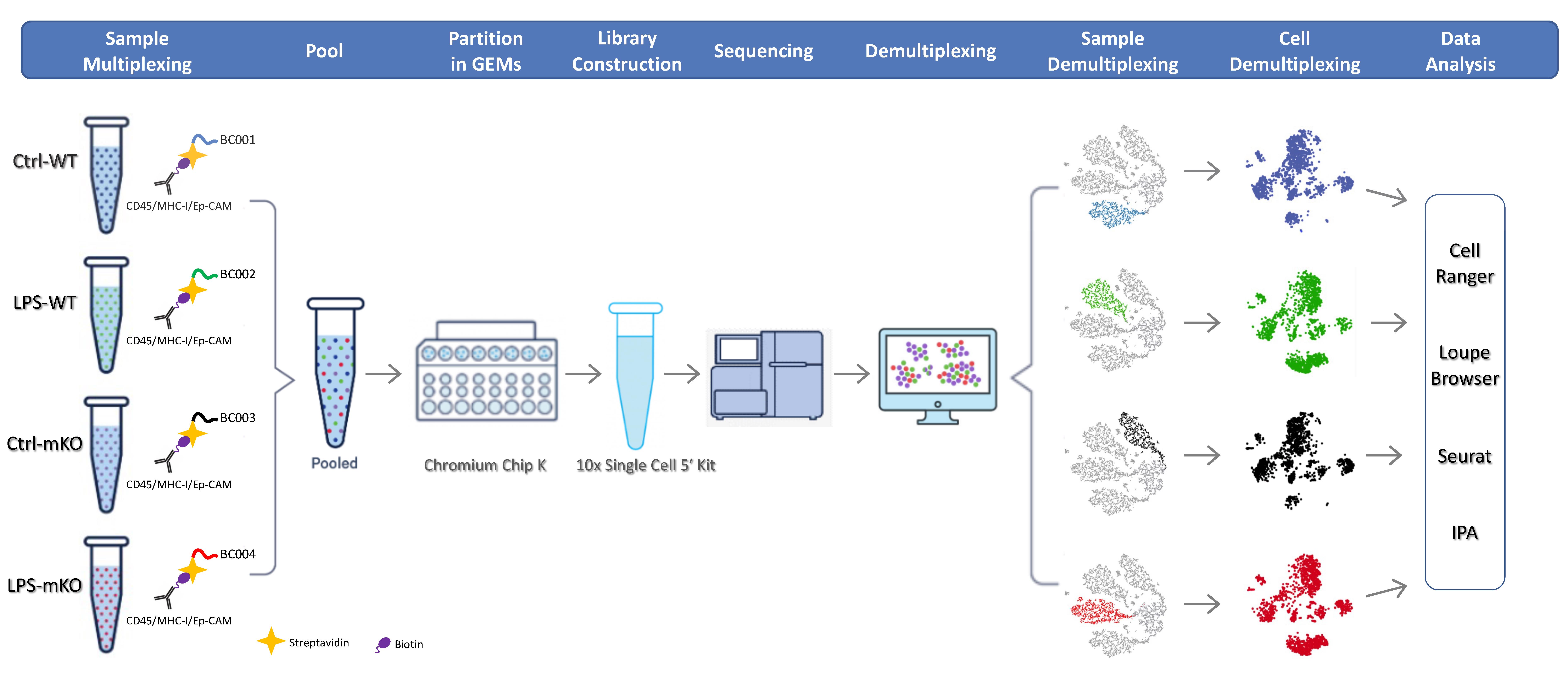
Single-cell RNA sequencing (scRNA-seq) protocol utilizing custom-designed multiplexing
Background
Since its inception in 2009, single-cell RNA sequencing (scRNA-seq) has become an essential technology for profiling the transcriptomic heterogeneity of complex multicellular systems [1]. However, when multiple biological samples are processed in parallel, technical challenges arise, including limited throughput, prohibitive reagent costs, and potential batch effects. Sample multiplexing via cell hashing has emerged as a robust solution to these challenges [2–6]. By labeling individual samples with unique molecular barcodes before pooling and compartmentalization, researchers can integrate multiple samples into a single run, significantly reducing costs while enabling the computational identification and removal of cell doublets or multiplets.
Cell hashing is typically achieved by labeling cell surfaces with oligonucleotide-barcoded reagents, such as oligo-conjugated antibodies targeting ubiquitous surface proteins or lipid/cholesterol-modified oligonucleotides (LMO/CMO) that anchor into the plasma membrane [2–6]. For mouse studies, antibody-based hashing often relies on commercial TotalSeq reagents from BioLegend, which utilize a cocktail of anti-CD45 and anti-MHC Class I (M1/42) antibodies. However, these “universal” reagents are incompatible with the FVB/N and several other mouse strains. Specifically, the M1/42 clone in the commercial mix lacks reactivity with the H-2q haplotype found in FVB/N mice, leading to labeling failure in non-immune (CD45-) populations. Furthermore, while alternative lipid-based multiplexing (e.g., 10× Genomics 3′ CellPlex) approaches are available, they are currently incompatible with single-cell 5′ chemistries, thereby precluding simultaneous profiling of T-cell and B-cell receptor (TCR/BCR) repertoires. The incompatibility of the M1/42 clone is not limited to the FVB/N strain; it also extends to other widely used models carrying the H-2q, H-2p, or H-2r haplotypes, such as the NOD, BUB/BnJ, and RIIIS/J strains. In these genetic backgrounds, standard antibody-based multiplexing often results in the loss of signal in non-immune cell populations, a critical hurdle for lung research where epithelial-immune crosstalk is paramount.
To address these limitations, we developed a versatile cell hashing protocol optimized for the FVB/N strain and compatible with 10× Genomics 5′ workflows [7]. Our method utilizes a customized cocktail of biotin-conjugated antibodies, including anti-CD45 for immune cells, anti-MHC Class I (H-2q) for broad nucleated cell coverage, and anti-CD326 (Ep-CAM) for epithelial and tumor cell enrichment, paired with sample-specific streptavidin-conjugated barcodes. This tripartite antibody approach ensures robust capture of both CD45+ immune infiltrates and CD45- parenchymal or malignant cells, the latter of which may exhibit MHC-I downregulation. Here, we provide a detailed, experimentally validated protocol for custom-designed multiplexing and cell hashing to enable high-resolution scRNA-seq on the lung tissues of wild-type (WT) or myeloid PDLIM2 deletion (mKO) FVB/N mice intratracheally instilled with lipopolysaccharide (LPS) or phosphate-buffered saline (PBS).
The choice of the PDLIM2-deficient model for this protocol validation is based on the gene’s critical role in lung homeostasis. PDLIM2 (PDZ-LIM domain-containing protein 2, also known as SLIM or mystique) is a ubiquitously expressed gene with the highest level in the lung [8–10]. At the cellular level, PDLIM2 is expressed abundantly in various epithelial and immune cells, particularly lung epithelial cells and myeloid cells, two functionally critical cell types in the lung [7,10–18]. Not surprisingly, PDLIM2 serves as a vital molecular checkpoint whose repression has been linked to various pathogenic conditions, especially in the lung, such as lung cancer, chronic obstructive pulmonary disease (COPD), interstitial lung disease (ILD)/pulmonary fibrosis (PF), lung infection, and infectious diseases [7,11–16,19–23]. Mechanistically, PDLIM2 acts as a novel ubiquitin ligase enhancer (E5) to stabilize and chaperone the E3 SCFβ-TrCP to promote the ubiquitination of nuclear RelA (the prototypical NF-κB member that is also known as p65) and possibly STAT3 and other proteins for proteasomal degradation [24–32]. NF-κB and STAT3 are physiologically vital transcription factors that are tightly regulated, and their persistent activation plays causative roles in various diseases, including lung cancer, COPD, ILD/PF, and infections [33–60]. While it complements existing approaches, the novel scRNA-seq using custom-designed multiplexing is far more advanced in studying NF-κB, STAT3, or any other genes and pathways [61–63].
Materials and reagents
Biological materials
1. Mice: PDLIM2flox/flox/lysozyme M-Cre+/- mice (mKO) mice were generated as previously described [11,14,16]. Wild-type (WT) control mice were purchased from The Jackson Laboratory (Bar Harbor, ME). All mice were maintained on a pure FVB/N genetic background. For the scRNA-seq experiment, one biological replicate was used per experimental condition (WT-Ctrl, WT-LPS, mKO-Ctrl, and mKO-LPS) and multiplexed into a single library using the described cell hashing protocol.
Reagents
1. Lung Dissociation kit, mouse (Miltenyi Biotec, catalog number: 130-095-927); contains buffer S (20× stock solution), enzyme D (lyophilized powder), and enzyme A (lyophilized powder)
Note: See Recipes for enzyme D and enzyme A preparation before use.
2. Dead Cell Removal kit (Miltenyi Biotec, catalog number: 130-090-101); contains dead cell removal microbeads and 20× binding buffer stock solution
3. Red blood cell lysis solution (10×) (Miltenyi Biotec, catalog number: 130-094-183)
4. Biotin anti-mouse CD45 (BioLegend, catalog number: 103103)
5. Biotin anti-mouse MHC Class I (eBioscience, catalog number: 13-5998-81; clone 34-1-2S), which specifically reacts with the H-2q haplotype of FVB/N mice, as well as b, s, r, and p haplotypes
6. Biotin anti-mouse Ep-CAM (BioLegend, catalog number: 118203)
7. TotalSeqTM-C0951 PE streptavidin (BioLegend, catalog number: 405261)
8. TotalSeqTM-C0952 PE streptavidin (BioLegend, catalog number: 405263)
9. TotalSeqTM-C0953 PE streptavidin (BioLegend, catalog number: 405265)
10. TotalSeqTM-C0954 PE streptavidin (BioLegend, catalog number: 405267)
11. Ethanol (Decon Labs, catalog number: 2701)
12. RPMI-1640 medium (Lonza, catalog number: 12-702F)
13. Sodium chloride (NaCl) (Sigma-Aldrich, catalog number: S9625)
14. Potassium chloride (KCl) (Sigma-Aldrich, catalog number: P9541)
15. Disodium hydrogen phosphate heptahydrate (Na2HPO4·7H2O) (Fisher Scientific, catalog number: BP331-500)
16. Potassium phosphate monobasic (KH2PO4) (Acros Organics, catalog number: 205925000)
17. Bovine serum albumin (BSA) (MP Biomedicals, catalog number: 199898)
18. Dual Index kit TT Set A 96 rxns (10× Genomics, Inc., catalog number: 1000215)
19. Dual Index kit TN Set A 96 rxns (10× Genomics, Inc., catalog number: 1000250)
20. Chromium Next GEM Single Cell 5’ kit v2, 4 rxns (10× Genomics, Inc., catalog number: 1000265)
21. Chromium Next GEM Chip K Single Cell kit, 16 rxns (10× Genomics, Inc., catalog number: 1000287)
22. 5’ Feature Barcode kit, 16 rxns (10× Genomics, Inc., catalog number: 1000256)
23. Trypan Blue (Sigma-Aldrich, catalog number: T8154)
24. Lipopolysaccharide (LPS) (Sigma-Aldrich, catalog number: L2880)
Solutions
1. Phosphate-buffered saline (PBS) (see Recipes)
2. Washing buffer (see Recipes)
3. Labeling buffer (see Recipes)
4. Buffer S (1× solution) (see Recipes)
5. Enzyme D (see Recipes)
6. Enzyme A (see Recipes)
7. Red blood cell lysis solution (1×) (see Recipes)
8. LPS solution (see Recipes)
Recipes
1. PBS
| Reagent | Final concentration | Quantity or volume |
|---|---|---|
| NaCl | 137 mM | 8 g |
| KCl | 2.7 mM | 0.2 g |
| Na2HPO4·7H2O | 10 mM | 2.16 g |
| KH2PO4 | 1.8 mM | 0.24 g |
| H2O | 1,000 mL |
To prepare 1 L of 1× PBS, begin by dissolving 8.0 g of NaCl, 0.2 g of KCl, 2.16 g of Na2HPO4·7H2O, and 0.24 g of KH2PO4 in approximately 800 mL of distilled or Milli-Q water while stirring with a magnetic stir bar. Once the salts are completely dissolved, calibrate a pH meter and adjust the solution to pH 7.4 using a few drops of concentrated HCl (to lower pH) or NaOH (to raise pH), as needed. Finally, transfer the solution to a volumetric flask or graduated cylinder and add distilled water until the total volume reaches exactly 1 L. Then, filter-sterilize (0.22 μm) or autoclave the buffer to prevent microbial growth.
2. Washing buffer
| Reagent | Final concentration | Quantity or volume |
|---|---|---|
| BSA | 0.04% | 0.2 g |
| PBS | 1× | 500 mL |
3. Labeling buffer
| Reagent | Final concentration | Quantity or volume |
|---|---|---|
| BSA | 1% | 2 g |
| PBS | 1× | 200 mL |
4. Buffer S (1×)
| Reagent | Final concentration | Quantity or volume |
|---|---|---|
| Buffer S (20×) | 1x | 2 mL |
| H2O | 38 mL |
To prepare 40 mL of 1× buffer S, perform a 1:20 dilution by measuring 2 mL of 20× buffer S and transferring it into a clean container. Add 38 mL of sterile, distilled water to reach the final volume of 40 mL. Mix the solution thoroughly to ensure homogeneity and store the resulting 1× buffer at 2–8 °C until use.
5. Enzyme D
| Reagent | Final concentration | Quantity or volume |
|---|---|---|
| Enzyme D | 1× | 1 vial |
| RPMI-1640 | 3 mL |
Reconstitute the lyophilized enzyme D powder in each vial with 3 mL of RPMI-1640 medium. Close the vial and invert gently for 5 min. Prepare aliquots and store at -20 °C, avoiding freeze/thaw cycles. This solution is stable for 6 months after reconstitution.
6. Enzyme A
| Reagent | Final concentration | Quantity or volume |
|---|---|---|
| Enzyme A | 1× | 1 vial |
| Buffer S (1×) | 1 mL |
Reconstitute the lyophilized enzyme A powder in the vial with 1 mL of 1× buffer S. Do not vortex. Prepare aliquots and store at -20 °C, avoiding freeze/thaw cycles. This solution is stable for 6 months after reconstitution.
7. Red blood cell lysis solution (1×)
| Reagent | Final concentration | Quantity or volume |
|---|---|---|
| Red blood cell lysis solution (10×) | 1× | 5 mL |
| H2O | 45 mL |
Dilute 5 mL of red blood cell lysis solution (10×) with 45 mL of sterile, distilled water.
8. LPS solution
| Reagent | Final concentration | Quantity or volume |
|---|---|---|
| LPS | 5 mg/mL | 10 mg |
| PBS (1×) | 2 mL |
Stock solutions of LPS at 5 mg/mL are prepared, aliquoted, and frozen at -80 °C to help standardize instillations. Mice are intratracheally instilled with LPS at 4 mg/kg body weight.
Laboratory supplies
1. GentleMACS C tubes (Miltenyi Biotec, catalog number: 130-093-237)
2. Cell strainers, 40 μm (Fisher, catalog number: 22-363-547)
3. Cell strainers, 70 μm (Fisher, catalog number: 22-363-548)
4. Petri dish (Falcon, catalog number: 351029)
5. 35 mm cell culture dish (Greiner Bio-One, catalog number: 627160)
6. MACS MS column (Miltenyi Biotec, catalog number: 130-042-201)
7. 15 mL conical tubes (TH Geyer, catalog number: 7696714)
8. 50 mL conical tubes (Greiner Bio-One, catalog number: 227261)
9. 150 mL vacuum filtration devices, pore 0.22 μm (Jet Biofil, catalog number: FCF010004)
10. 500 mL vacuum filtration devices, pore 0.22 μm (Jet Biofil, catalog number: FPE204500)
11. 2 mL cryovials (Maxxline, catalog number: MLC2B)
12. 5 mL serological pipettes (Greiner Bio-One, catalog number: 606180)
13. 10 mL serological pipettes (Greiner Bio-One, catalog number: 607180)
14. 25 mL serological pipettes (Greiner Bio-One, catalog number: 760160-TRI)
15. Sterile PES syringe filters (Thermo Fisher Scientific, catalog number: 15206869)
16. 50 mL 3-part syringes (Chirana T. Injecta, catalog number: CH03050LL)
17. 10 μL pipette tips (TH Geyer, catalog number: 7695881)
18. 20 μL pipette tips (TH Geyer, catalog number: 7695882)
19. 200 μL pipette tips (TH Geyer, catalog number: 7695884)
20. 1,250 μL pipette tips (TH Geyer, catalog number: 7695887)
21. Countess cell counting chamber slides (Thermo Fisher Scientific, Invitrogen, catalog number: C10283)
Equipment
1. CO2 chamber
2. Biosafety cabinet
3. Styrofoam board
4. Forceps (Roboz Surgical Instrument, catalog number: RS-5135)
5. Scissors (Roboz Surgical Instrument, catalog number: RS-6802)
6. GentleMACSTM Octo Dissociator with Heaters (Miltenyi Biotec, catalog number: 130-096-427)
Note: Alternatively, a standard GentleMACSTM Dissociator without heating units may be used; in this case, the programmed heating steps must be substituted with manual incubations in a 37 °C water bath for the durations specified in the lung dissociation protocol.
7. Centrifuge (Thermo Fisher Scientific, Thermo ScientificTM, model: IEC CL40R, catalog number: 11210927)
8. MACS MultiStand (Miltenyi Biotec, catalog number: 130-042-303)
9. MiniMACS Separator (Miltenyi Biotec, catalog number: 130-042-102) or OctoMACS Separator (Miltenyi Biotec, catalog number: 130-042-109) for use with MACS MS columns
Note: For higher throughput processing, the QuadroMACSTM Separator (supporting up to 4 columns) or the semi-automated MultiMACSTM Cell 24 Separator Plus (supporting up to 24 columns) may be used as alternatives to accommodate larger sample batches.
10. Countess II FL automated cell counter (Invitrogen, catalog number: AMQAF1000)
11. Chromium Controller & Next GEM Accessory kit (10× Genomics, catalog number: 1000202 or 1000204)
12. Thermocycler (Thermo Fisher Scientific, catalog number: 4375786)
Software and datasets
1. Cell Ranger (v7.0): Used for primary data processing, including alignment, filtering, and UMI counting. Available from 10× Genomics (https://support.10xgenomics.com/single-cell-gene-expression/software/pipelines/latest/what-is-cell-ranger)
2. Seurat (v5.0): An R package utilized for secondary analysis, including quality control, normalization, dimensional reduction, and clustering. Available via CRAN or GitHub (https://satijalab.org/seurat/)
3. Ingenuity Pathway Analysis (IPA, QIAGEN): Used for functional enrichment and metabolic pathway analysis of differentially expressed genes. Accessible via QIAGEN Digital Insights (https://digitalinsights.qiagen.com/products-overview/discovery-insights-portfolio/analysis-and-visualization/qiagen-ipa/)
4. Dataset availability: The raw and processed mouse scRNA-seq datasets generated in this study have been deposited in the NCBI Gene Expression Omnibus (GEO) and are publicly available under the accession number GSE249800
Procedure
登录/注册后免费查看全文
文章信息
稿件历史记录
提交日期: Feb 5, 2026
接收日期: Mar 23, 2026
在线发布日期: Apr 12, 2026
出版日期: May 5, 2026
版权信息
© 2026 The Author(s); This is an open access article under the CC BY-NC license (https://creativecommons.org/licenses/by-nc/4.0/).
如何引用
Gao, F., Liu, X., Sun, F., Xiao, Y., Xiao, G. and Qu, Z. (2026). An Advanced Single-Cell RNA Sequencing (scRNA-seq) Protocol Utilizing Custom-Designed Multiplexing. Bio-protocol 16(9): e5678. DOI: 10.21769/BioProtoc.5678.

 Download Citation in RIS Format
分类
分子生物学 > RNA > RNA 测序
细胞生物学 > 单细胞分析
系统生物学 > 转录组学 > RNA测序
您对这篇实验方案有问题吗?
在此处发布您的问题,我们将邀请本文作者来回答。同时,我们会将您的问题发布到Bio-protocol Exchange,以便寻求社区成员的帮助。
 发布问题
发布问题


