


Quantifying Epigenetic Changes Induced by Chemical Exposure Using the epi-TK Assay
基于 epi-TK 检测法定量评估化学暴露诱导的表观遗传改变

发布: 2026年04月20日第16卷第8期 DOI: 10.21769/BioProtoc.5671 浏览次数: 502
评审: Anonymous reviewer(s)
 PDF
PDF  Q&A
Q&A
参见作者原研究论文
The authors used this protocol in:

Mar 2025
Advertisement








Abstract
Epigenetic modifications play essential roles in regulating gene expression and maintaining cellular identity. Accumulating evidence suggests that chemical agents can contribute to carcinogenesis through epigenetic alterations, such as changes in DNA methylation and histone modifications, even in the absence of direct DNA damage. Here, we have developed a simple, cost-effective, and quantitative reporter assay, termed the epi-TK assay, to evaluate chemically induced epigenetic alterations. The assay is built upon the thymidine kinase (TK) gene mutation assay, a standardized and widely used in vitro genotoxicity assay for chemical safety evaluation. This system is based on an engineered human lymphoblastoid cell line (mTK6), in which the promoter region of the endogenous housekeeping TK gene is site-specifically methylated using epigenome-editing technology, resulting in stable transcriptional repression. Following chemical exposure, epigenetic perturbations at the TK locus are detected by culturing cells under hypoxanthine–aminopterin–thymidine selection and quantifying the frequency of TK revertant colonies, which reflects restoration of TK gene expression. Using the DNA methyltransferase 1 inhibitor GSK3484862 as a model compound, this protocol demonstrates that the epi-TK assay enables sensitive and quantitative detection of epigenetic state transitions. Importantly, this assay allows bi-directional detection of epigenetic changes, including DNA demethylation events and broader alterations in histone modification landscapes. Together, the epi-TK assay provides a practical and quantitative platform for evaluating epigenetic toxicity, with potential applications in chemical safety assessment frameworks.
Key features
• This protocol describes testing of the epigenetic effects of chemicals using the mTK6 cell line and a modified version of the TK gene-mutation assay.
• By employing a DNA-methylated housekeeping TK gene and colony formation as the readout, the assay enables quantitative epigenetic changes without the need for specialized equipment.
• The protocol offers a simple, quantitative, and cost-effective platform that is suitable for routine testing and comparative assessment of multiple compounds.
Keywords: Toxicology (毒理学) Epi-genotoxicity (表观遗传毒性)
Reporter assay (报告基因检测)
TK6 (TK6细胞)
TK gene (TK基因)
DNMT inhibitor (DNMT抑制剂)
Epi-genotoxicity (表观遗传毒性)
Reporter assay (报告基因检测)
TK6 (TK6细胞)
TK gene (TK基因)
DNMT inhibitor (DNMT抑制剂)
Graphical overview
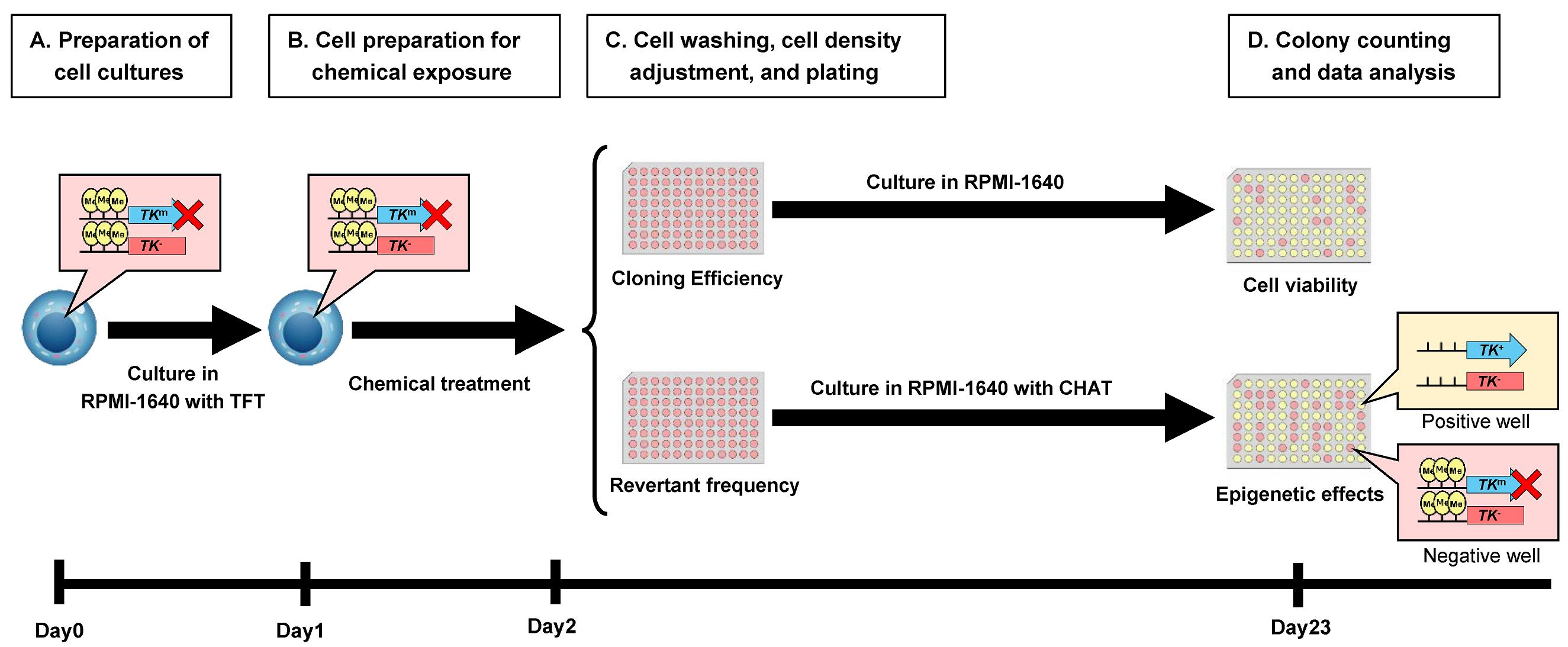
Experimental flow of epi-TK assay. The parental cell line TK6 is heterozygous at the TK gene locus, carrying one functional (TK+) allele and one non-functional (TK-) allele. In mTK6 cells, expression of the functional TK allele is suppressed by DNA methylation (indicated by yellow circles) in the promoter region (TKm). (A) mTK6 cells are cultured to logarithmic growth in the presence of TFT while maintaining DNA methylation-mediated suppression of the TK gene. (B) mTK6 cells are resuspended in fresh RPMI-1640 medium and exposed to the test compound or vehicle control for 24 h. (C) Following treatment of mTK6 cells with the test compound, cells are washed, adjusted to the appropriate density, plated onto revertant frequency (RF) and cloning efficiency (CE) plates with or without CHAT (2′-deoxyCytidine, hypoxanthine, aminopterin, and thymidine) selection, and incubated for 3 weeks for colony formation. (D) After 3 weeks of culture, colonies on CE and RF plates are counted, and TK revertant frequency, reflecting epigenetic changes in TK gene methylation status, is determined using Poisson distribution-based calculations.
Background
A broad range of chemical substances has been implicated in carcinogenesis through genetic, epigenetic, and metabolic mechanisms. Chemical-induced genetic alterations, collectively referred to as genotoxicity, arise from direct DNA damage or from disruption to DNA replication, repair, and damage response pathways. To evaluate these risks, standardized in vitro genotoxicity assays are widely applied in accordance with the recommendations of the Organization for Economic Co-operation and Development guidelines [1]. However, increasing evidence indicates that many chemical agents contribute to carcinogenesis not only through the induction of DNA mutations but also via perturbation of epigenetic regulation, including changes in DNA methylation and histone modifications. Current chemical safety testing frameworks rely primarily on in vitro mutation-based assays, such as the Ames test and mammalian cell gene mutation assays [2-4], which are unable to assess epigenetic alterations. Although high-resolution sequencing-based epigenome profiling technologies can generate detailed information, their high cost, technical complexity, and significant analytical burden limit their routine application in regulatory testing, highlighting the need for practical approaches to assess epigenetic toxicity. To address this need, we developed a practical approach for assessing epigenetic alterations at a housekeeping gene locus by adapting the TK gene mutation assay [2,5], which utilizes the human lymphoblastoid TK6 cell line widely employed in genotoxicity testing. The TK gene mutation assay quantifies mutant frequency by measuring colony formation according to the TK genotype. Trifluorothymidine (TFT) is used to select TK-deficient mutant colonies because cells expressing functional thymidine kinase convert TFT into a toxic nucleotide and undergo cell death, whereas TK-deficient cells survive in the presence of TFT. Conversely, cells carrying a functional or reverted TK gene can be selectively detected using hypoxanthine–aminopterin–thymidine medium, in which only cells with restored thymidine kinase activity survive through the salvage pathway under aminopterin-mediated inhibition of de novo nucleotide synthesis. Building upon standardized in vitro genotoxicity testing frameworks, we developed the epi-TK assay, a mammalian cell–based reporter system in which the promoter region of the housekeeping TK gene in human lymphoblast TK6 cells is site-specifically methylated using CRISPR/dCas9-based epigenome-editing technology [6]. The engineered reporter cell line enables quantitative and bidirectional detection of chemically induced epigenetic effects through changes in frequency of TK revertant colony formation. Following chemical exposure, epigenetic alterations at the TK locus are assessed using hypoxanthine–aminopterin–thymidine selection, with restoration of TK gene expression quantified as the frequency of TK revertant colonies. Because the methylated TK locus retains active histone acetylation marks, it exists in a transcriptionally suppressed but reversible state, allowing spontaneous TK reactivation to occur at a measurable baseline frequency during culture [6]. An increase in revertant frequency suggests inhibition of DNA methylation or promotion of DNA demethylation, whereas a decreased reversion may reflect changes in histone modifications associated with reduced chromatin accessibility. These trends can be further validated by locus-specific DNA methylation analysis, chromatin immunoprecipitation, or protein-level assessment, depending on the expected mode of action of the test compound. Using GSK3484862 as a model DNA methyltransferase 1 inhibitor [7], this protocol quantitatively evaluates its epigenetic effects using the epi-TK assay. As previously reported, the assay also enables quantitative assessment of inflammation-associated global reductions in histone acetylation [6], further highlighting its bidirectional capability to detect distinct modes of epigenetic alteration. Collectively, this assay provides a simple, cost-effective, and biologically relevant platform for evaluating epigenetic toxicity and offers a practical approach for routine chemical safety assessment.
Materials and reagents
Biological materials
1. Human lymphoblastoid mTK6 cell line (TK6-derived cell line originally established in a previous study [6]; available from the authors upon request), stored at -80 °C for short-term storage or in liquid nitrogen for long-term storage. Alternatively, equivalent cell lines can be established from the parental TK6 cell line or other cell types by isolating TFT-resistant clones generated via transient expression of dCas9-DNMT3A with guide RNAs targeting the TK gene promoter [6].
Reagents
1. RPMI-1640 medium (Nacalai Tesque, catalog number: 30264-56); store at 4 °C
2. Fetal bovine serum (FBS) (Nichirei Biosciences, catalog number: 174012-500ML); aliquot and store at -20 °C
3. 100 mM sodium pyruvate solution (Nacalai Tesque, catalog number: 06977-34); store at 4 °C
4. Penicillin-streptomycin solution (Nacalai Tesque, catalog number: 09367-34); store at 4 °C
5. Trifluorothymidine (TFT) (Sigma-Aldrich, catalog number: T2255-100MG)
6. 2′-Deoxycytidine hydrochloride (Sigma-Aldrich, catalog number: D0776)
7. Hypoxanthine (Sigma-Aldrich, catalog number: H9377)
8. Aminopterin Hybri-MaxTM (Sigma-Aldrich, catalog number: A5159)
9. Thymidine (Sigma-Aldrich, catalog number: T9250)
10. GSK-3484862 (CHEMIETEK, catalog number: CT-GSKMI)
11. Dimethyl sulfoxide (DMSO) (Nacalai Tesque, catalog number: 13408-64)
12. Hydrochloric acid (HCl) (Nacalai Tesque, catalog number: 18321-05)
Solutions
1. RPMI-1640 complete medium (see Recipes)
2. 1,000× TFT (see Recipes)
3. 1,000× 2′-deoxycytidine (see Recipes)
4. 1,000× hypoxanthine (see Recipes)
5. 1,000× thymidine (see Recipes)
6. 100× CHAT (2′-deoxyCytidine, hypoxanthine, aminopterin, and thymidine) (see Recipes)
Recipes
1. RPMI-1640 complete medium
| Reagent | Final concentration | Quantity or volume |
|---|---|---|
| RPMI-1640 | n/a | 500 mL |
| 100 mM sodium pyruvate solution | 1.8 mM | 10 mL |
| Penicillin-Streptomycin mixed solution | 88 units/mL | 5.0 mL |
| Heat-inactivated FBS | 8.8% v/v | 50 mL |
Store at 4 °C.
2. 1,000× TFT
| Reagent | Final concentration | Quantity or volume |
|---|---|---|
| Trifluorothymidine | 3.0 mg/mL | 15 mg |
| H2O | n/a | To 5 mL |
Sterilize by filtration through a Minisart 0.2-μm syringe filter and store at -20 °C.
3. 1,000× 2′-deoxycytidine
| Reagent | Final concentration | Quantity or volume |
|---|---|---|
| 2′-Deoxycytidine hydrochloride | 10 mM | 0.132 g |
| H2O | n/a | To 50 mL |
Sterilize by filtration through a Minisart 0.2-μm syringe filter and store at -20 °C.
4. 1,000× hypoxanthine
| Reagent | Final concentration | Quantity or volume |
|---|---|---|
| Hypoxanthine | 200 mM | 1.361 g |
| 1 M HCl | n/a | To 50 mL |
Sterilize by filtration through a Minisart 0.2-μm syringe filter and store at -20 °C.
5. 1,000× thymidine
| Reagent | Final concentration | Quantity or volume |
|---|---|---|
| Thymidine | 17.5 mM | 0.2119 g |
| H2O | n/a | To 50 mL |
Sterilize by filtration through a Minisart 0.2-μm syringe filter and store at -20 °C.
6. 100× CHAT
| Reagent | Final concentration | Quantity or volume |
|---|---|---|
| Aminopterin Hybri-MaxTM | 10 μM | 1 vial (lyophilized powder) |
| H2O | n/a | 14 mL |
| 1,000× 2′-deoxycytidine | 1 mM | 2.0 mL |
| 1,000× hypoxanthine | 20 mM | 2.0 mL |
| 1,000× thymidine | 1.75 mM | 2.0 mL |
Sterilize by filtration through a Minisart 0.2-μm syringe filter and store at -20 °C.
Laboratory supplies
1. 10-cm culture dish (AS ONE, catalog number: 1-7484-01)
2. 15-cm culture dish (SPL Life Science, catalog number: 20150)
3. 15- and 50-mL conical tubes (Greiner Bio-One, catalog numbers: 188271-N and 227261)
4. 1.5-mL microcentrifuge tube [Scientific Specialities Inc (SSI bio), catalog number: 1210-10]
5. Reagent reservoir (Greiner Bio-One, catalog number: J908305)
6. Serological pipettes (5, 10, 25, and 50 mL) (Greiner Bio-One, catalog numbers: 606180, 607180, 760180, and 768180)
7. 96-well flat-bottom plate (VIOLAMO, catalog number: 1-1610-06)
8. Minisart 0.2-μm syringe filter (Sartorius, catalog number: S7597FXOSK)
Equipment
1. Pipetman P10, P20, P200, and P1000 (Gilson, catalog numbers: F144055M, F144056M, F144058M, and F144059M)
2. P200L multichannel micropipette (Gilson, catalog number: FA10011)
3. Pipet-Aid XP (Drummond, catalog number: 4-040-101-J)
4. Inverted microscope (Carl Zeiss, catalog number: Primovert)
5. Stereomicroscope (Leica, catalog number: M205 C)
6. Biological safety cabinet (Panasonic Healthcare, catalog number: MHE-S1301A2)
7. CO2 incubator (Panasonic Healthcare, catalog number: MCO-19AIC)
8. Multitube refrigerated centrifuge (TOMY SEIKO, catalog number: AX-511)
9. Beckman Coulter Z2 particle counter (Beckman Coulter, catalog number: 6605700)
10. Counting chamber (Erma, catalog number: 10-2170-05)
Procedure
登录/注册后免费查看全文
文章信息
稿件历史记录
提交日期: Jan 29, 2026
接收日期: Mar 22, 2026
在线发布日期: Apr 2, 2026
出版日期: Apr 20, 2026
版权信息
© 2026 The Author(s); This is an open access article under the CC BY license (https://creativecommons.org/licenses/by/4.0/).
如何引用
Kuroki, S., Yamada, H., Odagiri, M., Sugiyama, K., Yasui, M. and Sassa, A. (2026). Quantifying Epigenetic Changes Induced by Chemical Exposure Using the epi-TK Assay. Bio-protocol 16(8): e5671. DOI: 10.21769/BioProtoc.5671.

 Download Citation in RIS Format
分类
癌症生物学 > 癌症生物化学 > 基因毒性
细胞生物学 > 基于细胞的分析方法 > 集落形成
您对这篇实验方案有问题吗?
在此处发布您的问题,我们将邀请本文作者来回答。同时,我们会将您的问题发布到Bio-protocol Exchange,以便寻求社区成员的帮助。
 发布问题
发布问题


