


Limited Proteolysis Mass Spectrometry to Identify Protein Structural Differences in Brain Tissue
利用有限蛋白水解质谱鉴定脑组织中的蛋白质结构差异

发布: 2026年05月05日第16卷第9期 DOI: 10.21769/BioProtoc.5670 浏览次数: 1090
评审: Ting MiaoDhananjay D ShindeElena A. Ostrakhovitch
 PDF
PDF  Q&A
Q&A
参见作者原研究论文
The authors used this protocol in:

Jul 2025
Advertisement








Abstract
Structural proteomics methods allow for the proteome-wide interrogation of protein structural differences between two different conditions. Limited proteolysis mass spectrometry (LiP-MS), as originally implemented by the Picotti lab, utilizes a promiscuous protease to cleave at solvent-exposed regions of a protein to encode structural information, which is then read out with mass spectrometry proteomics. Here, we present a protocol that details experimental steps and data analysis for a LiP-MS workflow. First, tissue is homogenized under native conditions and then subjected to limited proteolysis using proteinase K (PK). The samples are prepared for mass spectrometry, and data are acquired using either data-dependent acquisition (DDA) or data-independent acquisition (DIA). Raw data is processed using FragPipe, and raw ion abundances are processed in FragPipe Limited-Proteolysis Processor (FLiPPR). Proteins with structural changes between the two conditions are identified in a proteome-wide manner.
Key features
• Protocol describes how to perform limited proteolysis mass spectrometry to identify proteins in brain tissue with structural changes proteome-wide between two experimental conditions.
• Includes context for how to ensure results are reliable, using permutation analyses.
• Utilizes tools (FragPipe and FLiPPR) that are free and open source.
• Sample preparation can be performed in two days, not including mass spec acquisition and data analysis.
Keywords: Limited proteolysis mass spectrometry (有限蛋白水解质谱) Structural proteomics (结构蛋白质组学)
Brain (脑组织)
FragPipe (FragPipe)
FLiPPR (FLiPPR)
Structural proteomics (结构蛋白质组学)
Brain (脑组织)
FragPipe (FragPipe)
FLiPPR (FLiPPR)
Graphical overview
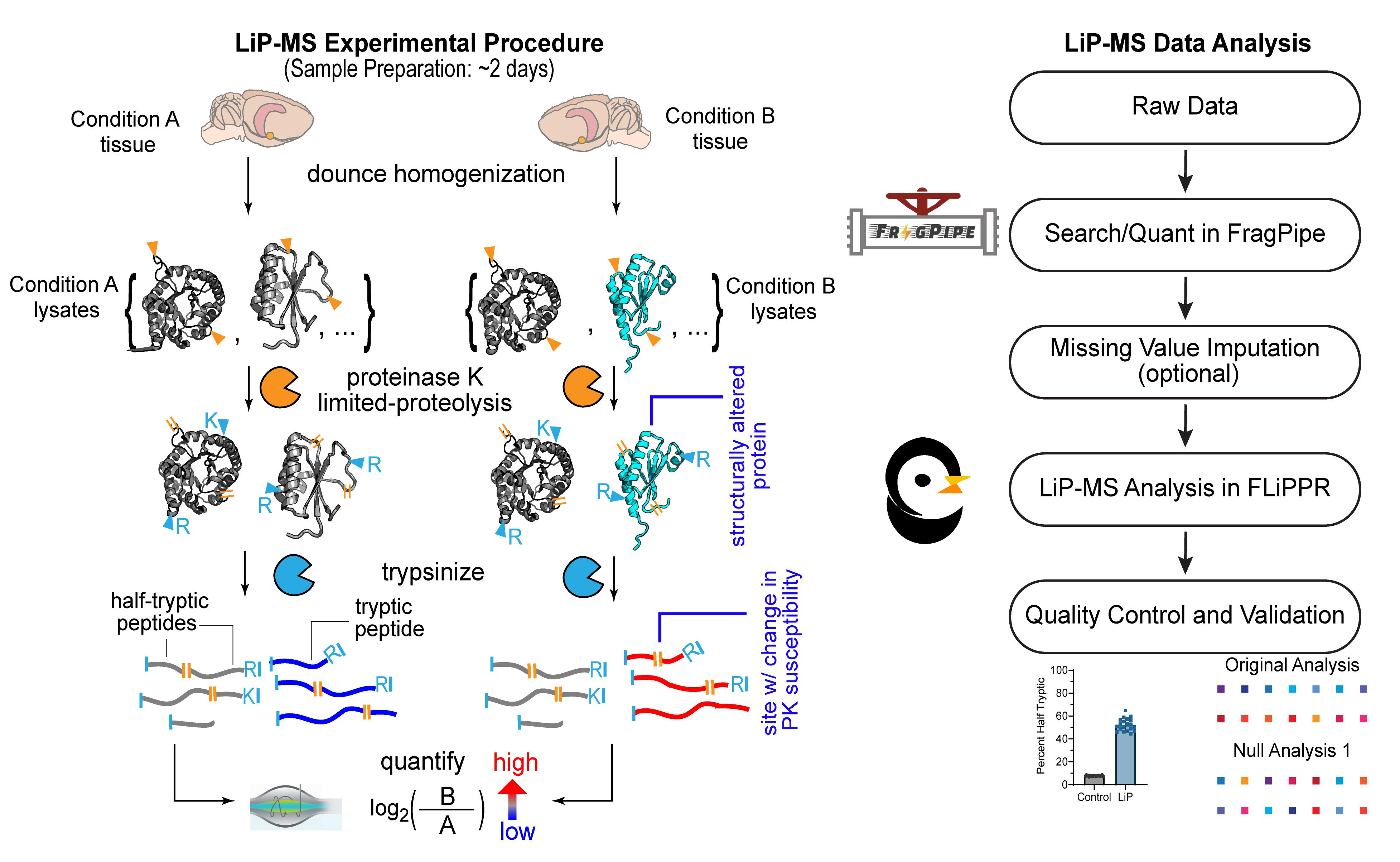
Limited proteolysis mass spectrometry (LiP-MS) experimental workflow and data analysis pipeline
Background
The study of protein structures is a cornerstone of molecular biology, as most proteins must be correctly folded to perform their cellular functions. However, many structural methods are low-throughput, studying one protein at a time [e.g., cryo-electron microscopy (cryo-EM), nuclear magnetic resonance (NMR), and x-ray crystallography]. In many systems, such as the environment of the aging brain, protein misfolding is known to occur, such as amyloid-β aggregation, seen in Alzheimer’s disease [1], or α-synuclein, seen in Parkinson’s disease [2]. However, such extreme misfolding events are uncommon, and only a few proteins are known to aggregate so dramatically. In an aging context, more subtle protein conformational changes have been demonstrated in multiple tissues [3–5]. Additionally, it is generally thought that the proteostasis network, which is responsible for ensuring that proteins are properly synthesized, correctly folded, and degraded when they become misfolded, declines with age [6–8]. Therefore, the conditions are present for potential widespread protein structural changes in a system such as the aged brain.
Limited proteolysis mass spectrometry (LiP-MS) is a technique to detect protein structural changes proteome-wide, with one modern incarnation developed by the Picotti group [9,10]. In LiP-MS, protein lysates undergo pulse proteolysis using proteinase K (PK), a protease that cleaves preferentially at solvent-exposed/flexible areas. This encodes structural information: if a protein is in a different conformation under a different condition, PK will cut at different places in the protein. Proteins then undergo complete digestion with trypsin, which cuts after arginine and lysine residues, and the resultant peptides are quantified using mass spectrometry. Differential cleavage by PK results in different peptide abundances between the two conditions, which can then be used to determine which proteins have different structures between the two conditions. Structural changes that can be detected by LiP-MS include conformational changes, differences in soluble oligomerization state, different binding to other proteins/ligands, etc. LiP does not reveal the exact nature of the structural change compared to a high-resolution method such as cryo-EM, but it has the advantage of being a proteome-wide technique. LiP-MS has already been used in a variety of contexts, such as to find protein structural changes in cerebrospinal fluid (CSF) between young and old mice [11], between young and old yeast extracts [12], between native bacterial lysates and refolded bacterial lysates [13], and between CSF of healthy and Parkinson’s disease patients [14].
Analysis of LiP-MS data can be performed using the free tools FragPipe [15] and FragPipe Limited-Proteolysis Processor (FLiPPR) [16]. However, care must be taken to ensure that analysis is done appropriately and does not induce false positives, especially in highly variable systems like tissue from outbred animals or from patient cohorts. Here, we describe how to perform LiP experiments on fresh-frozen rat brain tissue, how to prepare the samples for liquid chromatography–mass spectrometry (LC-MS), and how to acquire the raw data. We then describe how to analyze the data, first using FragPipe to process the raw LC-MS-MS data, and then using FLiPPR to process the FragPipe outputs. We also describe how to impute for missing values if needed, how to perform permutation analyses to ensure false positives are not being induced by the data analysis and/or imputation parameters, and how to assess experimental quality.
Materials and reagents
Biological materials
1. Long Evans rats (Charles River, catalog number: Crl:LE) or fresh-frozen brain tissue
Reagents
1. E-64 (Thermo Scientific, catalog number: 78434)
2. Dimethyl sulfoxide (DMSO) (Fisher Chemical, catalog number: D159-4)
3. Bestatin (Alfa Aesar, CAS number: 58970-76-6)
4. Phenylmethylsulfonyl fluoride (PMSF) (Acros Organics, CAS number: 329-98-6)
5. Deoxyribonuclease 1 (DNase) (Sigma-Aldrich, catalog number: DN25-1G)
6. Tris HCl (Millipore Sigma, catalog number: T15760)
7. Tris base (Millipore Sigma, catalog number: T1503)
8. Hydrochloric acid (HCl) (Sigma-Aldrich, CAS number: 7647-01-0)
9. Potassium chloride (KCl) (Sigma-Aldrich, catalog number: P3911)
10. Sodium chloride (NaCl) (Sigma-Aldrich, catalog number: S9888)
11. Magnesium chloride hexahydrate (MgCl2) (Fisher Chemical, catalog number: M33)
12. Glycerol (Fisher Chemical, catalog number: G33)
13. Proteinase K (PK) (Thermo Scientific, catalog number: 17916)
14. DL-dithiothreitol (DTT) (Sigma-Aldrich, catalog number: D0632)
15. Iodoacetamide (IAA) (Acros Organics, catalog number: 12227)
16. Ammonium bicarbonate (Thermo Scientific, catalog number: 393210050)
17. Ultrapure MS-grade water (Fisher Chemical, catalog number: W6-4)
18. MS-grade trifluoroacetic acid (TFA) (Fisher Chemical, catalog number: A116)
19. Ultrapure MS-grade acetonitrile (Fisher Chemical, catalog number: A955)
20. BCA Protein Assay kit (Thermo Scientific, catalog number: A55860)
21. Mineral oil (Thermo Scientific, catalog number: 415080025)
22. Urea, ultrapure, 99% (Thermo Scientific, catalog number: J65769.A4)
23. Trypsin, mass spectrometry grade (New England Biolabs, catalog number: P8101S)
24. Ultrapure MS-grade formic acid (Fisher Chemical, catalog number: A117-50)
Solutions
1. 100× E-64 Protease Inhibitor Stock (100× E-64) (see Recipes)
2. 100× Bestatin protease inhibitor stock (100× Bestatin) (see Recipes)
3. 100× Phenylmethylsulfonyl fluoride protease inhibitor stock (100× PMSF) (see Recipes)
4. 100× DNase stock (see Recipes)
5. 1 M Tris pH 8 Stock (see Recipes)
6. 1 M KCl stock (see Recipes)
7. 5 M NaCl stock (see Recipes)
8. 1 M MgCl2 stock (see Recipes)
9. Lysis buffer (see Recipes)
10. 20% glycerol solution (see Recipes)
11. PK stock (see Recipes)
12. DTT solution (see Recipes)
13. IAA solution (see Recipes)
14. Ammonium bicarbonate solution (AmBic solution) (see Recipes)
15. Buffer A (see Recipes)
16. Buffer B (see Recipes)
Recipes
1. 100× E-64
Prepare stocks of 1.5 mM E-64 in DMSO. Aliquot and store at -20 °C. Stocks can be thawed multiple times.
2. 100× Bestatin
Prepare stocks of 5 mM Bestatin in DMSO. Aliquot and store at -20 °C. Stocks can be thawed multiple times.
3. 100× PMSF
Prepare stocks of 50 mM PMSF in DMSO. Aliquot and store at -20 °C. Stocks can be thawed multiple times.
4. 100× DNase stock
Prepare a 10 mg/mL stock of DNase in Millipore water (MPW). Aliquot and store at -20 °C. Stocks can be thawed several times but should be kept on ice.
5. 1 M Tris pH 8 stock
| Reagent | Final concentration | Quantity or volume |
|---|---|---|
| Tris HCl | 88.8 g/L | 44.4 g |
| Tris base | 53 g/L | 26.5 g |
| MPW (to total volume) | n/a | 500 mL |
To approximately 400 mL of MPW, add 44.4 g of Tris acid and 26.5 g of Tris base. Adjust pH to 8 with HCl. Add MPW to a final volume of 500 mL. Autoclave to sterilize. Store at room temperature.
6. 1 M KCl stock
To approximately 400 mL of MPW, add 37.275 g of KCl and stir until dissolved. Add MPW to a final volume of 500 mL. Autoclave to sterilize. Store at room temperature.
7. 5 M NaCl stock
To approximately 400 mL of MPW, add 146.1 g of NaCl and stir until dissolved. Add MPW to a final volume of 500 mL. Autoclave to sterilize. Store at room temperature.
8. 1 M MgCl2 stock
To approximately 80 mL of MPW, add 20.33 g of magnesium chloride hexahydrate. Stir until dissolved, then add MPW to a final volume of 100 mL. Autoclave to sterilize. Store at room temperature.
9. Lysis buffer
| Reagent | Final concentration | Quantity or volume |
|---|---|---|
| 1 M Tris pH 8 stock | 20 mM | 1 mL |
| 1 M KCl stock | 150 mM | 7.5 mL |
| 5 M NaCl stock | 10 mM | 100 μL |
| 1 M MgCl2 stock | 2 mM | 100 μL |
| MPW | n/a | To 50 mL |
10. 20% glycerol solution
Prepare a 20% v/v solution of glycerol in MPW. Stir. Autoclave to sterilize.
11. PK stock
Measure an appropriate mass of PK and dissolve in a solution of 1:1 (v/v) 20% glycerol (Recipe 10; final glycerol concentration of 10%) and lysis buffer (Recipe 9) to a final PK concentration of 1 μg/μL. Aliquot into 1.5 mL tubes, flash freeze in liquid nitrogen, and store at -20 °C. Only thaw aliquots once.
12. DTT solution
Prepare a 700 mM DTT solution in MPW. The solution must be prepared fresh.
13. IAA solution
Prepare a 700 mM IAA solution in MPW. Protect from light. The solution must be prepared fresh.
14. AmBic solution
Prepare a 100 mM stock of ammonium bicarbonate (AmBic) in MPW. The solution must be prepared fresh.
15. Buffer A
| Reagent | Final concentration | Quantity or volume |
|---|---|---|
| Ultrapure MS-grade water | n/a | 200 mL |
| MS-grade TFA | 0.5% (v/v) | 1 mL |
Add TFA to the ultrapure water dropwise. Buffer A can be stored at room temperature for one week.
16. Buffer B
| Reagent | Final concentration | Quantity or volume |
|---|---|---|
| Ultrapure MS-grade water | 20% (v/v) | 20 mL |
| Ultrapure MS-grade acetonitrile | 80% (v/v) | 80 mL |
| MS-grade TFA | 0.5% (v/v) | 500 μL |
Add TFA to the ultrapure water and acetonitrile dropwise. Buffer B can be stored at room temperature for one week.
Laboratory supplies
1. 1.5 mL tubes (USA Scientific, catalog number: 1615-5500)
2. 2 mL tubes (Fisher Scientific, catalog number: 05-408-138)
3. C18 cartridges (Sep-Pak Vac 1cc) for desalting (Waters, catalog number: WAT054955)
4. 15 mL conical tubes (Sarstedt, catalog number: 62.554.100)
5. Mass spec vials (Thermo Scientific, catalog number: 6ERV11-03PPC)
6. Mass spec vial caps (Thermo Scientific, catalog number: 6ARC11ST1)
7. Disposable glass pipette (Fisher Scientific, catalog number: 13-678-20A)
8. 10 μL pipette tips (USA Scientific, catalog number: 1111-3700)
9. 200 μL pipette tips (USA Scientific, catalog number: 1111-0706)
10. 1,000 μL pipette tips (USA Scientific, catalog number: 1111-2721)
11. Gloves (Halyard, catalog number: 55082)
12. Kimtech Science Kimwipes (Kimberly-Clark Professional, catalog number: 34155)
Equipment
1. MilliQ water purification system (Millipore Sigma, model: EQ 7000)
2. Dounce homogenizer (Fisher Scientific, catalog number: 06-434)
3. Centrifuge (Eppendorf, model: 5430 R)
4. Temperature-controlled hot plate with stirring (IKA, model: C-MAG HS 7 control)
5. Glass container for oil bath (Pyrex, catalog number: 3140)
6. Metal grate for oil bath, taken from the bottom part of the water bath tube rack (United States Plastic Corp, catalog number: 96866)
7. Centrifuge (Eppendorf, model: 5425)
8. Vortex mixer (Benchmark, model: BenchMixer V2)
9. Shaking heat block (Benchmark, model: Multi-Therm)
10. Shaking heat block tube adaptor (Benchmark, model: H5000-15)
11. Analytical balance (Accuris Instruments, model: Series Dx)
12. Vacuum manifold (Zymo Research, catalog number: S7000)
13. Vacuum pump (Cole-Parmer, model: Air Admiral 79202-30)
14. Vacuum centrifuge (Eppendorf, model: Vacufuge plus)
15. -80 °C freezer (Thermo Scientific, model: Revco RLE series)
16. Centrifuge (Eppendorf, model: 5910 R)
17. Water bath sonicator (Rovsun, model: 230HT)
18. Microvolume UV-Vis spectrophotometer (Thermo Scientific, model: NanoDrop OneC)
19. UHPLC system (Thermo Scientific, model: UltiMate 3000)
20. Mass spectrometer (Thermo Scientific, model: Q Exactive HF-X)
21. Acclaim PepMap 100 Trap column, C18, 75 μm × 2 cm, 3 μm particle size (Thermo Scientific, catalog number: 164946)
22. Acclaim PepMap 100 Analytical Column, C18, 75 μm × 25 cm, 2 μm particle size (Thermo Scientific, catalog number: 164941)
23. Nanospray Flex ion source (Thermo Scientific, catalog number: ES071)
24. pH meter (Mettler Toledo, model: FiveEasy Plus FP20)
25. Pipettes (Eppendorf, catalog number: 2231300004)
26. 4 °C fridge (Fisher Scientific, catalog number: FBV05RPSA)
27. -20 °C freezer (VWR, catalog number: 10819-894)
Software and datasets
Note: All software is free to use.
1. FragPipe (https://fragpipe.nesvilab.org/, v23.1)
2. Anaconda Navigator (Anaconda Inc., v2.7.0)
3. VS Code (Microsoft, v1.105.1)
4. Missing value imputation script prefilter_impute_ion.ipynb (https://zenodo.org/records/15103402)
5. FLiPPR (https://github.com/FriedLabJHU/FragPipe-Limited-Proteolysis-Processor, v0.2.2)
6. Sample LiP data (https://www.ebi.ac.uk/pride/archive/projects/PXD052770)
Procedure
登录/注册后免费查看全文
文章信息
稿件历史记录
提交日期: Feb 3, 2026
接收日期: Mar 23, 2026
在线发布日期: Apr 1, 2026
出版日期: May 5, 2026
版权信息
© 2026 The Author(s); This is an open access article under the CC BY-NC license (https://creativecommons.org/licenses/by-nc/4.0/).
如何引用
Readers should cite both the Bio-protocol article and the original research article where this protocol was used:
- Tarbox, H. E. and Fried, S. D. (2026). Limited Proteolysis Mass Spectrometry to Identify Protein Structural Differences in Brain Tissue. Bio-protocol 16(9): e5670. DOI: 10.21769/BioProtoc.5670.
- Tarbox, H. E., Branch, A. and Fried, S. D. (2025). Proteins with cognition-associated structural changes in a rat model of aging exhibit reduced refolding capacity. Sci Adv. 11(28): eadt3778. https://doi.org/10.1126/sciadv.adt3778

 Download Citation in RIS Format
分类
神经科学 > 基础技术
生物化学 > 蛋白质 > 结构
生物信息学与计算生物学
您对这篇实验方案有问题吗?
在此处发布您的问题,我们将邀请本文作者来回答。同时,我们会将您的问题发布到Bio-protocol Exchange,以便寻求社区成员的帮助。
 发布问题
发布问题


