


Quantitative Analysis of Splenic Natural Killer Cells of Mice Using Imaging Flow Cytometry
基于成像流式细胞术的小鼠脾脏自然杀伤细胞定量分析方法



发布: 2026年04月20日第16卷第8期 DOI: 10.21769/BioProtoc.5666 浏览次数: 544
评审: Sucheta ChopraSanjay Kumar KureelAnonymous reviewer(s)
 PDF
PDF  Q&A
Q&A
参见作者原研究论文
The authors used this protocol in:
Aug 2022






Abstract
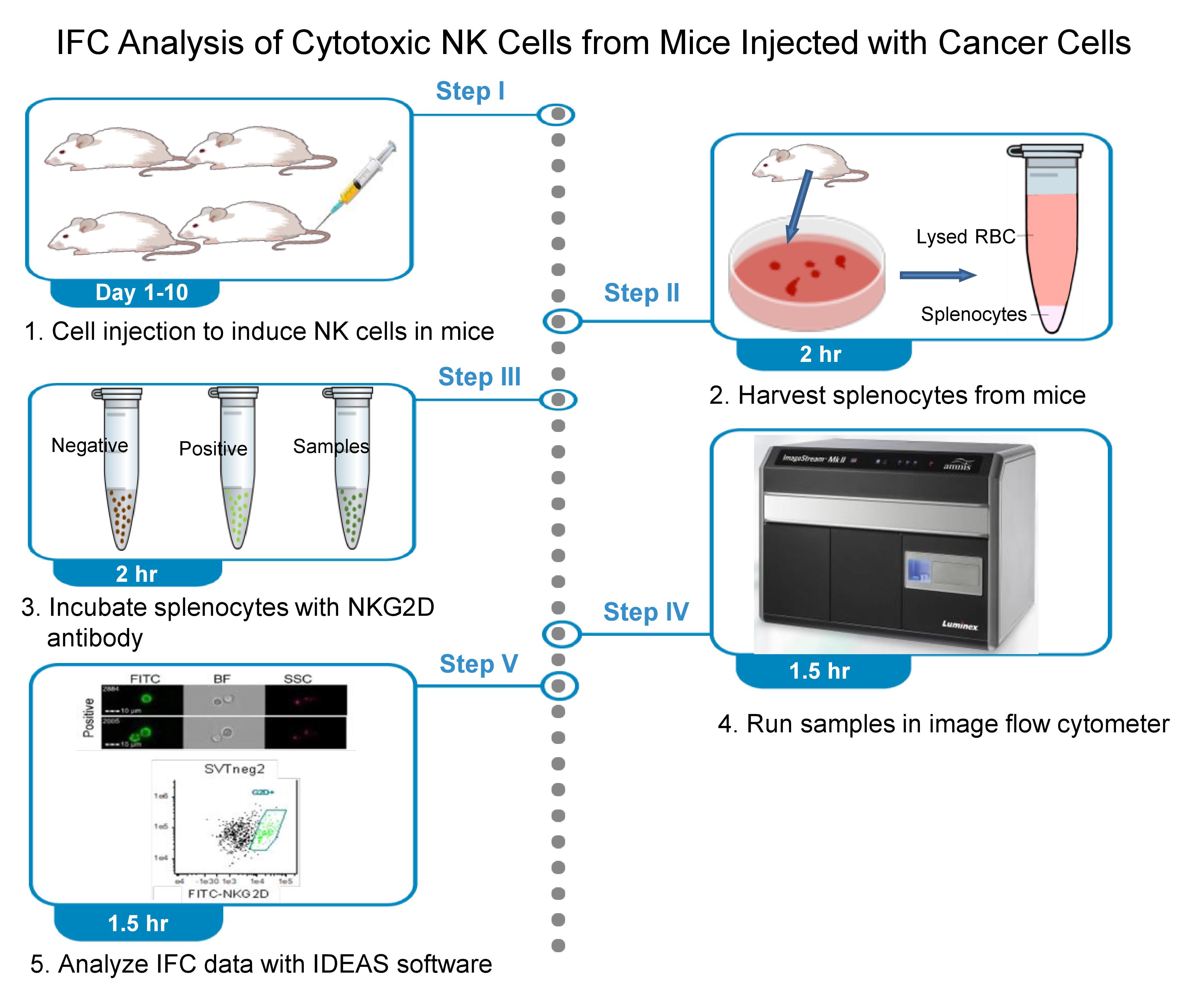
Natural killer (NK) cells are crucial innate immune effectors, mediating cytotoxicity against cancer and infected cells through receptors such as NKG2D. Reliable quantification of NK cell subsets is essential for evaluating NK cell-based immune responses in cancer research. Unlike other assays, including traditional flow cytometry used in assessing NK cells, imaging flow cytometry (IFC) is a simple and direct method for quantitative analysis of NK cells. This protocol describes the necessary procedures, including harvesting splenocytes, acquiring these cells labeled with NKG2D antibodies, and analyzing IFC data with IDEAS® software. We applied this protocol to quantitatively assess the number of splenic NKG2D+ NK cells in mice injected with SVTneg2 cancer cells (which carry the p53 G242A missense mutation) and compared them to mice injected with EMT6 cancer cells (which have wild-type p53) or normal fibroblasts. We found that the SVTneg2 cancer cells significantly decreased the number of NKG2D+ NK cells in mice by approximately 2-fold (933 cells vs. 2360 cells, p < 0.001) compared with mice injected with EMT6 cancer cells. This IFC protocol can be applied to directly quantify NK cells in vivo. This quantitative protocol allows novices to quickly handle the analysis of cytotoxic NK cells with a single NKG2D marker. Further multicolor flow cytometry and cytokine assay may be required to precisely define the subtypes and effects of NK cells in anticancer immunity.
Key features
• A simple and direct assay using imaging flow cytometry (IFC) to quantify cytotoxic NKG2D NK cells against breast cancer cells in mice.
• Simultaneously collect the flow cytometry characters and each cell image of NK cells and other populations.
• Step-by-step identification of interested NK cells mainly relying on image gating (focus, size, morphology).
• Highly reliable and applicable to analyze other immune cell subsets or tumor-associated populations with corresponding conjugate antibodies.
Keywords: Natural killer cells (自然杀伤细胞) Imaging flow cytometry (成像流式细胞术)
Immune cytotoxicity (免疫细胞毒性)
Cancer cells (癌细胞)
Analyzing IFC data (IFC 数据分析)
Imaging flow cytometry (成像流式细胞术)
Immune cytotoxicity (免疫细胞毒性)
Cancer cells (癌细胞)
Analyzing IFC data (IFC 数据分析)
Graphical overview
Background
Natural killer (NK) cells are key effectors for innate immunity that play a pivotal role in immune surveillance and cytotoxicity against cancer [1]. Recent works highlighted the potential of NK cells in cancer immunotherapy, owing to their directed cytotoxic capabilities and their roles in orchestrating broader immune responses to cancer cells [2]. With surface receptors recognizing other cells expressing low levels of major histocompatibility complex (MHC) class I molecules, NK cells can mount potent assaults on cancerous or infected cells and destroy those cells by secreting various cytokines [3]. NK group member 2D (NKG2D, also known as CD314) is one of the most common activating receptors on NK cells. By the binding of NKG2D receptors to the ligands on cancer cells, NK cells can be activated in immune recognition [4]. However, many cancer cells are enabled to aberrantly express the NKG2D ligands, thus avoiding recognition so as to inactivate and even destroy NK cells [5,6]. p53 protein acts as a powerful transcriptional factor that upregulates the stimulatory NKG2D ligands, including retinoic acid early transcript 1 (RAET1), histocompatibility antigen 60 (H60), and mouse UL16-binding protein-like transcript 1 (Mult1) of murine cancer cells, to activate NK cells against tumors [7,8]. Missense mutants of p53 proteins are frequently detected in cancer cells and exhibit gain-of-function to promote tumor progression [9]. Both loss of the function of wild-type p53 protein and gain-of-function of p53 mutants can alter NKG2D ligands, either by decreasing stimulatory ligands (such as Mult1, ULPB1) or by increasing inhibitory ligands (such as H60a, RAET1E2), and then inactivate NK cells [6,8,10,11]. In an effort to garner evidence on p53 and NK cell activation, we designed a method to quantitatively analyze cytotoxic NK cells using imaging flow cytometry (IFC)—a technology that combines multiparametric flow cytometry and fluorescence microscopy [8,12].
A wide variety of NK cell subsets exist in a variable tissue microenvironment, reflecting different maturation stages with functional capabilities. The NKG2D+ subset of NK cells is the major phenotype of NK cells and is highly cytotoxic to cancer cells [13–16]. It has been estimated that NKG2D+ splenic cells included only approximately 3% of γδ T cells, αβ CD8+ T cells, and macrophages [14–17]. IFC combines the high-event-rate nature of flow cytometry with the advantages of single-cell image acquisition associated with microscopy [18].
Tissue NK cells can be quantitatively analyzed using several methods, including traditional flow cytometry and advanced analysis techniques, such as multicolor flow cytometry or high-parameter cytometry, single-cell proteo-genomics, and molecular imaging. However, these often lack the detailed morphological information needed to distinguish true positive cells from nonspecific staining or background noise for precise quantification [19]. We developed IFC as a tool to measure the cytotoxic subtype of NK cells with the NKG2D marker through quantitative comparison with high-resolution confocal microscopy. We show that IFC can be used as a simple and direct measure of cytotoxic NK cells against tumors, as this technology possesses greater dimensionality than standard flow cytometry. This cell-imaging dimensionality significantly improves discrimination of true NKG2D+ NK cells from nonspecific staining or background noise using double-gating, including brightfield and fluorescence. IFC enables the analysis of spatial and structural details of cell populations with greater precision, which is critical for identifying cell features that conventional flow cytometry is unable to reach.
This protocol describes the steps to quantify cytotoxic NK cells (NKG2D+) of mice after injections of breast cancer cells. With minor modifications, such as replacing the conjugated CD8 antibody, we also used it to analyze CD8+ T cells [8]. This protocol can be applied to analyze cytotoxic NK cells with the NKG2D receptor in cell culture and animal tissues. Further analysis, such as multicolor IFC and cytokine assay, is required to precisely characterize the subsets and effects of NK cells in immunity [20].
Materials and reagents
Biological materials
1. Mouse: BALB/c, female, 4–5 weeks of age (Charles River, strain code: 028, BALB/cAnNCrl)
2. EMT6 cells (mouse, mammary carcinoma, BALB/cKa strain, wild-type Trp53) (American Type Culture Collection, CRL-2755)
3. SVTneg2 cells (mouse, mammary carcinoma, BALB/c strain, Trp53 G242A) (Charitè Universitätsmedizin Berlin, Dr. Andreas Klein, Institute of Biochemistry)
4. Fibroblasts (BALB/c mouse, lung) (Cell Biologics, catalog number: BALB-5013)
Note: Fibroblasts can also be isolated from BALB/c mice.
Reagents
1. FITC anti-mouse CD314 (NKG2D) antibody, clone C7, 0.5 mg/mL in 100 μL (BioLegend, catalog number: 115711)
2. FITC anti-p53 antibody, mouse IgG2b, clone DO-7 (BioLegend, catalog number: 645804)
3. HBSS supplemented with 10% FBS (Fisher Scientific, catalog number: 24020117)
4. Ketamine hydrochloride (Sigma-Aldrich, catalog number: K2753)
5. Xylazine hydrochloride (Sigma-Aldrich, catalog number: X1251-1G)
6. Bovine serum albumin (BSA) (Sigma-Aldrich, catalog number: A9418)
7. Normal mouse serum (NMS) (Fisher Scientific, catalog number: 10410)
8. DPBS 10× (500 mL) (Fisher Scientific, catalog number: AAJ61917K3)
9. DMEM base medium (no glucose) (Fisher Scientific, catalog number: 11-966-025)
10. RPMI 1640 base medium (Fisher Scientific, catalog number: 11-875-093)
11. Penicillin-streptomycin-glutamine (100×) (Fisher Scientific, catalog number: 10-378-016)
12. Trypsin-EDTA (0.25%) (Fisher Scientific, catalog number: 25-200-056)
13. ACK (ammonium-chloride-potassium) lysing buffer (Fisher Scientific, catalog number: A1049201)
14. Sheath fluid (Millipore, catalog number: BSS-1006-B)
15. Cleanser (Beckman Coulter, catalog number: 8546929)
16. 5% sodium hypochlorite (sterilizer) (VWR, catalog number: JT9416-1)
17. 70% isopropanol (debubbler) (Sigma-Aldrich, catalog number: 67-63-0)
18. Deionized water (Milli-Q water) (Millipore Milli-Q water purified, catalog number: ZRXQ005US)
Solutions
1. DMEM growth medium (see Recipes)
2. RPMI 1640 growth medium (see Recipes)
3. Euthanasia solution (see Recipes)
4. 1× phosphate buffer saline (PBS) (see Recipes)
5. 2% NMS in PBS (see Recipes)
Recipes
1. DMEM growth medium
| Reagent | Final concentration | Volume for 20 mice |
|---|---|---|
| Fetal bovine serum (FBS) | 10% | 50 mL |
| Penicillin/streptomycin/glutamine | 1% | 5 mL |
| DMEM base medium | 445 mL | |
| Total | 500 mL |
Keep at 4 °C for up to one month.
2. RPMI 1640 growth medium
| Reagent | Final concentration | Volume for 20 mice |
|---|---|---|
| FBS | 10% | 50 mL |
| Penicillin/streptomycin/glutamine | 1% | 5 mL |
| RPMI 1640 medium | 445 mL | |
| Total | 500 mL |
Keep at 4 °C for up to one month.
3. Euthanasia solution
| Reagent | Final concentration | Volume for 20 mice |
|---|---|---|
| Ketamine hydrochloride (100 mg/mL) | 10 mg/mL | 2 mL |
| Xylazine hydrochloride (20 mg/mL) | 1 mg/mL | 1 mL |
| PBS 1× | n/a | 17 mL |
| Total | n/a | 20 mL |
Critical: This stock has to be sterile; prepare it in a laminar flow hood.
Keep at 4 °C for up to three days.
4. 1× PBS
| Reagent | Final concentration | Volume for 20 mice |
|---|---|---|
| Sterile PBS 10× | 1× | 5 mL |
| MilliQ water (autoclaved) | 45 mL | |
| Total | 50 mL |
Critical: This stock has to be sterile; prepare it in a laminar flow hood.
Keep at 4 °C for up to one week.
5. 2% NMS in PBS
| Reagent | Final concentration | Volume for 20 mice |
|---|---|---|
| Sterile PBS 10× | 1× | 5 mL |
| MilliQ water (autoclaved) | 44 mL | |
| NMS | 2% | 1 mL |
| Total | 50 mL |
Critical: This stock has to be sterile; prepare it in a laminar flow hood.
Keep at 4 °C for up to one week.
Laboratory supplies
1. Conical centrifuge tubes, 25 mL (Fisher Scientific, catalog number: 05-413-921)
2. Microcentrifuge tubes, 1.5 mL (Fisher Scientific, catalog number: 05-408-129)
3. Standard dissecting scissors (straight) (Fisher Scientific, catalog number: 08-951-20)
4. Dissecting dressing forceps (Fisher Scientific, catalog number: 13-812-40)
5. Syringe and needle (0.3 mL, 30-G) (Fisher Scientific, catalog number: 50-209-2910)
6. Syringe (3 mL, 21-G) (Fisher Scientific, catalog number: 14-823-55)
7. Cell strainers, 70 μm, sterile (Fisher Scientific, catalog number: 07-201-431)
Equipment
1. Imaging flow cytometer (EMD Millipore, model: ImageStreamX Mark II)
2. CO2 incubator for cell culture (Thermo Fisher Scientific, model: 3310)
3. Refrigerator (4 °C)
4. Centrifuge (Eppendorf, model: 5804R, 15 amp version)
Software and datasets
1. IDEAS software (CYTEK, https://cytekbio.com/pages/imagestream)
Procedure
登录/注册后免费查看全文
文章信息
稿件历史记录
提交日期: Jan 9, 2026
接收日期: Mar 17, 2026
在线发布日期: Apr 1, 2026
出版日期: Apr 20, 2026
版权信息
© 2026 The Author(s); This is an open access article under the CC BY-NC license (https://creativecommons.org/licenses/by-nc/4.0/).
如何引用
Amin, M. N., Mostaq, M. S., Uddin, M. B. and Liu, Y. (2026). Quantitative Analysis of Splenic Natural Killer Cells of Mice Using Imaging Flow Cytometry. Bio-protocol 16(8): e5666. DOI: 10.21769/BioProtoc.5666.

 Download Citation in RIS Format
分类
免疫学 > 免疫细胞染色 > 流式细胞术
细胞生物学 > 单细胞分析 > 流式细胞术
癌症生物学 > 肿瘤免疫学 > 细胞生物学试验
您对这篇实验方案有问题吗?
在此处发布您的问题,我们将邀请本文作者来回答。同时,我们会将您的问题发布到Bio-protocol Exchange,以便寻求社区成员的帮助。
 发布问题
发布问题


