


Preparation and Assembly of the Axial Invasion Chamber for Live-Cell Invadopodia Imaging
用于活细胞侵袭性伪足成像的轴向侵袭小室制备与组装

发布: 2026年04月20日第16卷第8期 DOI: 10.21769/BioProtoc.5658 浏览次数: 385
评审: Gururaj KidiyoorAnonymous reviewer(s)
 PDF
PDF  Q&A
Q&A
参见作者原研究论文
The authors used this protocol in:

Dec 2025
Advertisement








Abstract
Metastasis is initiated by cell invasion of the basement membrane, facilitating cell migration and colonization at a secondary tumor site. Cancer cells remodel the cytoskeleton to form ventral protrusions, termed invadopodia, that traffic and deliver matrix metalloproteases to degrade the extracellular matrix. Traditional efforts have utilized immunolabeling to measure protein localization within invadopodia, an approach limited by reduced temporal resolution, logistical challenges in orienting invadopodia within the focal plane of the objective lens, and impaired ability to reconstitute physiological conditions. Here, we describe a protocol for constructing and utilizing the axial invasion chamber (AIC) to perform live-cell 3D visualization of mature elongating invadopodia under physiological conditions. The AIC is simple to build, using standard 35 mm glass-bottom dishes that suit most microscope stage holders. A polyester membrane is used to uniformly orient and promote invadopodia formation and restrict cell migration. The AIC extracellular matrix is composed of readily available reagents that have been optimized to facilitate cell adhesion and invadopodia maturation. Critical advances of the AIC include imaging and measurements of protein localization without immunolabeling, imaging of live cell invadopodia using conventional inverted microscopes, and production of a fully operational apparatus within 28 h from initial assembly. While the protocol has been used for live-cell invadopodia protein localization and structure, it provides an opportunity to interchange components of the polyester membrane and/or the extracellular matrix to optimize the device for a variety of different cell types and cell invasion studies.
Key features
• Enables high-resolution live-cell invadopodia imaging along the axial plane and visualization of protein localization and length of protrusion.
• Live-cell imaging with transient transfection of fluorescent proteins and interchangeable components to study various aspects of cell invasion and migration.
Keywords: Invadopodia (侵袭性伪足) Live-cell imaging (活细胞成像)
Confocal fluorescence microscopy (共聚焦荧光显微镜)
Cell invasion (细胞侵袭)
Matrix degradation (基质降解)
Actin-rich protrusions (富含肌动蛋白的突起)
Live-cell imaging (活细胞成像)
Confocal fluorescence microscopy (共聚焦荧光显微镜)
Cell invasion (细胞侵袭)
Matrix degradation (基质降解)
Actin-rich protrusions (富含肌动蛋白的突起)
Graphical overview
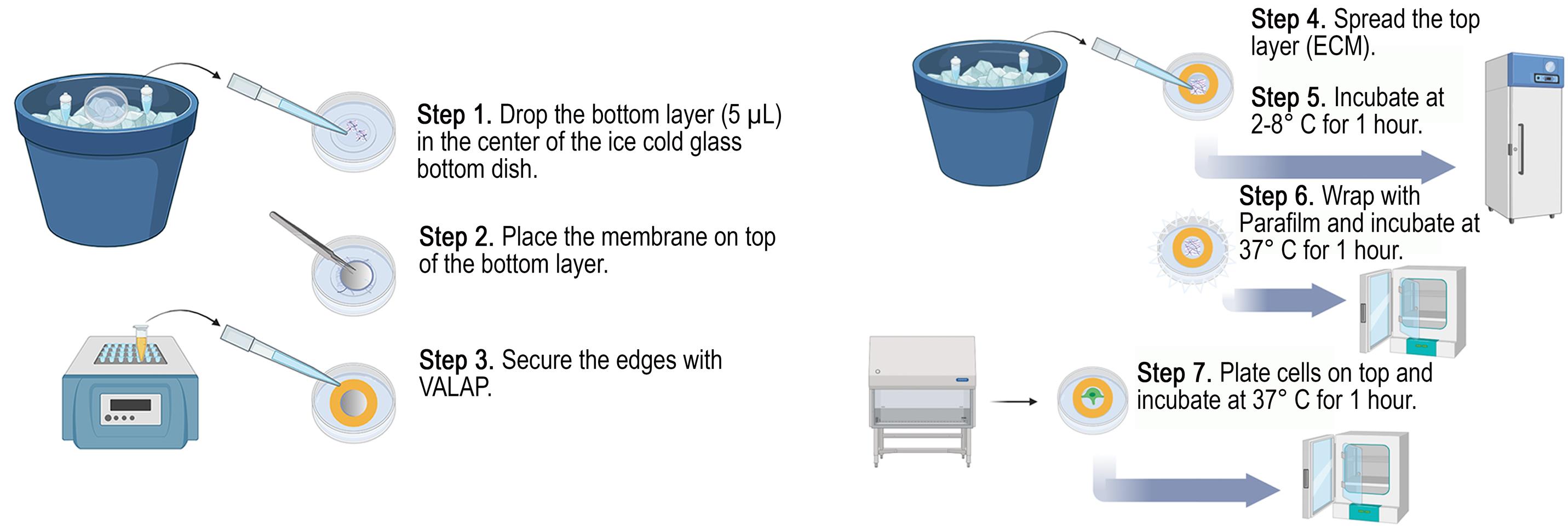
Schematic of axial invasion chamber assembly. Steps to build the axial invasion chamber (AIC). Steps 1–3: building the base layers; steps 4–6: constructing the top layers and polymerizing both layers (top and bottom) of the extracellular matrix (ECM).
Background
Hallmarks of cancer that include epithelial–mesenchymal transition (EMT), increased cell motility, immune system evasion, and unlimited cell proliferation, among others, contribute to the cancer cell phenotype for sustained tumor progression [1]. Notably, all hallmarks are shared between malignant and benign tumors, except for cell invasion and metastasis, which is considered highly critical and the most elusive [2]. Despite these findings, clinical research targeting cancer cell metastasis and secondary lesions remains far less prevalent than studies aimed at inhibiting the primary tumor cell hallmarks. Furthermore, tumor cell dissemination and migration to another colonizing site is a major cause of death in cancer patients [3]. Thus, it is imperative to investigate the molecular mechanisms of metastasis to enable the development of therapies with greater impact on patient survival.
The process of establishing secondary tumor sites is known as the metastatic cascade, being initiated by cell invasion and degradation of the extracellular matrix (ECM) beyond the basement membrane [3–5]. Cancer cells remodel the cytoskeleton to form an actin-rich ventral protrusion that facilitates trafficking and delivery of matrix metalloproteases (MMPs), termed invadopodia [6–9]. Proteomic analyses have identified more than 70 proteins that localize to invadopodia; however, the functional roles of many of these components remain largely unknown [10]. Thus, there is a clear gap and need for a research tool to study the dynamic behaviors and functional roles of these proteins during cancer cell invasion. Previous research methods to study protein localization within invadopodia used cells cultured on a thin layer of fluorescent matrix to search for areas of ECM degradation and to co-localize fluorescent biomarkers [11,12], while studies including tumor spheroid imaging and observing cells embedded within matrices have also been attempted [13–16]. However, prior investigations are limited by fixed immunolabeling, inconsistent orientations of cancer cell protrusions that create a logistical challenge to image cells within the focal plane of the objective, and cell culture systems using atypical physiological conditions.
Here, we created an axial invasion chamber (AIC) that is capable of live-cell imaging with transient expression of proteins, a consistent orientation for invadopodia, and optimized for physiological conditions of the ECM (Figure 1). The AIC consists of a bottom chemoattractant ECM layer, with an adhesive-secured transparent 3.0 μm pore size membrane placed on top, followed by coating and polymerization of the membrane with ECM. Cells are then cultured on top of the coated membrane for 24 h to invade, followed by imaging. Limitations of the AIC consist of decreased resolution in the Z-plane due to fluorescent light scattering through the ECM, along with rapid photobleaching, a limited cell culture area defined by the bottom well dish diameter, and the required use of a long working distance objective lens. The AIC provides experimental design opportunities via interchangeable components, including various membrane pore sizes and ECM compositions, useful for cell invasion studies, such as blood–brain barrier and invasion-mediated whole cell migration.
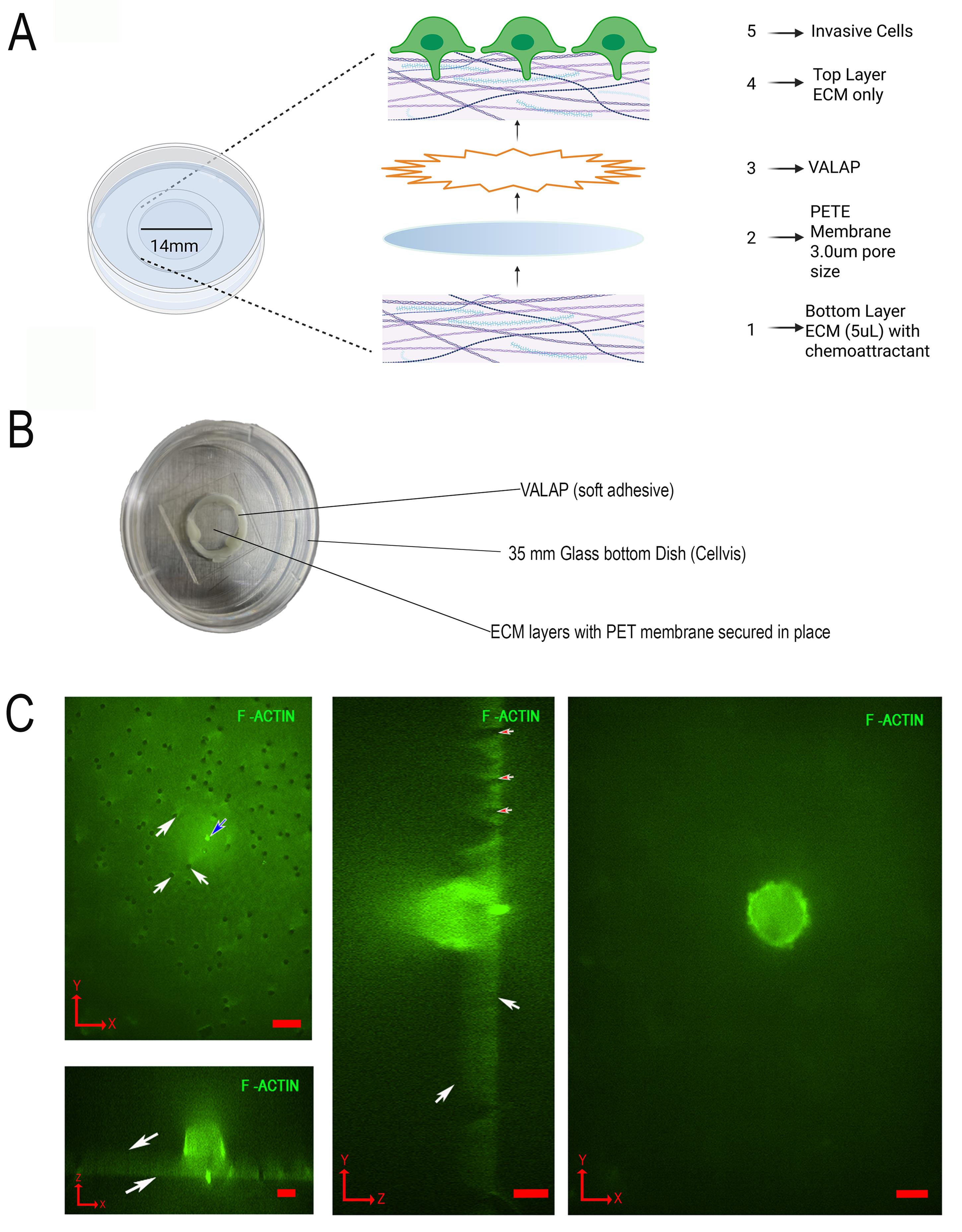
Figure 1. Design and construction of the axial invasion chamber (AIC). (A) Zoomed out view of all layers constructed during assembly from bottom to top (numbered 1–5). (B) Picture of assembled AIC. (C) Representative coordinate plane images of an invading MDA-MB-231 cell cultured in the AIC. The cell is expressing GFP-F-Actin, and images were acquired by spinning disk confocal fluorescence microscopy with 60× magnification. (Top left) XY image of the bottom of the PETE membrane, showing invadopodia protrusion through the 3.0 μm pore (blue arrow) and non-protruding pores (white arrows). (Bottom left) XZ image showing the top and bottom of the PETE membrane (white arrows). (Middle) YZ image showing pores (red arrows) and the top and bottom of the membrane (white arrows). (Right) XY image of a cell viewed from the top of the membrane. All scale bars, 10 μm. Red arrows in the bottom left corners indicate the axis orientation as pictured (X, Y, and Z). Panels A and B, adapted from Garewal et al. [17] MBoC, licensed from CC BY 4.0.
Materials and reagents
Biological materials
1. Human breast cancer metastatic cell line (MDA-MB-231; human origin; catalog number: ATCC HTB-26)
Reagents
1. Dulbecco’s modified Eagle’s medium (DMEM), sterile, high glucose with L-glutamine, without phenol red, sodium pyruvate, liquid (Cytiva, catalog number: SH30284.01)
2. Sodium pyruvate solution, sterile-filtered, 100 mM (Cytiva, catalog number: SH30239.01)
3. Fetal bovine serum, 500 mL, regular, USDA-approved origin (heat inactivated) (Corning, catalog number: 35-011-CV)
4. BioReagent penicillin-streptomycin, sterile, 100×, 10,000 U/mL penicillin, 10 mg/mL streptomycin, solution stabilized, suitable for cell culture, liquid (Sigma-Aldrich, catalog number: P4333-100ML)
5. Lipofectamine 3000 transfection reagent (Thermo Fisher Scientific, catalog number: L3000008)
6. Trypsin EDTA 1× (Corning, catalog number: 25-053-Cl)
7. GeltrexTM LDEV-free reduced growth factor basement membrane matrix (Thermo Fisher Scientific, catalog number: A1413202)
8. BioReagent Laminin, 1–2 mg/mL, sterile, from Engelbreth–Holm–Swarm murine sarcoma basement membrane, suitable for cell culture (Sigma-Aldrich, catalog number: L2020)
9. hEGF, EGF, recombinant, expressed in E. coli, lyophilized powder, suitable for cell culture (Sigma-Aldrich, catalog number: E9644)
10. Petroleum jelly (Medline, catalog number: CUR0053457)
11. Paraffin (Sigma-Aldrich, catalog number: 708860)
12. Lanolin (TagetMol, catalog number: T3238)
13. Dulbecco's phosphate-buffered saline, modified (DPBS), w/o calcium chloride and magnesium chloride, filter-sterilized (Sigma-Aldrich, catalog number: D8537)
14. HEPES solution,1 M, pH 7.0–7.6, filter-sterilized (Sigma-Aldrich, catalog number: H0887-20M)
Solutions
1. Cell culture media mix (see Recipes)
2. ECM bottom layer mix 20× stock solution (see Recipes)
3. ECM top layer mix 6× stock solution (see Recipes)
4. VALAP 1:1:1 mix (see Recipes)
Recipes
1. Cell culture media mix
| Reagent | Final concentration | Quantity or volume |
|---|---|---|
| DMEM | 88.5% | 500 mL |
| Sodium pyruvate | 0.88% | 5 mL |
| Fetal bovine serum | 8.85% | 50 mL |
| Penicillin-streptomycin | 1.77% | 10 mL |
| Total | 100% | 565 mL |
Store at 2–8 °C for up to 1 month.
2. ECM bottom layer mix 20× stock solution
| Reagent | Final concentration | Quantity or volume |
|---|---|---|
| Geltrex:Laminin 1:1 mix | 10% | 10 μL |
| hEGF | 9% | 9 μL |
| Cell culture media mix | 8% | 8 μL |
| Fetal bovine serum | 73% | 73 μL |
| Total | 100% | 100 μL |
Aliquot and store at -20 °C for up to 6 months.
3. ECM top layer mix 6× stock solution
| Reagent | Final concentration | Quantity or volume |
|---|---|---|
| Geltrex:Laminin 1:1 mix | 10% | 30 μL |
| Cell culture media mix | 90% | 270 μL |
| Total | 100% | 300 μL |
Aliquot in 300 μL and store at -20 °C for up to 6 months.
4. VALAP 1:1:1 mix
| Reagent | Final concentration | Quantity or volume |
|---|---|---|
| Petroleum jelly | 33.3% | 30 g |
| Paraffin | 33.3% | 30 g |
| Lanolin | 33.3% | 30 g |
| Total | ~100% | 90 g |
Heat on a hot plate until in a liquid state. Aliquot in 1.5 mL Eppendorf tubes to place in the biosafety cabinet (BSC) and store at room temperature for up to 2 years.
Laboratory supplies
1. 35 mm glass-bottom dishes (Cellvis, catalog number: D35-14-1.5-N)
2. Polyester track etch (PETE) membrane filters, transparent, 3.0 µm, 12 µm thickness, 6 × 10⁵ pores/cm2, 13 mm, 100/pack (Steriltech, catalog number: 1300025)
3. 225 cm2 polystyrene tissue culture treated flasks, sterile, vent cap, canted neck (Celltreat, catalog number: 229371)
4. 12-well polystyrene tissue culture treated multiple well plates, sterile, individually wrapped, with lid (Fablab, catalog number: FL7111)
5. 5 mL serological pipette, individual plastic/plastic wrapper packed in bags, sterile (Celltreat, catalog number: 229205A)
6. 10 mL polystyrene serological pipettes, sterile, individually paper/plastic wrapped, color coded orange (Celltreat, catalog number: 667210B)
7. Corning® 1–200 μL universal fit racked pipette tips, natural, sterile (Corning, catalog number: 4864)
8. Corning® 100–1,000 μL universal fit racked pipette tips, blue, sterile (Corning, catalog number: 9032)
9. Razor blades
10. 15 mL polypropylene centrifuge tubes, sterile, bulk bag (Celltreat, catalog number: 667015B)
11. Eppendorf safe-lock tubes, 1.5 mL, colorless (Eppendorf, catalog number: 022363204)
12. Pyrex® Petri dish bottom only, O.D. × H 100 mm × 20 mm
13. Hemacytometer
14. Parafilm
Equipment
1. Biosafety cabinet (BSC)
2. TiE microscope with stage, laser, camera, computer, etc. (Nikon, catalog number: 12831)
3. Tweezers
4. Cell culture incubator at 37 °C, 5% CO2, 90% relative humidity (Thermo Fisher Scientific, catalog number: 51030401)
5. Water bath 37 °C (Thermo Fisher Scientific, catalog number: FSGPD20)
6. Benchtop centrifuge for 15 mL conical tubes (Benchmark Scientific, catalog number: WBB3113837)
7. Freezer -20 °C
8. Refrigerator 2–8 °C
9. Large diameter >10” ice container
10. Hotplate (Thermo Fisher Scientific, catalog number: 1152049H)
11. Digital heat block with holes to accommodate the diameter of 1.5 mL Eppendorf tubes (VWR, catalog number: 15259-050)
Software and datasets
1. NIS elements version 4.3; license needed
Procedure
登录/注册后免费查看全文
文章信息
稿件历史记录
提交日期: Jan 30, 2026
接收日期: Mar 11, 2026
在线发布日期: Mar 22, 2026
出版日期: Apr 20, 2026
版权信息
© 2026 The Author(s); This is an open access article under the CC BY license (https://creativecommons.org/licenses/by/4.0/).
如何引用
Readers should cite both the Bio-protocol article and the original research article where this protocol was used:
- Garewal, M. and Myers, K. A. (2026). Preparation and Assembly of the Axial Invasion Chamber for Live-Cell Invadopodia Imaging. Bio-protocol 16(8): e5658. DOI: 10.21769/BioProtoc.5658.
- Garewal, M., Ramos, P. and Myers, K. A. (2025). A structured framework of cytoskeletal proteins and noncentrosomal microtubules promotes the initiation and elongation of invadopodia. Mol Biol Cell. 36(12): ee25–07–0313. https://doi.org/10.1091/mbc.e25-07-0313

 Download Citation in RIS Format
分类
癌症生物学 > 侵袭和转移 > 细胞生物学试验 > 细胞侵袭
细胞生物学 > 细胞成像 > 活细胞成像
细胞生物学 > 细胞成像 > 共聚焦显微镜
您对这篇实验方案有问题吗?
在此处发布您的问题,我们将邀请本文作者来回答。同时,我们会将您的问题发布到Bio-protocol Exchange,以便寻求社区成员的帮助。
 发布问题
发布问题


