


Isolation, Culture, and Differentiation of Bovine Muscle Resident Stem Cells
牛肌肉驻留干细胞的分离、培养与分化方法

发布: 2026年04月05日第16卷第7期 DOI: 10.21769/BioProtoc.5647 浏览次数: 925
评审: Samantha HallerNeha SaxenaAnonymous reviewer(s)
 PDF
PDF  Q&A
Q&A
参见作者原研究论文
The authors used this protocol in:

May 2025
Advertisement







Abstract
Bovine muscle satellite cells (MuSC) and fibro-adipogenic progenitor cells (FAP) are muscle resident stem cells that are responsible for postnatal muscle growth, intramuscular fat deposition, and extracellular matrix generation. These cells are of increasing interest for the cultivated meat community due to their ability to generate all the major components of meat; additionally, these cells are of interest to conventional animal science research to elucidate mechanisms to improve meat quality. To use these cells for these goals, efficient and accurate cell isolation, culture, and differentiation are essential to evaluate their cell fate decisions and behaviors. In this protocol, we detail a simultaneous isolation of both MuSCs and FAPs with multiple intermediate stopping points, allowing for flexibility for day-of time constraints. We also detail improved growth conditions to maximize cell expansion and procedures to assess cell differentiation. This protocol provides a flexible isolation procedure that is compatible with sampling in modern slaughterhouses or from biopsies. Additionally, the differentiation procedures provide improved differentiation but still allow in vitro treatment and assessment.
Key features
• This protocol offers a flexible in-lab procedure to isolate bovine FAPs and MuSCs from tissue collected post-slaughter with multiple pause points.
• The protocol demonstrates successful conditions to grow, expand, and differentiate bovine FAPs with an optimized adipogenic differentiation medium.
• Strategies for planning your primary cell isolation, choosing the sampling location, and characterizing differentiation of bovine FAPs and MuSCs are included.
Keywords: Bovine muscle resident stem cells (牛肌肉驻留干细胞) Fibro-adipogenic progenitor cell (成纤维-脂肪生成祖细胞)
Muscle satellite cell (肌卫星细胞)
Primary cell isolation (原代细胞分离)
Adipogenic differentiation (脂肪分化)
Fibro-adipogenic progenitor cell (成纤维-脂肪生成祖细胞)
Muscle satellite cell (肌卫星细胞)
Primary cell isolation (原代细胞分离)
Adipogenic differentiation (脂肪分化)
Graphical overview
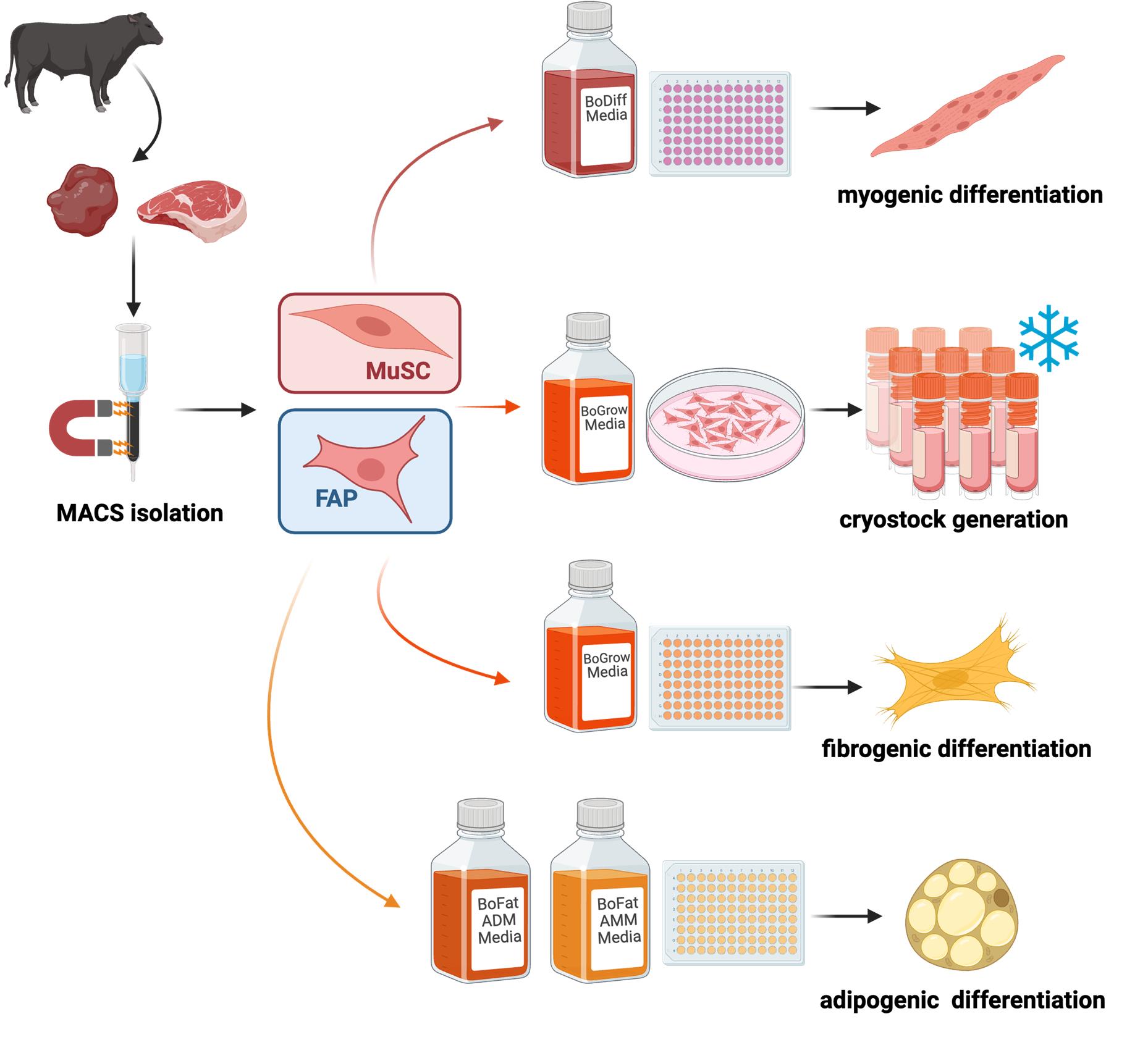
Isolation and differentiation of bovine muscle resident stem cells. MuSC, muscle satellite cells; FAP, fibro-adipogenic progenitor cells.
Background
Skeletal muscle tissue is the predominant form of tissue consumed as meat [1]. Critical muscle structures for meat include contractile myofibers, which provide the majority of protein, lipids stored as fat, which provide flavor, and the extracellular matrix, which is integral to texture [2–6]. The development and growth of skeletal muscle rely on stem cell differentiation into these muscle structures. Each of these structures arises from muscle resident stem cells, with muscle stem cells (MuSC) or satellite cells as a progenitor to myofibers and fibro-adipogenic progenitors (FAP) which are capable of adipogenic differentiation into adipocytes or activation into prolific extracellular matrix-producing myofibroblasts [7,8]. Thus, knowledge of how these stem cells respond to a variety of stimuli is essential to understanding the adaptations of muscle tissue and thus meat quality [9,10]. The isolation of these stem cells provides the opportunity to explicitly control their environment or alter the cells using the plethora of cell biology tools available [10,11]. While many of these tools have focused on biomedical applications, isolation and culture of MuSCs and FAPs from agriculturally relevant species is vital to meat science [12,13]. Particularly, in the emerging field of cultivated meat, which utilizes in vitro methods to create meat, investigation of MuSCs and FAPs is essential [14,15].
Beef is the most studied meat, with many factors such as marbling impacting meat quality [16]. Thus, bovine MuSCs and FAPs are among the most commonly studied in meat science. Although these cells are the focus of this protocol, it may also serve as a template for other meats such as pork, poultry, or fish. Isolation of bovine MuSCs has been performed extensively, using a variety of protocols, while FAP isolation is increasingly being performed [17–21]. Pre-plating techniques that rely on differential adhesion dynamics of stem cell types allow enrichment of MuSCs in a simple, low-resource, intensive manner [22]. Fluorescence-activated cell sorting (FACS) alternatively provides high-purity stem cell populations but is resource-intensive and stressful to primary cells [23]. Magnetic activated cell sorting (MACS) offers a compromise to more selectively sort cells than pre-plating techniques while also being more accessible and gentler to cells than FACS [24]. Following isolation, these adherent cell types must be expanded in vitro. Many factors of the in vitro environment, from media composition to adhesion substrate, can impact cell behavior. The differentiation of stem cells is a key behavior, and a number of different media are used for proliferation or myogenic differentiation of MuSCs or adipogenic differentiation of FAPs [25–28]. Here, we provide a protocol for the simultaneous isolation and in vitro investigation of bovine MuSCs and FAPs. These protocols include coating procedures and multi-stage media compositions for differentiation that have been optimized for bovine stem cells [18]. These protocols establish a reproducible framework for in vitro studies of bovine stem cell contributions to meat development and quality.
Materials and reagents
Biological materials
1. Bovine primary fibro-adipogenic progenitor cells (generated as part of this protocol)
2. Bovine primary muscle satellite cells (generated as part of this protocol)
Reagents
1. F10 media (Fisher Scientific, catalog number: 11550043)
2. Debris removal solution (Miltenyi Biotec, catalog number: 130-109-398)
3. CD140a (PDGFRα) Microbead kit, mouse (Miltenyi Biotec, catalog number: 130-101-547)
4. Satellite Cell Isolation kit, mouse (Miltenyi Biotec, catalog number: 130-104-268)
5. 0.25% Trypsin-EDTA (Fisher Scientific, catalog number: 25-200-056)
6. Collagenase type II powder (Fisher Scientific, catalog number: 17101015)
7. EDTA (Affymetrix, catalog number: 15694)
8. EDTA salt (Fisher Scientific, catalog number: BP120 500)
9. Bovine serum albumin (BSA) (Fisher Scientific, catalog number: 9048-46-8)
10. Penicillin-Streptomycin (10,000 U/mL) (Thermofisher Scientific, catalog number: 15140122)
11. Amphotericin B (Thermofisher Scientific, catalog number: 15290018)
12. Gentamicin solution (Thermofisher Scientific, catalog number: 15710064)
13. DMEM, high glucose (Thermofisher Scientific, catalog number: 11-965-092)
14. Ammonium chloride salt (Thermofisher Scientific, catalog number: 012361-36)
15. Sodium bicarbonate (Thermofisher Scientific, catalog number: 50-287-33)
16. Fibronectin solution (Advanced Biomatrix, catalog number: 5050)
17. PBS pH 7.2 (Thermofisher Scientific, catalog number: 20012027)
18. FBS (Biowest, catalog number: S1620)
19. FGF2 (ThermoFisher, catalog number: PHG6015)
20. DMSO (Thermofisher Scientific, catalog number: BP231-100)
21. GlutaMAX (Thermofisher Scientific, catalog number: 35050061)
22. Troglitazone (Thermofisher Scientific, catalog number: 501150786)
23. Insulin, human recombinant (Millipore Sigma, catalog number: 91077C-100MG)
24. IBMX (VWR, catalog number: 102516-252)
25. Dexamethasone (Thermofisher Scientific, catalog number: 1126100)
26. Trypan blue (Thermofisher Scientific, catalog number: C10228)
27. Paraformaldehyde (PFA), 16% w/v aq. soln., methanol free (Thermofisher Scientific, catalog number: 043368.9M)
28. Perilipin-1 (D1D8) rabbit monoclonal antibody (Cell Signaling, catalog number: 9349)
29. Actin, smooth muscle, Ab-1 mouse monoclonal antibody, EprediaTM (Fisher Scientific, catalog number: MS113P1)
30. Hoechst 33342, trihydrochloride trihydrate (Invitrogen, catalog number: H3570)
31. Acti-stain 555 phalloidin (Cytoskeleton, Inc., catalog number: PHDH1)
32. Desmin antibody (B-7) (Santa Cruz Biotechnology, catalog number: sc-271677)
33. Myogenin polyclonal antibody (Invitrogen, catalog number: PA5-116750)
34. Donkey anti-rabbit IgG (H+L) highly cross-adsorbed secondary antibody, Alexa FluorTM Plus 647 (Invitrogen, catalog number: A32795)
35. Donkey anti-mouse IgG (H+L) highly cross-adsorbed secondary antibody, Alexa FluorTM Plus 488 (Invitrogen, catalog number: A32766)
36. Nile Red (ThermoFisher, catalog number: N1142)
37. Oil Red O (ThermoFisher, catalog number: A12989.22)
38. 70% v/v denatured ethanol (ThermoFisher, catalog number: BP82031GAL)
39. Triton X-100 (Fisher Scientific, catalog number: BP151-100)
Solutions
1. PEB (see Recipes)
2. Collagenase (see Recipes)
3. PBS antibiotic (see Recipes)
4. Erythrocyte lysis buffer (ACK) 10× (see Recipes)
5. Fibronectin coating solution (see Recipes)
6. BoFreeze media (see Recipes)
7. BoGrow media (see Recipes)
8. BoFat adipogenic differentiation media (ADM) (see Recipes)
9. BoFat adipogenic maturation media (MM) (see Recipes)
10. BoDiff media (see Recipes)
Recipes
1. PEB
| Reagent | Stock concentration | Final concentration | Quantity or volume |
|---|---|---|---|
| BSA | n/a | 5 g/L | 0.2 g |
| PBS | 1× | n/a | 40 mL |
| EDTA | 0.5 M | 2 mM | 160 mL |
| Total | n/a | n/a | 40 mL |
Weigh out BSA and transfer to a sterile 50 mL conical tube. On the day of use, in a biosafety cabinet, add the EDTA and PBS and vortex to combine.
Note: Some foam will form. You may allow 1–3 h at 2–8 °C for it to settle or aspirate the foam and use the liquid immediately. The BSA must be solubilized on the day of use, and the solution must always be made fresh.
Store at 2–8 °C for up to 16 h and let it rise to room temperature before use.
2. Collagenase
| Reagent | Stock concentration | Final concentration | Quantity or volume |
|---|---|---|---|
| Collagenase | Variable, >125 U/mg | 10,000 units | Variable |
| DMEM | n/a | n/a | 5 mL |
| Pen/Strep | 10,000 U/mL | 2% | 100 mL |
| Total | n/a | n/a | 5 mL |
Weigh out collagenase and transfer it to a sterile 15 mL conical tube. Adjust collagenase weight based on your reagent’s units/g specification.
Bring this into a biosafety cabinet and add pen/strep and DMEM. Homogenize via vortex or pipetting up and down until all solids are dissolved. Once dissolved, centrifuge at 300× g for 5 min. If solids remain after centrifugation, repeat homogenization and centrifugation as needed. Store at 2–8 °C for up to 2 weeks, and heat using a 37 °C water bath before use.
Note: This recipe makes 5 mL of solution, which is sufficient for 10 g of tissue (0.5 mL/1 g). Scale up or down as needed.
3. PBS antibiotic
| Reagent | Stock concentration | Final concentration | Quantity or volume |
|---|---|---|---|
| PBS | 1× | n/a | 49 mL |
| Pen/Strep | 10,000 U/mL | 1% | 500 mL |
| Gentamicin | 10 mg/mL | 0.5% | 250 mL |
| Amphotericin B | 250 μg/mL | 0.4% | 200 mL |
| Total | n/a | n/a | 50 mL |
Add from largest to smallest volume using sterile technique in a biosafety cabinet. Store at 2–8 °C for up to 3 months.
4. ACK (10×)
| Reagent | Stock concentration | Final concentration | Quantity or volume |
|---|---|---|---|
| DMEM | n/a | n/a | ~50 mL |
| Ammonium chloride | n/a | 80.2 g/L | 4.01 g |
| Sodium bicarbonate | n/a | 8.4 g/L | 0.42 g |
| EDTA salt | n/a | 3.7 g/L | 0.185 g |
| Total | n/a | n/a | 50 mL |
Weigh out all solids and transfer them into a 50 mL conical tube. Bring this into a biosafety cabinet and add DMEM up to the 50 mL line. Use a vortex or pipette up and down to mix.
Note: Solids are resistant to dissolution and may require time overnight to fully dissolve. Additionally, bubbles will form once DMEM is added due to the bicarbonate.
Filter-sterilize through a 0.22 mm filter and store at 2–8 °C for up to 3 months. Once ready to use, dilute this solution 1:9 with PBS to create a 1× solution.
5. Fibronectin coating solution
| Reagent | Stock concentration | Final concentration | Quantity or volume |
|---|---|---|---|
| PBS | 1× | n/a | 1 mL |
| Fibronectin | 0.5 mg/mL | 5 mg/mL | 10 mL |
| Total | n/a | n/a | 1.01 mL |
Thaw a fibronectin aliquot at room temperature in a biosafety cabinet. Once thawed, combine the described volume with PBS, flushing out the pipette tip by pipetting up and down 2–3× when adding the fibronectin into your working solution. Vortex to homogenize and use immediately. To coat, in a biosafety cabinet, apply the working solution to your culture vessel to completely cover the vessel bottom and incubate at room temperature for 1 h; see Table 1 for suggested coating volumes. Aspirate the working solution, with care not to touch the bottom of the culture vessel, and wash 2–3 times with 1× PBS. Alternatively, the remaining solution may be collected, stored at -20 °C for 3 months, and reused up to 3×. Coated plates should be covered with ample amounts of 1× PBS, sealed with parafilm, and stored at 2–8 °C for up to 3 months. Discard plates if they dry out. Scale up the recipe as needed to thoroughly cover the bottom of your culturing vessel. This achieves a 1.56 mg/cm2 coating density.
Table 1. Recommended seeding densities and media volumes for culture
| Vessel | Growth area (cm2) | Cells to seed for growth | Wash, trypsin, or coating solution volume (mL) | Media volume (mL) |
|---|---|---|---|---|
| 96-well plate | 0.32/well | 1,650 | 0.05/well | 0.100/well |
| 6-well plate | 9.6/well | 40,000 | 0.5/well | 2/well |
| T25 | 25 | 125,000 | 1 | 5 |
| T75 | 75 | 375,000 | 3 | 15 |
| T182 | 182 | 900,000 | 5 | 25 |
6. BoFreeze media
| Reagent | Stock concentration | Final concentration | Quantity or volume |
|---|---|---|---|
| FBS | n/a | 90% | 9 mL |
| DMSO | >99.7% | 10% | 1 mL |
| Total | n/a | n/a | 10 mL |
Add the FBS and then DMSO into a sterile 15 mL conical tube in a biosafety cabinet; invert 3× to homogenize. Store at 2–8 °C for up to 3 months, and warm to room temperature before use.
7. BoGrow media
| Reagent | Stock concentration | Final concentration | Quantity or volume |
|---|---|---|---|
| F-10 media | n/a | n/a | 39mL |
| FBS | n/a | 20% | 10 mL |
| Pen/Strep | 10,000 U/mL | 1% | 500 mL |
| Glutamax | 100× | 1% | 500 µL |
| Gentamicin | 10 mg/mL | 50 mg/mL | 250 mL |
| Amphotericin B | 250 μg/mL | 1 μg/mL | 200 µL |
| FGF2 | 2,500 ng/mL | 5 ng/mL | 100 mL |
| Total | n/a | n/a | 50 mL |
Combine all reagents from largest to smallest volume in a 50 mL conical tube in a biosafety cabinet. Invert three times to homogenize, and optionally filter-sterilize with a 0.22 mm filter. Store at 2–8 °C for up to 3 months, and warm in a 37 °C water bath prior to use. This recipe may be scaled up to make larger stocks of media, but aliquots no larger than 50 mL should be made to reduce temperature exposure of the whole stock.
8. BoFat ADM
| Reagent | Stock concentration | Final concentration | Quantity or volume |
|---|---|---|---|
| BoGrow | n/a | n/a | 4.864 mL |
| Insulin | 2 mg/mL | 5.258 mM | 76.34 mL |
| Troglitazone | 2.5 mg/mL | 5 mM | 44.15 mL |
| Dexamethasone | 10 mM | 1 mM | 5 mL |
| IBMX | 250 mM | 500 mM | 10 mL |
| Total | n/a | n/a | 5 mL |
Thaw all adipogenic-inducing reagents in a 37 °C water bath and then transfer to a biosafety cabinet. Add reagents from largest to smallest volume in a sterile 5 or 15 mL conical tube and vortex or invert. Some precipitate may form, which will require time and heat to resolubilize. Use the media the next day or heat in a 37 °C water bath for ~30 min to encourage solubilization. Filter sterilization through a 0.22 mm filter may be optionally conducted after all precipitate is resolubilized. Store at 2–8 °C for up to 3 months.
9. BoFat AMM
| Reagent | Stock concentration | Final concentration | Quantity or volume |
|---|---|---|---|
| BoGrow | n/a | n/a | 4.879 mL |
| Insulin | 2 mg/mL | 5.258 mM | 76.34 mL |
| Troglitazone | 2.5 mg/mL | 5 mM | 44.15 mL |
| Total | n/a | n/a | 5 mL |
Thaw all adipogenic-inducing reagents in a 37 °C water bath and then transfer to a biosafety cabinet. Add reagents from largest to smallest volume in a sterile 5 or 15 mL conical tube and vortex or invert. Some precipitate may form, which will require time and heat to resolubilize. Use the media the next day or heat in a 37 °C water bath for ~30 min to encourage solubilization. Filter sterilization through a 0.22 mm filter may be optionally conducted after all precipitate is resolubilized. Store at 2–8 °C for up to 3 months.
10. BoDiff media
| Reagent | Stock concentration | Final concentration | Quantity or volume |
|---|---|---|---|
| DMEM | n/a | n/a | 9.7 mL |
| FBS | n/a | 2% | 200 µL |
| P/S | 10,000 U/mL | 1% | 100 µL |
| Total | n/a | n/a | 10 mL |
Combine all reagents from largest to smallest volume in a 15 mL conical tube in a biosafety cabinet. Invert three times to homogenize, and optionally filter-sterilize with a 0.22 μm filter. This recipe may be scaled up to make larger stocks of media, but aliquots should be no larger than 50 mL. Store at 2–8 °C for up to 3 months, and warm in a 37 °C water bath prior to use.
Laboratory supplies
1. 15 mL conical centrifuge tubes, polypropylene, sterile (Genesee, catalog number: 28-101)
2. 50 mL conical centrifuge tubes (Genesee, catalog number: 28-106)
3. Olympus Premium 0.6 mL snap cap microcentrifuge tubes (Genesee, catalog number: 24-272)
4. Petri dish 150 mm × 22 mm (Genesee, catalog number: 32-106)
5. P10–P1000 tips, sterile (USA Scientific, catalog numbers: 1112-1720, 1110-1700, 1110-3700)
6. Serological pipettes, 5–50 mL, sterile (Genesee, catalog numbers: 12-102, 12-104, 12-106, 12-107)
7. TC plates of various sizes: 96 well, 6 well, T25, T75 (Genesee, catalog numbers: 25-109, 25-105, 25-207, 25-209)
8. MACS C Tube (Miltenyi, catalog number: 130-093-237)
Equipment
1. Biological Safety Cabinet 1300 Clas II Type A2 (Fisher Scientific, catalog number: 1911310)
2. Water bath capable of reaching 37 °C
3. Freezer (-20 °C)
4. Refrigerator (2–8 °C)
5. Liquid nitrogen tank
6. Serological pipettes and micropipettes (standard sizes, sterile)
7. Incubator capable of maintaining 37 °C and 5% CO2
8. Microscope capable of 10×, 20×, and 40× magnification in brightfield
9. Hemocytometer
10. Countess (ThermoFisher) or other automated cell counter
11. Warming oven capable of reaching 37 °C
12. MACSmix tube rotator (Miltenyi Biotec, catalog number: 130-090-753)
13. Mini & MidiMACS Starting kit (Miltenyi Biotec, catalog number: 130-042-501)
14. gentleMACS dissociator (Miltenyi Biotec, catalog number: 130-093-235)
Software and datasets
1. FIJI (ImageJ2, version 2.16.0/1.54p); FIJI is ImageJ with many plugins already added. This is a free-to-use, open-source software that can be downloaded at www.imagej.net/software/fiji/downloads
Procedure
登录/注册后免费查看全文
文章信息
稿件历史记录
提交日期: Dec 23, 2025
接收日期: Feb 24, 2026
在线发布日期: Mar 5, 2026
出版日期: Apr 5, 2026
版权信息
© 2026 The Author(s); This is an open access article under the CC BY license (https://creativecommons.org/licenses/by/4.0/).
如何引用
Gish, P., Stewart, M. W., Tsonov, M., Khuu, B., Espinoza, R., Vahmani, P. and Smith, L. R. (2026). Isolation, Culture, and Differentiation of Bovine Muscle Resident Stem Cells. Bio-protocol 16(7): e5647. DOI: 10.21769/BioProtoc.5647.

 Download Citation in RIS Format
分类
干细胞 > 成体干细胞 > 肌肉干细胞
细胞生物学 > 细胞分离和培养 > 细胞分化
干细胞 > 成体干细胞 > 维持和分化
您对这篇实验方案有问题吗?
在此处发布您的问题,我们将邀请本文作者来回答。同时,我们会将您的问题发布到Bio-protocol Exchange,以便寻求社区成员的帮助。
 发布问题
发布问题


