

Quantifying Lysosomal Degradation of Extracellular Proteins With a Fluorescent Protein-Based Internalization Assay
(*contributed equally to this work) 发布: 2026年03月05日第16卷第5期 DOI: 10.21769/BioProtoc.5619 浏览次数: 36
评审: David PaulAnonymous reviewer(s)
参见作者原研究论文
The authors used this protocol in:

Mar 2023


Abstract
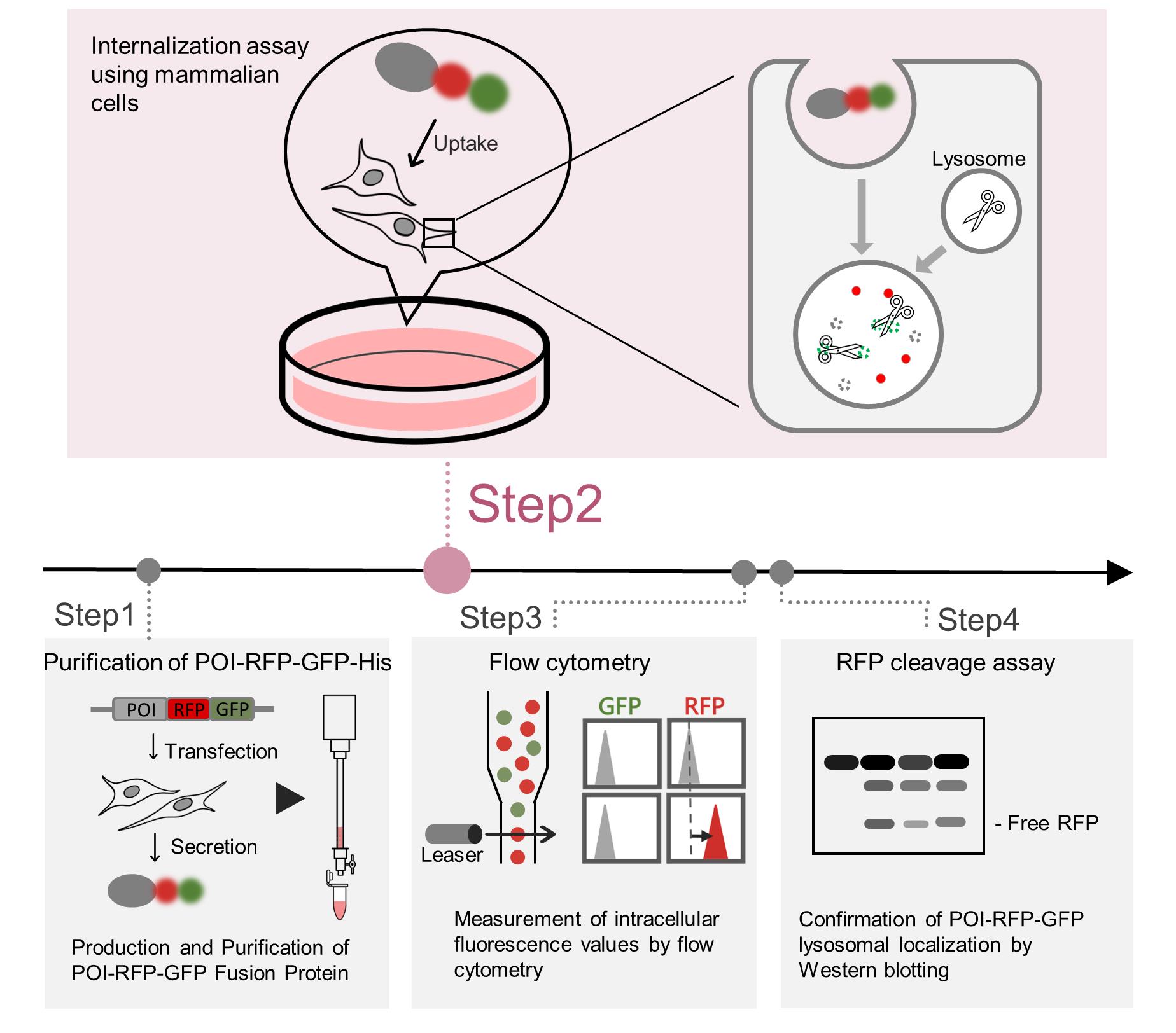
Endocytosis is an essential membrane transport mechanism that is indispensable for the maintenance of life. It is responsible for the selective internalization and subsequent degradation or recycling of specific extracellular proteins and nutrients, thereby facilitating cellular nutrient supply, modulation of receptor signaling, and clearance of foreign substances. However, methods for the quantitative analysis of lysosomal degradation of extracellular proteins via endocytosis remain limited. This protocol describes a method for purifying the protein-of-interest (POI)–red fluorescent protein (RFP)–green fluorescent protein (GFP) fusion protein, which is modified with specific mammalian cell glycans or other modifications, from the conditioned medium of mammalian cell cultures. Subsequently, the protocol details a quantitative approach for evaluating its internalization and lysosomal degradation within cells using the RFP–GFP tandem fluorescent reporter. Following the addition of POI-RFP-GFP to the medium, cells can be subjected to cell biological assays, such as flow cytometry, as well as biochemical analyses, such as immunoblotting. This protocol is broadly applicable to studies of the internalization of extracellular proteins.
Key features
• Purification of secreted GFP-RFP-fused POI from mammalian cell culture supernatant.
• Quantification of POI-RFP-GFP internalization through measurement of GFP and RFP signals using flow cytometry.
• Confirmation of lysosomal degradation of POI-RFP-GFP by immunoblotting.
Keywords: LysosomeGraphical overview
Background
Extracellular proteins, including those circulating in the bloodstream, are continuously secreted, internalized by mammalian cells, and subsequently degraded in lysosomes [1,2]. Proteins such as transferrin and immunoglobulins are recycled via the endosomes, whereas lipoproteins are constitutively degraded within lysosomes [3,4]. In contrast, various diseases, for example, Alzheimer’s disease, are associated with abnormal accumulation of extracellular proteins [5–7]. Accurate quantification of internalization is therefore essential in understanding extracellular proteostasis, pathological protein accumulation, and chaperone-dependent internalization pathways. However, methods for quantitatively tracking extracellular protein internalization and lysosomal degradation have long been limited, posing significant challenges for such analyses. Covalent fluorescent labeling approaches rely on random conjugation of fluorophores to proteins of interest (POIs), which can interfere with protein structure or function. Moreover, increases in cellular fluorescence often fail to distinguish whether the signal reflects simple binding of the POI to the cell surface or enhanced internalization. The differences between the red fluorescent protein (RFP)–green fluorescent protein (GFP) fusion system and covalent fluorescent labeling are summarized in Table 1.
Tandem RFP-GFP reporters have traditionally been used to monitor intracellular lysosomal transport processes, such as autophagy [8]. We have, for the first time, adapted this system to monitor the lysosomal degradation of extracellular proteins. The internalization reporter system, in which a POI is fused to an RFP–GFP tandem fluorescent tag, was specifically developed to quantify this extracellular protein endocytosis and lysosomal transport [9–11]. GFP is quenched in the acidic environment of lysosomes, whereas RFP retains fluorescence; thus, GFP serves as an indicator of cell-surface binding or internalized but not yet transported to lysosomes, while an increase in RFP reflects accumulation at both the cell surface and within lysosomes. Therefore, an increase in RFP fluorescence without a corresponding increase in GFP fluorescence indicates that the POI-RFP-GFP fusion protein has undergone lysosomal degradation. This reporter enables sensitive, quantitative, and spatial analysis of both internalization and lysosomal transport of the POI.
This protocol describes an internalization assay using POI-RFP-GFP fusion proteins and presents a comprehensive workflow that integrates protein purification, flow cytometry, and immunoblotting. Together, these approaches enable robust evaluation of lysosomal degradation of extracellular protein.
The protocol has been used in Tomihari et al. (2023) [9], where it contributed to elucidating the mechanisms of extracellular protein internalization and lysosomal degradation.
Table 1. Comparison of conventional methods and the red fluorescent protein (RFP)–green fluorescent protein (GFP) tandem reporter
| Method | Principle | Advantages | Limitations | Target detection |
|---|---|---|---|---|
Covalent fluorescence labeling | Chemical conjugation of fluorescent dyes to Lys/Cys residues | Applicable to numerous proteins | Random labeling may interfere with protein function; increased fluorescence does not always reflect internalization | Cell surface and lysosome (indistinguishable by flow cytometry; distinguishable by microscopy) |
| GFP-only reporter | Fusion of GFP tag to the N- or C-terminus of POI | Smaller molecular weight than the tandem reporter | Since GFP is degraded in the lysosomes, lysosomal transport cannot be detected via the GFP signal | Cell surface |
| RFP-only reporter | Fusion of RFP tag to the N- or C-terminus of POI | Smaller molecular weight than the tandem reporter; the RFP cleavage assay enables biochemical analysis of lysosomal transport | Cell surface binding and lysosomal transport are indistinguishable by flow cytometry | Cell surface and lysosome (indistinguishable by flow cytometry; distinguishable by RFP cleavage assay and microscopy) |
| RFP-GFP tandem reporter (current protocol) | Fusion of RFP-GFP tag to the N- or C-terminus of POI | Simultaneous evaluation of cell surface and lysosomal transport; the RFP cleavage assay enables biochemical analysis of lysosomal transport | Large tag may affect the function of certain POIs | Cell surface and lysosome (distinguishable by flow cytometry, RFP cleavage assay, and microscopy) |
Materials and reagents
Biological materials
1. Human: Flp-In T-REx 293 cell line (Thermo Fisher, catalog number: R78007)
2. Human: 293FT cell line (Thermo Fisher, catalog number: R70007)
3. Human: HeLa cells (ATCC, catalog number: CCL-2)
Plasmids
1. pcDNA5/FRT/TO vector (Thermo Fisher, catalog number: V601020)
2. pOG44 Flp-recombinase expression vector (Thermo Fisher, catalog number: V600520)
3. pcDNA5/FRT/TO–α2M–RG–His (RIKEN BioResource Research Center, catalog number: RDB20287)
Reagents
1. Dulbecco’s modified Eagle medium (DMEM), high glucose (Nacalai Tesque, catalog number: 16971-55)
2. Advanced DMEM/F-12 (Thermo Fisher, catalog number: 12634010)
Note: Reduced serum medium (DMEM/Ham’s F-12) (Nacalai Tesque, catalog number: 21906-55) is also compatible with this protocol.
3. Fetal bovine serum (FBS) (MERCK, catalog number: 12003C)
4. Penicillin-streptomycin (Nacalai Tesque, catalog number: 09367-34)
5. Opti-MEM (Thermo Fisher, catalog number: 31985070)
6. MiniPrep kit (Favorgen, catalog number: FAPDE 001-1)
7. Polyethylenimine (Polysciences, catalog number: 24765-2)
8. Hygromycin (Fujifilm, catalog number: 085-06153)
9. Doxycycline (Sigma, catalog number: D9891)
10. 2.5 g/L trypsin, 1 mmol/L EDTA solution (Nacalai Tesque, catalog number: 32777-44)
11. Bafilomycin A1 (Cayman, catalog number: 11038)
12. Ni-NTA agarose resin (Fujifilm, catalog number: 141-09683)
13. Imidazole (Fujifilm, catalog number: 099-00013)
14. Sodium chloride (NaCl) (Fujifilm, catalog number: 191-01665)
15. di-sodium hydrogenphosphate dodecahydrate (Na2HPO4·12H2O) (Nacalai Tesque, catalog number: 31722-45)
16. Potassium dihydrogenphosphate (KH2PO4) (Nacalai Tesque, catalog number: 28721-55)
17. Potassium chloride (KCl) (Kanto Kagaku, catalog number: 32326-00)
18. D-PBS without Ca and Mg, powder (Nacalai Tesque, catalog number: 07269-84)
19. Triton X-100 (Nacalai Tesque, catalog number: 35501-02)
20. Polyoxyethylene (20) sorbitan monolaurate (100% Tween20) (Fujifilm, catalog number: 166-21115)
21. Glycerol (Nacalai Tesque, catalog number: 17017-35)
22. 2-Mercaptoethanol (Fujifilm, catalog number: 198-15781)
23. Coomassie Brilliant Blue R-250 (Fujifilm, catalog number: 031-17922)
24. Skim milk (Nacalai Tesque, catalog number: 31149-75)
25. DAPI (Nacalai Tesque, catalog number: 11034-14)
26. Tris(hydroxymethyl)aminomethane (Tris) (Nacalai Tesque, catalog number: 35406-91)
27. Methanol (Nacalai Tesque, catalog number: 21915-93)
28. Acetic acid (Nacalai Tesque, catalog number: 00212-43)
29. Hydrochloric acid (HCl) (Fujifilm, catalog number: 080-01066)
30. 100× protease inhibitor cocktail (Nacalai Tesque, catalog number: 03969-21)
31. Phenylmethanesulfonyl fluoride (PMSF) (MP Biomedicals, catalog number: MPB195381-5)
32. Signal enhancer HIKARI for Western Blotting and ELISA (Nacalai Tesque, catalog number: 02267-41)
33. Rabbit polyclonal anti-mCherry (homemade), mouse monoclonal anti-RFP (MBL, catalog number: M204-3), or other mCherry antibodies
34. Mouse monoclonal anti-α tubulin (Fujifilm , catalog number: 071-25031)
35. Anti-mouse IgG, HRP-linked antibody (CST, catalog number: 7076S)
36. Anti-rabbit IgG, HRP-linked antibody (CST, catalog number: 7074S)
37. Recombinant luciferase (Promega, catalog number: E1702)
38. Purified recombinant His-Alfa-S-formylglutathione hydrolase (ESD) (homemade; see [9])
39. Purified recombinant Clusterin-mCherry-sfGFP-His (homemade; see [10])
Solutions
1. Column wash buffer (see Recipes)
2. Column elution buffer (see Recipes)
3. DMEM complete medium (see Recipes)
4. Trypsin solution (see Recipes)
5. FCM buffer (see Recipes)
6. 10× lysis buffer (see Recipes)
7. Lysis buffer (see Recipes)
8. 1 M imidazole (see Recipes)
9. Coomassie Brilliant Blue (CBB) stain solution (see Recipes)
10. Coomassie Brilliant Blue (CBB) bleaching (see Recipes)
11. 25× TBS (see Recipes)
12. 5% Tween20 (see Recipes)
13. TBST (see Recipes)
14. 25× PBS (see Recipes)
15. PBS (see Recipes)
Recipes
1. Column wash buffer
| Reagent | Final concentration | Quantity |
|---|---|---|
| 5 M NaCl | 500 mM | 100 mL |
| 1 M Imidazole | 10 mM | 10 mL |
| 25× PBS | 1× | 40 mL |
| H2O | N/A | 850 mL |
| Total | N/A | 1,000 mL |
Store at 4 °C.
2. Column elution buffer
| Reagent | Final concentration | Quantity |
|---|---|---|
| 5 M NaCl | 150 mM | 12 mL |
| 1 M Imidazole | 200 mM | 80 mL |
| 25× PBS | 1× | 16 mL |
| H2O | N/A | 292 mL |
| Total | N/A | 400 mL |
Store at 4 °C.
3. DMEM complete medium
| Reagent | Final concentration | Quantity |
|---|---|---|
| DMEM | N/A | 500 mL |
| FBS | 10% | 55 mL |
| Penicillin-streptomycin | 1% | 5.5 mL |
| Total | N/A | 555 mL |
Store at 4 °C.
4. Trypsin solution
| Reagent | Final concentration | Quantity |
|---|---|---|
| 2.5 g/L trypsin and 1 mmol/L EDTA solution | 0.5 g/L trypsin and 0.2 mmol/L EDTA | 10 mL |
| PBS | N/A | 40 mL |
| Total | N/A | 50 mL |
Store at 4 °C.
5. FCM buffer
| Reagent | Final concentration | Quantity |
|---|---|---|
| FBS | 5% | 750 μL |
| 1 mg/mL DAPI | 1 μg/mL | 15 μL |
| PBS | N/A | 14.25 mL |
| Total | N/A | 15 mL |
Store at 4 °C. Prepare immediately before use.
6. 10× lysis buffer
| Reagent | Final concentration | Quantity |
|---|---|---|
| 1 M Tris-HCl pH 7.5 | 500 mM | 50 mL |
| 5 M NaCl | 1500 mM | 30 mL |
| 0.5 M EDTA pH 8.0 | 10 mM | 2 mL |
| Triton X-100 | 10% | 10 mL |
| H2O | N/A | 8 mL |
| Total | N/A | 100 mL |
Store at 4 °C.
7. Lysis buffer
| Reagent | Final concentration | Quantity |
|---|---|---|
| 10× lysis buffer | 1× | 100 μL |
| 100× protease inhibitor cocktail | 1× | 10 μL |
| 0.1 M PMSF | 1 mM | 10 μL |
| H2O | N/A | 880 μL |
| Total | N/A | 1,000 μL |
Store at 4 °C. Prepare immediately before use.
8. 1 M imidazole
1 M imidazole in H2O, with pH adjusted to 8.0 using HCl. Store at 20 °C.
9. Coomassie Brilliant Blue (CBB) stain solution
| Reagent | Final concentration | Quantity |
| Coomassie Brilliant Blue R-250 | 0.1% | 1 g |
| Methanol | 40% | 400 mL |
| Acetic acid | 10% | 100 mL |
| H2O | N/A | 500 mL |
| Total | N/A | 1,000 mL |
Store at 20 °C.
10. Coomassie Brilliant Blue (CBB) bleaching solution
| Reagent | Final concentration | Quantity |
| Methanol | 40% | 400 mL |
| Acetic acid | 10% | 100 mL |
| H2O | N/A | 500 mL |
| Total | N/A | 1,000 mL |
Store at 20 °C.
11. 25× TBS
| Reagent | Final concentration | Quantity |
| Tris | 0.5 M | 605 g |
| NaCl | 3.42 M | 2,000 g |
| H2O | N/A | Bring the final volume to 10 L |
Adjust the pH to 7.6 with HCl. Store at 20 °C.
12. 5% Tween 20
| Reagent | Final concentration | Quantity |
| 100% Tween20 | 5% | 25 mL |
| H2O | N/A | 475 mL |
| Total | N/A | 500 mL |
Pipette until well mixed, then autoclave. Store at 20 °C.
13. TBST
| Reagent | Final concentration | Quantity |
| 25× TBS | 1× | 100 mL |
| 5% Tween20 | 0.02% | 50 mL |
| H2O | N/A | 2,350 mL |
| Total | N/A | 2,500 mL |
Store at 20 °C.
14. 25× PBS
| Reagent | Final concentration | Quantity |
| NaCl | 3.42 M | 1,000 g |
| Na2HPO4·12H2O | 0.2 M | 362.5 g |
| KH2PO4 | 0.037 M | 25 g |
| KCl | 0.067 M | 25 g |
| H2O | N/A | Bring the final volume to 5 L |
Store at 20 °C.
15. PBS
| Reagent | Final concentration | Quantity |
| D-PBS without Ca and Mg, Powder | N/A | 48 g |
| H2O | N/A | Bring the final volume to 5 L |
After mixing with a stirrer until dissolved, autoclave. Store at 20 °C.
Laboratory supplies
1. 60-mm dish (VIOLAMO, catalog number: 2-8590-02)
2. 100-mm dish (VIOLAMO, catalog number: 2-8590-03)
3. 12-well plates (VIOLAMO, catalog number: 2-8588-02)
4. 24-well plates (VIOLAMO, catalog number: 2-8588-03)
5. Econo-column chromatography columns, 0.7 × 10 cm (Bio-Rad, catalog number: 7370712B02)
6. Econo-column funnels (Bio-Rad, catalog number: 7310003)
7. Microsep Advance Centrifugal Devices with Omega Membrane 30 K (PALL, catalog number: MCP030C46)
8. Easy strainer 70 μm (Greiner, catalog number: 542070)
Equipment
1. Flow cytometer (Beckman Coulter, model: CytoFLEX S)
Procedure
文章信息
稿件历史记录
提交日期: Dec 10, 2025
接收日期: Jan 28, 2026
在线发布日期: Feb 6, 2026
出版日期: Mar 5, 2026
版权信息
© 2026 The Author(s); This is an open access article under the CC BY-NC license (https://creativecommons.org/licenses/by-nc/4.0/).
如何引用
Bun, S., Kamikawa, K., Matsuura, A. and Itakura, E. (2026). Quantifying Lysosomal Degradation of Extracellular Proteins With a Fluorescent Protein-Based Internalization Assay. Bio-protocol 16(5): e5619. DOI: 10.21769/BioProtoc.5619.
分类
癌症生物学 > 通用技术 > 生物化学试验 > 蛋白质分析
细胞生物学 > 基于细胞的分析方法 > 流式细胞术
您对这篇实验方法有问题吗?
在此处发布您的问题,我们将邀请本文作者来回答。同时,我们会将您的问题发布到Bio-protocol Exchange,以便寻求社区成员的帮助。