

Non-Enzymatic Isolation of Cancer-Associated Fibroblasts From Human Prostate Tumor Explants
(*contributed equally to this work) 发布: 2026年03月05日第16卷第5期 DOI: 10.21769/BioProtoc.5614 浏览次数: 38
评审: Vinit SharmaMartin V KolevAnonymous reviewer(s)
参见作者原研究论文
The authors used this protocol in:
Apr 2022


Abstract
Prostate carcinoma (PCa) progression is strongly influenced by the surrounding tumor microenvironment, where cancer-associated fibroblasts (CAFs) represent the most abundant and functionally relevant stromal population. Despite their importance, the lack of stable cell lines representing CAF phenotypes limits the study of stromal–tumor interactions. To address this limitation, we provide an optimized protocol for isolating CAFs from fresh human PCa biopsies based on a mechanical procedure exploiting the specific CAF ability to migrate out from the tumor explants. This approach preserves tissue architecture and maintains CAF viability and phenotype. The resulting ex vivo CAF cultures provide a suitable model to investigate CAF biology within the tumor microenvironment.
Key features
• The protocol provides an optimized workflow for isolating CAFs from prostate tumor explants by exploiting their selective outgrowth.
• It is an optimized, enzymatic-free procedure that minimizes fibroblast cell stress while preserving cell phenotypic features.
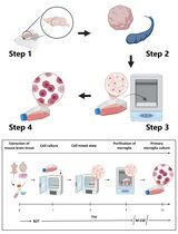
Keywords: Prostate cancerGraphical overview
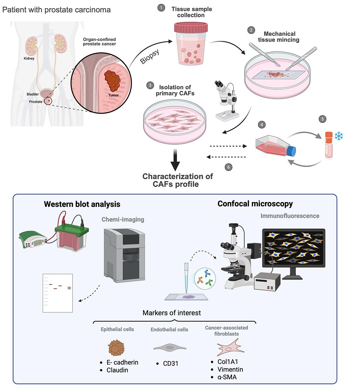
Workflow for cancer-associated fibroblasts (CAFs) isolation
Background
Prostate carcinoma (PCa) arises from the malignant transformation of epithelial cells residing within the prostatic acini. Although the genetic background of transformed cells is crucial for tumor initiation [1], increasing evidence indicates that PCa progression is strongly influenced by interactions with both resident and recruited cell populations of the surrounding microenvironment [2]. Under physiological conditions, the prostate stroma is composed of several cell types, including fibroblasts, endothelial cells, and a small number of immune cells. When stromal homeostasis is disrupted by PCa onset, many of these cells become “educated” by cancer cells, acquiring pro-tumorigenic features. Among pro-tumoral stromal components, cancer-associated fibroblasts (CAFs) constitute the predominant population across different PCa stages [3]. Recently, distinct CAF phenotypic subtypes, including myofibroblastic CAFs (myCAFs) and/or inflammatory CAFs (iCAFs), have been identified according to their specific expression profiles and spatial localization/interactions within the tumor mass [4–6]. Briefly, the dynamic crosstalk with the neighboring tumor cells results in profound phenotypic, transcriptional, and functional alterations compared with the normal fibroblasts populating the non-cancerous areas. In particular, tumor-induced activation of normal fibroblasts into a myCAF population results in higher contractility, primarily supported by increased expression of the alpha-smooth muscle actin (α-SMA) [7] and a metabolic shift toward enhanced glycolysis [8]. These CAF features are crucial to establish a crosstalk with epithelial tumor cells, ultimately promoting enhanced cell invasion, immune evasion, and resistance to therapy insults, key processes driving metastatic progression of tumors, including PCa [9–11].
Given the key role of CAFs in supporting PCa progression, developing in vitro models that accurately recapitulate CAF-tumor interactions is essential for research. The absence of cell lines recapitulating the CAF phenotype represents a limitation in the study of CAF pathobiology. In this regard, the establishment of CAF cultures ex vivo is an actual need, as it provides a physiologically relevant model to study this crosstalk in vitro. Indeed, fibroblast standard isolation procedures are mainly based on enzymatic dissociation procedures, followed by flow cytometry cell sorting or magnetic bead–based isolation [12, 13]. However, while these procedures are likely proper for single-cell transcriptomics analysis, they show restricted applicability for cell culture, as they may alter the phenotype of the isolated cells due to the exposure to enzymatic cocktails and to prolonged maintenance in non-adherent conditions. For a proper isolation of CAFs for cell culture applications, we optimized a protocol for mechanically isolating myCAFs from minced human fresh PCa biopsies by exploiting the intrinsic motility of these cells toward a nutrient-rich environment. This approach has the advantage of preserving tissue architecture, maintaining CAF viability and intrinsic phenotypic characteristics, providing a more physiologically relevant model for in vitro studies. Here, we provide a step-by-step procedure to selectively separate activated fibroblasts (i.e., myCAFs) from fresh human PCa biopsies; we also describe validation methods to assess the purity of the resulting primary cultures, as well as the acquisition of phenotypic and metabolic CAF-specific markers compared to normal fibroblasts (e.g., derived from benign prostatic hyperplasia). Applying the protocol described here allows the preparation of ex vivo fibroblast populations suitable for studying the mechanisms underlying fibroblast activation and for elucidating their role in the tumor microenvironment, especially focusing on PCa. These primary CAFs can be co-cultured with tumor cells, allowing a more faithful recapitulation of tumor physiology in vitro.
Materials and reagents
Biological materials
1. Human surgical explants from patients affected by benign prostatic hyperplasia (BPH) for (not-activated) healthy prostate fibroblasts (HPF) isolation
2. Human surgical explants from patients subjected to surgical intervention for prostate cancer (Gleason Group > 3) for CAFs isolation
3. Human epithelial PCa cell line DU145 (RRID: CVCL 0105) obtained from ATCC and routinely tested for Mycoplasma contamination using the MycoAlert Mycoplasma Detection kit (Lonza, #LOLT07710)
4. Endothelial colony-forming cells (ECFCs) isolated from umbilical cord blood
Notes: DU145 cells and ECFCs can be replaced with other similar commercially available cell lines to be used as epithelial and endothelial controls, respectively.
Reagents
1. Physiological saline solution, NaCl, 0.9% (B. Braun, catalog number: 030902391)
2. Phenol-red high glucose Dulbecco’s modified Eagle’s medium (DMEM) (Euroclone, catalog number: ECB7501L)
3. Fetal bovine serum (FBS) (Euroclone, catalog number: ECS5000L)
4. L-Glutamine (Merck Sigma, catalog number: G7513-100 ML)
5. 10,000 units penicillin/10 mg streptomycin solution in 0.9% (Merck Sigma, catalog number: P0781-100 ML)
6. Kanamycin (Merck, catalog number: 246933-9)
7. Amphotericin B (Fungizone) (Euroclone, catalog number: ECM0009D)
8. Trypsin (Sigma-Aldrich, catalog number: T4049)
9. Dimethyl sulfoxide (DMSO) (Sigma-Aldrich, catalog number: 472301)
10. RIPA lysis buffer (Thermo Fisher Scientific, catalog number: 8990)
11. Protease inhibitors (Sigma-Aldrich, catalog number: P8340)
12. Phosphatase inhibitors (Sigma-Aldrich, catalog number: P0044)
13. BCA Protein Assay kit (Sigma-Aldrich, catalog number: 1003579336)
14. 4%–20% acrylamide precast SDS-PAGE gels (Bio-Rad, catalog numbers: 4568093 and 4568096)
15. PVDF membranes (Bio-Rad, catalog number: 1704157)
16. Phosphate buffered saline (PBS) (Euroclone, catalog number: ECB4004L)
17. Tween 20 (Sigma-Aldrich, catalog number: P1379)
18. Non-fat dry milk (Regilait Ecreme 750gr)
19. Primary antibodies (Table 1)
Table 1. Primary antibodies list
| Target | Catalog number | Dilution | Secondary antibodies |
|---|---|---|---|
| E-Cadherin | Cell Signaling Technology, 24E10 | 1:1,000 | Rabbit |
| Claudin | Cell Signaling Technology, D5H1C | 1:1,000 | Rabbit |
| CD31 | Cell Signaling Technology, 89C2 | 1:1,000 | Mouse |
| Col1a1 | Cell Signaling Technology, 72026 | 1:1,000 | Rabbit |
| Vimentin | Cell Signaling Technology, D21H3 | 1:1,000 | Rabbit |
| Alpha-SMA | Merck Sigma, SAB5700835 | 1:1,000 | Mouse |
| LDHA | Santa Cruz Biotechnology, D0220 | 1:1,000 | Mouse |
| MCT4 | Santa Cruz Biotechnology, 376140 | 1:1,000 | Mouse |
| HSP90 | Santa Cruz Biotechnology, sc-69703 | 1:1,000 | Mouse |
20. Secondary antibodies: Anti-rabbit HRP (Santa Cruz, catalog number: sc-2357) and anti-mouse HRP (Santa Cruz, catalog number: sc-516102)
21. ECL substrates: Clarity Western ECL (Bio-Rad, catalog number: 1705061) or Clarity Max ECL (Bio-Rad, catalog number: 1705062)
22. Formaldehyde (Sigma-Aldrich, catalog number: 252549-1L)
23. Methanol (Sigma-Aldrich, catalog number: 34860-2.5L)
24. DAPI (Thermo Fisher Scientific, catalog number: D3571)
25. Triton X-100 (Sigma-Aldrich, catalog number: T8787-250ML)
26. Alexa Fluor-488 rabbit secondary antibody (1:1,000) (Thermo Fisher Scientific, catalog number: A-11008)
27. α-SMA antibody for immunofluorescence analysis (1:100) (Abcam, catalog number: ab5694)
28. Glass mountant Pro-Long (Invitrogen, catalog number: P36980)
29. Horse serum (Euroclone, catalog number: ECS0091L)
Solutions
1. Starvation medium (see Recipes)
2. Complete medium (see Recipes)
3. Freezing medium (see Recipes)
4. Non-fat dry milk (see Recipes)
Recipes
1. Starvation medium
| Reagent | Final concentration | Quantity or volume |
|---|---|---|
| DMEM high glucose | - | 400 mL |
| Penicillin-streptomycin (stock 100×) | 2× | 10 mL |
| L-Glutamine (stock 100×) | 2 mM | 5 mL |
2. Complete medium
| Reagent | Final concentration | Quantity or volume |
|---|---|---|
| DMEM high glucose | - | 400 mL |
| FBS | 20% (v/v) | 100 mL |
| Penicillin-streptomycin (stock 100×) | 2× | 10 mL |
| L-Glutamine (stock 100×) | 2 mM | 5 mL |
| Kanamycin (stock 100×) | 100 μg/mL | 5 mL |
| Fungizone (Amphotericin B stock 100×) | 2.5 μg/mL | 5 mL |
3. Freezing medium
| Reagent | Final concentration | Quantity or volume |
|---|---|---|
| FBS | 90% (v/v) | 9 mL |
| DMSO | 10% (v/v) | 1 mL |
4. Non-fat dry milk
| Reagent | Final concentration | Quantity or volume |
|---|---|---|
| Non-fat dry milk | 5% (w/v) | 5 g |
| T-PBS (PBS 1× + Tween 20 1%) | - | 100 mL |
Laboratory supplies
1. Cell culture plates/dishes [Euroclone, catalog numbers: ET2100 (p100) and ET2060 (p60)]
2. Tubes [Euroclone, catalog numbers: ET5015B (15 mL) and ET5050B (50 mL)]
3. Microcentrifuge (Euroclone catalog number: ET3415)
4. Pipettes (micropipettes, serological pipettes) [Euroclone, catalog numbers: EPS02N (2 mL), EPS05N (5 mL), EPS10N (10 mL), and EPS25N (25 mL)]; pipette controller Primo® Mate (Euroclone, catalog number: ECP2000); micropipettes P10, P20, P100, P200, and P1000 (Gilson MyPipetman); micropipette tips [Gilson, catalog numbers: DF10ST (0.1–20 μL), DF200ST (2–200 μL), and DF1000ST (100–1,000 μL)]
5. Glass coverslips (BioSigma, catalog number: VBS636)
Equipment
1. Tweezers (DIMART, catalog number: DIM.2120/14) sterilized in an oven at 180 °C for 3–4 h
2. Sterile scalpel (Feather, catalog number: 530050)
3. Microscope slides (BioSigma, catalog number: VBS654), individually packed in aluminum and sterilized as above
4. Laminar flow hood (CELLBIO, model: MARS)
5. CO2 cell culture incubator (Thermo Scientific, model: Forma Direct Heat)
6. Refrigerated centrifuge (Sigma, model: 1-14K)
7. Inverted microscope (Leica Microsystem, model: DMi1)
8. SDS-PAGE gel tank (Bio-Rad, model: Mini-PROTEAN Tetra System) and power supply (Bio-Rad, model: PowerPAC Basic)
9. Trans-Blot Unit (Bio-Rad, model: Trans-Blot Turbo)
10. Confocal microscope (Leica Microsystems, model: TCS SP8)
11. ChemiDoc MP imaging system (Bio-Rad)
12. 4 °C fridge (Gorenje), -20 °C freezer (Gorenje), and -80 °C freezer
13. Liquid nitrogen tank
Software and datasets
1. LAS-AF image acquisition software (Leica Microsystems)
Procedure
文章信息
稿件历史记录
提交日期: Dec 1, 2025
接收日期: Jan 22, 2026
在线发布日期: Feb 6, 2026
出版日期: Mar 5, 2026
版权信息
© 2026 The Author(s); This is an open access article under the CC BY-NC license (https://creativecommons.org/licenses/by-nc/4.0/).
如何引用
Gangarossa, G., Grillo, C., Roccabianca, S., Pranzini, E., Iozzo, M., Venditti, G., Bertoli, G., Ippolito, L., Giannoni, E., Comito, G. and Chiarugi, P. (2026). Non-Enzymatic Isolation of Cancer-Associated Fibroblasts From Human Prostate Tumor Explants. Bio-protocol 16(5): e5614. DOI: 10.21769/BioProtoc.5614.
分类
癌症生物学 > 通用技术 > 肿瘤微环境
细胞生物学 > 细胞分离和培养 > 细胞分离
您对这篇实验方法有问题吗?
在此处发布您的问题,我们将邀请本文作者来回答。同时,我们会将您的问题发布到Bio-protocol Exchange,以便寻求社区成员的帮助。