

Quick and Cheap: Optimized Purification and Concentration of Bacteriophages Produced in Rich Culture Media
快速又省钱:富营养培养基中噬菌体的优化纯化与浓缩方法

发布: 2026年02月20日第16卷第4期 DOI: 10.21769/BioProtoc.5608 浏览次数: 57
评审: Anonymous reviewer(s)


Abstract
This protocol describes an easy, quick, cheap, and effective method for the purification and concentration of bacteriophages (phages) produced in rich culture media, meeting the quality criteria required for structural analyses. It is based on a tube dialysis system that replaces the classical but expensive and tedious density gradient ultracentrifugation step. We developed this protocol for the Oenococcus oeni bacteriophage OE33PA from its amplification to imaging by negative stain electron microscopy (NS-EM). The host bacterium, O. oeni, is a lactic acid bacterium that lives in harsh oenological ecosystems and grows only in rich and complex media such as Man–Rogosa–Sharpe (MRS) or fruit juice-based media in laboratory conditions. This raises experimental challenges in pure and concentrated phage preparations for further uses such as structure-function studies.
Key features
• Simple, rapid, and cheap, this method provides a fast and easy approach for efficient bacteriophage purification and concentration.
• The method meets the structural study requirements of phage particles.
• The method is compatible with bacteria that grow only in complex and rich culture media.
• This protocol is also compatible with the purification of phages that produce low-titer lysates.
Keywords: Phage purification (噬菌体纯化)Graphical overview
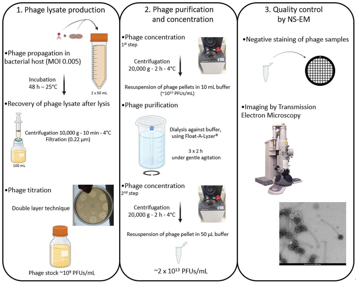
Flowchart illustrating the production, purification, and concentration protocols of phages produced in rich culture media before observation by negative stain electron microscopy (NS-EM). MOI, Multiplicity of infection; PFU, plaque forming unit.
Background
Bacteriophages (phages, viruses of bacteria) are major players in all ecosystems, whether they are natural or anthropic [1]. They affect their bacterial hosts in different ways: they regulate bacterial populations via predation, improve their host fitness (i.e., lysogenic conversion), and are drivers of bacterial evolution [2]. Moreover, given their potential to be used as molecular tools [3], they are increasingly studied in a wide range of disciplines, from medicine to ecology or biotechnology.
Phages, like all viruses, have a “nanoscopic” size and are unable to propagate on their own. However, they can be seen by transmission electron microscopy (TEM), and their propagation can be followed through cell lysis of their host. It is worth pointing out that the existence of phages, as biological entities, was definitively proven by TEM imaging more than 20 years after their discovery [4].
The description of virion structure is part of the basic characterization of phages. Yet, it is not uncommon to come across poor-quality TEM micrographs in the literature, showing phage preparations contaminated by components of the bacterial culture medium. In most studies, bacteriophages are purified using cesium chloride (CsCl) density gradient ultracentrifugation, which separates particles by buoyant density [5]. While this method yields highly pure phage preparations, it is labor-intensive, requires expensive ultracentrifugation equipment, and involves CsCl, which is toxic for operators and poses handling risks. To avoid using this type of gradient, we previously developed a simple protocol for the preparation of the GC1 phage amplified upon infection of its host, the acetic-acid bacterium Gluconobacter cerinus in poor media, which is efficient enough to observe virion ultrastructure using cryo-electron microscopy (cryo-EM) [6,7]. Here, we present an improved protocol for the preparation of phages whose hosts grow in complex and rich media based on the use of a tube dialysis system.
This protocol was developed using the Oenococcus oeni phage OE33PA, an ex-temperate phage (i.e., a mutant only able to perform lytic infections) previously isolated from wine [8]. The host, O. oeni, has a fastidious growth that occurs only in rich culture media such as Man–Rogosa–Sharpe (MRS) or fruit juice-based media, which makes the purification of its phages complicated for later uses, such as structural analyses. In the present article, we show that our optimized protocol is simpler and more efficient than the one we published in 2020 [7] to produce high-quality GC1 phage particles.
Materials and reagents
Biological materials
1. Oenococcus oeni S277 [9]
2. OE33PA phage suspension [8]
Reagents
1. Tris-HCl pH 7.5 (Fisher Scientific, catalog number: 10123722)
2. Sodium chloride (NaCl) (Fisher Scientific, catalog number: 10055850)
3. Magnesium sulfate heptahydrate (MgSO4·7H2O) (Fisher Scientific, catalog number: 10135453)
4. Calcium chloride dihydrate (CaCl2·2H2O) (Fisher Scientific, catalog number: 10158280)
5. Hydrochloric acid (HCl), 33% (w/v) aqueous solution (Fisher Scientific, catalog number: 11331588)
6. Granulated agar (Dutscher, catalog number: 214510)
Note: All above chemicals, which are used in media recipes, are reagent grade (ACS grade).
7. Uranyl acetate dihydrate 98% (Sigma-Aldrich, withdrawn from the market)
8. MRS (Difco, BD, Fisher Scientific, catalog number: 13689811713553)
Solutions
1. Man–Rogosa–Sharpe (MRS) broth (see Recipes)
2. MRS and MRSΦ solid medium (see Recipes)
3. MRSΦ soft agar medium (see Recipes)
4. 10× phage buffer (ΦB) (see Recipes)
Recipes
1. MRS broth
| Reagent | Final concentration | Quantity or volume |
|---|---|---|
| MRS powder | 55 g/L | 55 g |
a. Make up the volume to 1 L with distilled water.
b. Adjust the pH to pH 4.8 with HCl 33.33% (w/v) aqueous solution.
c. When needed, add 1.77 g of CaCl2·2H2O and 1.8 g of MgSO4·7H2O (e.g., for MRSΦ, used for phage propagation).
d. Sterilize by autoclaving.
e. Store at 4 °C for up to 6 months.
2. MRS and MRSΦ solid medium
| Reagent | Final concentration | Quantity or volume |
|---|---|---|
| MRS powder | 55 g/L | 55 g |
| Granulated agar | 20 g/L | 20 g |
a. Make up the volume to 1 L with distilled water.
b. Adjust the pH to pH 4.8 with HCl 33.33% (w/v) aqueous solution.
c. Sterilize by autoclaving.
d. Store at 4 °C for up to 6 months.
3. MRSΦ soft agar medium
| Reagent | Final concentration | Quantity or volume |
|---|---|---|
| MRS powder | 55 g/L | 55 g |
| Granulated agar | 6 g/L | 20 g |
| CaCl2·2H2O | 16 mM | 1.77 g |
| MgSO4·7H2O | 15 mM | 1.8 g |
a. Make up the volume to 1 L with distilled water.
b. Dissolve agar by heating.
c. Aliquot 5 mL of the medium in approximately 200 glass tubes (20 mL).
d. Sterilize by autoclaving.
e. Store at 4 °C for up to 6 months.
4. Phage buffer 10× (ΦB)
| Reagent | Final concentration | Quantity or volume |
|---|---|---|
| NaCl | 1 M | 58 g |
| MgSO4·7H2O | 0.08 M | 20 g |
| 1 M Tris-HCl pH 7.5 | 0.5 M | 500 mL |
| Distilled water | n/a | 500 mL |
| Total | n/a | 1000 mL |
a. Sterilize by autoclaving.
b. Store at room temperature for up to 6 months.
c. To prepare 1× ΦB, dilute 10 mL of 10× ΦB with 90 mL of sterile distilled water.
Laboratory supplies
1. 50 mL centrifuge tubes (Dutscher, catalog number: 352070)
2. 100 mL SHOTT bottle (Dutscher, catalog number: 090441A)
3. 250 mL SHOTT bottle (Dutscher, catalog number: 090346)
4. 1.6 mL spectrophotometer cuvettes (Dutscher, catalog number: 030101)
5. 0.22 μm PES membrane syringe filters (Fisher Scientific, catalog number: SLGP033NK)
6. 50 mL sterile disposable plastic syringes (Fisher Scientific, catalog number: 10119350)
7. 1.5 mL polypropylene microcentrifuge tubes (Fisher Scientific, catalog number: 10154671)
8. Sterile 90 × 13 mm Petri dishes (Dutscher, catalog number: 076084E)
9. 20 mL autoclavable glass tubes (Dutscher, catalog number: 045209)
10. Float-A-Lyzer® G2 100 kDa 10 mL (Fisher Scientific, catalog number: 11561170)
11. 300 mesh 3 mm carbon-coated copper grids (TAAB, catalog number: C267/100)
12. Whatman filter paper grade 5 (Dutscher, catalog number: 1005055)
Equipment
1. Spectrophotometer (Shimadzu, model: UV-1280)
2. Incubator set to 25 °C
3. Water bath set to 55 °C
4. High-speed refrigerated centrifuge (Hitachi, model: CR 22N) and R15A-0688 rotor
5. pH meter (Hanna, model: pH211)
6. Magnetic stirrer
7. Glow discharge unit (Electron Microscopy Sciences, model: GloQube Plus)
8. Transmission electron microscope (Tecnai, model: G2 Spirit T12 TEM)
9. CCD camera (Olympus, Germany, model: Veleta 2k × 2k Side-Mounted TEM CCD Camera Solution)
Procedure
文章信息
稿件历史记录
提交日期: Nov 1, 2025
接收日期: Jan 16, 2026
在线发布日期: Feb 11, 2026
出版日期: Feb 20, 2026
版权信息
© 2026 The Author(s); This is an open access article under the CC BY license (https://creativecommons.org/licenses/by/4.0/).
如何引用
Chaïb, A., Schmitt, L., Goulet, A. and Le Marrec, C. (2026). Quick and Cheap: Optimized Purification and Concentration of Bacteriophages Produced in Rich Culture Media. Bio-protocol 16(4): e5608. DOI: 10.21769/BioProtoc.5608.
分类
微生物学 > 体内实验模型 > 病毒
生物化学 > 病毒 > 分离和纯化
细胞生物学 > 细胞分离和培养 > 病毒分离
您对这篇实验方法有问题吗?
在此处发布您的问题,我们将邀请本文作者来回答。同时,我们会将您的问题发布到Bio-protocol Exchange,以便寻求社区成员的帮助。