

Quantitative Proteomics of Nitrosylated Proteins in Melanoma Using the Biotin-Switch Technique Combined With Tandem Mass Tag Labeling
结合生物素开关技术与 TMT 标记的黑色素瘤蛋白硝基化定量蛋白组学分析
发布: 2025年12月05日第15卷第23期 DOI: 10.21769/BioProtoc.5535 浏览次数: 1619
评审: Willy R Carrasquel-UrsulaezRamya VisvanathanAnonymous reviewer(s)
参见作者原研究论文
The authors used this protocol in:

Jun 2025
Advertisement


Abstract
Protein S-nitrosylation is a critical post-translational modification that regulates diverse cellular functions and signaling pathways. Although various biochemical methods have been developed to detect S-nitrosylated proteins, many suffer from limited specificity and sensitivity. Here, we describe a robust protocol that combines a modified biotin-switch technique (BST) with streptavidin-based affinity enrichment and quantitative mass spectrometry to detect and profile nitrosylated proteins in cultured cells. The method involves blocking free thiols, selective reduction of nitrosothiols, biotin labeling, enrichment of biotinylated proteins, and identification by tandem mass tag (TMT)-based quantitative mass spectrometry. Additionally, site-directed mutagenesis is employed to generate “non-nitrosylable” mutants for functional validation of specific nitrosylation sites. This protocol provides high specificity, quantitative capability, and versatility for both targeted and global analysis of protein nitrosylation.
Key features
• Specific thiol blocking and labeling: Free thiols are blocked with N-ethylmaleimide, followed by selective reduction and biotinylation of S-nitrosothiols for precise nitrosylation detection.
• Quantitative proteomics: TMT-labeling with high-resolution LC-MS/MS enables multiplexed, accurate quantification and comprehensive nitrosylome profiling with faster data acquisition and fewer missing values than label-free proteomics.
• Functional mutagenesis: Site-directed mutagenesis of cysteine residues generates “non-nitrosylable” mutants to study nitrosylation’s impact on protein function.
• Versatile application: The protocol is adaptable for both targeted protein analysis and global nitrosylation profiling across diverse cell types and experimental conditions.
Keywords: Protein S-nitrosylation (蛋白 S-硝基化)Graphical overview

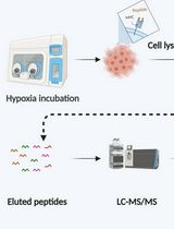
Schematic overview of the biotin switch assay for detection of protein S-nitrosylation. The biotin switch technique (BST) selectively detects S-nitrosylated (SNO) cysteine residues in proteins. Free thiol groups (–SH) are first blocked with N-ethylmaleimide (NEM) to prevent nonspecific labeling. Next, ascorbate selectively reduces S-nitrosothiols (SNO) to free thiols, which are then labeled with biotin-HPDP (N-[6-(biotinamido) hexyl]-3’-(2’-pyridyldithio) propionamide) forming stable disulfide bonds. Biotinylated proteins can be detected either directly by SDS-PAGE (20 μg protein/lane) and immunoblotting using anti-biotin antibodies (1:1,000) and anti-GAPDH (1:3,000, MW: 37 kDa) or enriched by streptavidin bead pulldown for further analysis. In this representative blot, YUGASP cells were pretreated with NOSi (1 mM L-NAME + 200 μM 1400 w acetate) and GSNO (250 μM) for 16 h before lysate preparation and BST procedure.
Background
Snitrosylation, the reversible covalent attachment of nitric oxide (NO) to cysteine thiols (–SH), forming Snitrosothiols (SNO), is a fundamental post-translational modification that regulates protein conformation, activity, localization, and interactions across cellular signaling networks [1]. It influences processes such as mitochondrial quality control, neuroprotection, and vascular tone, and dysregulated Snitrosylation is implicated in diseases including cardiovascular disorders, neurodegeneration, and cancer [2,3]. However, studying Snitrosylation is technically demanding: SNO bonds are labile and exist in low abundance, complicating direct detection. To address this, Jaffrey and Snyder introduced the biotin-switch technique (BST) in 2001. BST employs a three-step workflow: (1) irreversible blocking of free thiols [e.g., with N-ethylmaleimide or MMTS (S-methyl methanethiosulfonate)]; (2) selective reduction of SNO to free thiols using ascorbate; and (3) biotinylation of the newly exposed thiols with biotinHPDP, enabling detection by western blot or affinity enrichment [4,5]. Following its introduction, BST became widely adopted and continues to be the gold standard for both targeted and global Snitrosoproteome analysis. Variants such as SNO-SID, SNORAC, and iodoTMTswitch methods have further enhanced sensitivity and resolution, enabling site-specific identification via mass spectrometry. Complementing these experimental advances, computational tools like GPSSNO 1.0 offer predictive insights for potential Snitrosylation sites. Trained on hundreds of experimentally validated cysteines, GPSSNO achieves approximately 76% accuracy and 54% sensitivity, aiding targeted mutagenesis strategies to confirm functional roles of specific SNO sites [6]. Our current protocol integrates the classic BST with streptavidin-based protein and peptide enrichment, TMT-labeled LC–MS/MS quantitative proteomics, and GPSSNO guided site-directed mutagenesis to generate “nonnitrosylable” protein variants. This combination delivers specificity, quantitative depth, and functional validation, enabling comprehensive investigation of nitrosylation-mediated regulation in diverse cellular systems.
Materials and reagents
Biological materials
1. WM-1366 melanoma cells
2. YUDOSO, YUTICA, and YUGASP patient-derived melanoma cells
Reagents
1. RPMI 1640 medium (Thermo Fisher Scientific, catalog number: 11875-093)
2. FBS (Atlas Biologicals, catalog number: F-0500D)
3. Penicillin-Streptomycin (Thermo Fisher Scientific, catalog number: 15140-122)
4. DPBS (Thermo Fisher Scientific, catalog number: 14190144)
5. Protease inhibitor cocktail (PIC) (Thermo Fisher Scientific, catalog number: 78429)
6. Phosphatase inhibitor cocktail (PHIC) (Thermo Fisher Scientific, catalog number: 78420)
7. HENS buffer, pH ≤ 6.5 (Thermo Fisher Scientific, catalog number: 90106)
8. HEPES solution (Millipore, catalog number: H0887)
9. S-nitroso-L-glutathione (GSNO) (Cayman Chemicals, catalog number: 82240)
10. Diethylenetriamine/DETA-NONOate (Cayman Chemicals, catalog number: 82120)
11. BCA Protein Assay kit (Thermo Fisher Scientific, catalog number: 23227)
12. N-Ethylmaleimide (NEM) (Millipore, catalog number: E1271)
13. Acetone (Sigma, catalog number: 179124)
14. NaCl (Millipore, catalog number: S9888)
15. EDTA (Millipore, catalog number: E4884)
16. L-NAME (Tocris, catalog number: 0665)
17. 1400W dihydrochloride (Selleckchem, catalog number: S8337)
18. L-NIO (Cayman Chemical, catalog number: 80320)
19. L-NIL (Cayman Chemical, catalog number: 80310)
20. CPTIO (Cayman Chemical, catalog number: 81540)
21. Ultrapure water (Millipore, catalog number: DC01L-CS)
22. Biotin-HPDP (Cayman Chemicals, catalog number: 16459)
23. Sodium ascorbate (Sigma, catalog number: PHR1279)
24. Laemmli buffer (Bio-Rad, catalog number: 1610747)
25. Pre-cast 4%–20% SDS-PAGE gels (Bio-Rad, catalog number: 4561091)
26. PVDF membranes (Bio-Rad, catalog number: 162-0184)
27. Non-fat dry milk (Millipore, catalog number: MB9696)
28. 20× TBS buffer (Millipore, catalog number: 20845-M)
29. Tween-20 (Millipore, catalog number: P9416)
30. Triton X-100 (Millipore, catalog number: T8787)
31. HRP-tagged anti-biotin antibodies (Thermo Fisher Scientific, catalog number: 03-3720)
32. Streptavidin magnetic beads (Promega, catalog number: Z548C)
33. Streptavidin agarose resin (Thermo Fisher Scientific, catalog number: 20359)
34. Super Signal West Pico Chemiluminescent Substrate (Thermo Fisher Scientific, catalog number: 34080)
35. Total ERK (Cell Signaling Technology, catalog number: 9102S)
36. DUSP4 (Cell Signaling Technology, catalog number: 5149S)
37. RhoC (Cell Signaling Technology, catalog number: 3430S)
38. RSK-1 (Cell Signaling Technology, catalog number: 8408S)
39. GAPDH (Cell Signaling Technology, catalog number: 2118)
40. RASGAP (Proteintech, catalog number: 12935-1-AP)
41. 2-Mercaptoethanol (Millipore, catalog number: M3148)
42. LCMS-grade water (Millipore, catalog number: 1.15333)
43. Dithiothreitol (DTT) (Millipore, catalog number: 3483-12-3)
44. SOLAμTM SPE plates (Thermo Fisher Scientific, catalog number: 60209-001)
45. Trifluoroacetic acid (Millipore, catalog number: 900518)
46. Acetonitrile (ACN) (Millipore, catalog number: 271004)
47. Ammonium bicarbonate (Millipore, catalog number: A6141)
48. Iodoacetic acid (Millipore, catalog number: I4386)
49. Trypsin gold, mass spectrometry grade (Promega, catalog number: V5280)
50. Tandem mass tag (TMT) systems (Thermo Fisher Scientific, catalog number: A40000839)
51. Reversed-phase peptide fractionation cartridge (Thermo Fisher Scientific, catalog number: 84868)
52. Standard human cell line tryptic digest peptide mixture (Promega, catalog number: V6951)
53. Stable isotope-labeled standard peptides (pierce retention time calibration peptide mixture, PRTC) (Thermo Fisher Scientific, catalog number: 88320)
54. Formic acid (Millipore, catalog number: F0507)
Solutions
1. Ascorbate solution (see Recipes)
2. NEM solution (see Recipes)
3. Biotin-HPDP stock (see Recipes)
4. GSNO solution (see Recipes)
5. Neutralization buffer (see Recipes)
6. Wash buffer (see Recipes)
7. Elution buffer (see Recipes)
Recipes
1. Ascorbate solution
Prepare a fresh 50 mM solution of sodium ascorbate in deionized water.
2. NEM solution
Prepare a fresh 400 mM solution of NEM in ethanol.
3. Biotin-HPDP stock
Prepare biotin-HPDP as a 50 mM suspension in DMSO. Freeze at -20 °C until needed.
4. GSNO solution
Prepare a fresh 10 mM GSNO stock solution in PBS, protected from light.
5. Neutralization buffer
| Reagent | Final concentration | Quantity or volume |
|---|---|---|
| HEPES (pH 7.7) | 20 mM | 10 mL |
| NaCl | 150 mM | 4.383 g |
| EDTA | 1 mM | 146.12 mg |
| Triton X-100 | 0.5% | 2.5 mL |
| Water (to make up total volume) | n/a | 500 mL |
6. Wash buffer
| Reagent | Final concentration | Quantity or volume |
|---|---|---|
| HEPES (pH 7.7) | 20 mM | 10 mL |
| NaCl | 600 mM | 17.532 g |
| EDTA | 1 mM | 146.12 mg |
| Triton X-100 | 0.5% | 2.5 mL |
| Water (to make up total volume) | n/a | 500 mL |
7. Elution buffer
| Reagent | Final concentration | Quantity or volume |
|---|---|---|
| HEPES (pH 7.7) | 20 mM | 2 mL |
| NaCl | 100 mM | 584.4 mg |
| EDTA | 1 mM | 29.224 mg |
| β-mercaptoethanol | 5% | 5 mL |
| Water (to make up total volume) | n/a | 100 mL |
8. TBST (Tris-buffered saline and Tween 20)
| Reagent | Final concentration | Quantity or volume |
|---|---|---|
| Tris | 20 mM | 24.23 g |
| NaCl | 150 mM | 80.06 g |
| Tween 20 | 0.1% | 1 mL |
| Water (to make up total volume) | n/a | 1,000 mL |
Laboratory supplies
1. 100 × 20 mm cell culture dishes (Thermo Fisher Scientific, catalog number: 174903)
2. 25 mL sterile reservoirs (Thermo Fisher Scientific, catalog number: 95128095)
3. Cell scrapers (Thermo Fisher Scientific, catalog number: 179707PK)
4. 15 mL conical tubes (Greiner Bio-One, catalog number: 188261)
5. 50 mL conical tubes (Greiner Bio-One, catalog number: 227261)
6. 5 mL serological pipettes (Greiner Bio-One, catalog number: 606160)
7. 10 mL serological pipettes (Greiner Bio-One, catalog number: 607160)
8. 25 mL serological pipettes (Greiner Bio-One, catalog number: 760160)
9. 1.5 mL microcentrifuge tubes (Greiner Bio-One, catalog number: 616201)
10. Countess cell counting chamber slides (Thermo Fisher Scientific, catalog number: C10283)
Equipment
1. Sonicator (20% amplitude) (Thermo Fisher Scientific, catalog number: FB120110)
2. Heat block (Fisher Scientific, catalog number: 11-686-731)
3. Vortex mixer (Fisher Scientific, catalog number: 02-215-414)
4. UV-visible spectrophotometer (Fisher Scientific, catalog number: NC2027422)
5. BioTek microplate reader (Agilent Technologies, model: Synergy Neo2)
6. Class II biological safety cabinet (Baker, model: SterilGARD® e3)
7. Tissue culture incubator (Fisher Scientific, catalog number: 13-998-124)
8. Inverted microscope (Fisher Scientific, catalog number: LMI3PH4)
9. Tabletop microcentrifuge with rotors for 1.5 mL tubes (Eppendorf, model: 5425/5425 R)
10. Tabletop centrifuge with rotors for 15 mL and 50 mL tubes (Eppendorf, model: 5804/5804 R)
11. Water bath (37 °C) (Fisher Scientific, catalog number: FSSWB27)
12. Countess II FL automated cell counter (Fisher Scientific, model: AMQAF2000)
13. Freezer (-20 °C and -80 °C)
14. SDS-PAGE apparatus (Bio-Rad Mini-PROTEAN Tetra system, catalog number: 1658000)
15. ChemiDoc imaging system (Bio-Rad, model: ChemiDocTM Touch Imaging System)
16. Positive pressure-96 processor (Waters, Milford, MA, catalog number: 186006961)
17. Nanoflow ultra-high-performance liquid chromatograph (RSLCnano, Thermo, San Jose, CA)
18. Electrospray benchtop hybrid quadrupole orbitrap mass spectrometer (Q Exactive HF-X, Thermo, San Jose, CA)
19. SpeedVac, vacuum concentrator (Thermo Scientific, catalog number: 13-875-333)
Software and datasets
1. ImageJ (https://imagej.net/ij/)
2. GraphPad Prism (Dotmatics, https://www.graphpad.com/)
3. Thermo XCalibur or vendor equivalent
4. Skyline for Internal Standard Quantification (https://skyline.ms/project/home/software/Skyline/begin.view) [7].
5. MaxQuant for Database Searching and Quantification (https://maxquant.org/) [8].
6. Mascot Database Searching (Matrix Science)
7. Scaffold (Proteome Software)
Procedure
文章信息
稿件历史记录
提交日期: Aug 4, 2025
接收日期: Oct 19, 2025
在线发布日期: Dec 2, 2025
出版日期: Dec 5, 2025
版权信息
© 2025 The Author(s); This is an open access article under the CC BY-NC license (https://creativecommons.org/licenses/by-nc/4.0/).
如何引用
Yadav, V. K., Fang, B., Koomen, J. M., Srivastava, J. and Premi, S. (2025). Quantitative Proteomics of Nitrosylated Proteins in Melanoma Using the Biotin-Switch Technique Combined With Tandem Mass Tag Labeling. Bio-protocol 15(23): e5535. DOI: 10.21769/BioProtoc.5535.
分类
生物化学 > 蛋白质 > 定量
系统生物学 > 蛋白质组学
生物科学 > 生物技术 > 质谱
您对这篇实验方法有问题吗?
在此处发布您的问题,我们将邀请本文作者来回答。同时,我们会将您的问题发布到Bio-protocol Exchange,以便寻求社区成员的帮助。