

Selective Enrichment and Identification of Cerebrospinal Fluid-Contacting Neurons In Vitro via PKD2L1 Promoter-Driven Lentiviral System
基于 PKD2L1 启动子驱动的慢病毒系统在体外选择性富集与鉴定脑脊液接触神经元
(*contributed equally to this work) 发布: 2025年11月20日第15卷第22期 DOI: 10.21769/BioProtoc.5518 浏览次数: 1343
评审: Oneil Girish BhalalaBaeksun KimAnonymous reviewer(s)
参见作者原研究论文
The authors used this protocol in:

Mar 2025


Abstract
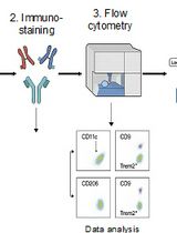
Cerebrospinal fluid-contacting neurons (CSF-cNs) are a specialized group of multifunctional neurons located around the central canal of the spinal cord. They play critical roles in motor regulation, postural maintenance, and spinal cord injury repair. However, the molecular mechanisms underlying the multifunctionality of CSF-cNs remain poorly understood, partly due to the lack of established in vitro methods for their efficient selection and purification, which significantly hinders mechanistic investigations. In this study, we describe a standardized method using a PKD2L1 promoter-driven lentiviral system, which enables effective enrichment and identification of CSF-cNs in vitro through GFP labeling and puromycin selection. This protocol includes key steps such as construction of the PKD2L1 promoter-driven lentiviral vector, spinal cord tissue collection and digestion from neonatal mice, lentiviral infection, antibiotic selection, and immunofluorescence-based identification of CSF-cNs. Our method provides a reliable platform for obtaining high-purity CSF-cNs (>99%), which facilitates their functional and mechanistic studies for regenerative approaches in vitro.
Key features
• Enables specific labeling and selection of CSF-cNs using a constructed PKD2L1 promoter-driven lentiviral vector.
• Combines GFP-based fluorescence tracing and puromycin selection for efficient dual-mode enrichment of high-purity CSF-cNs.
• Provides a simple and reproducible workflow that facilitates in vitro isolation and validation of CSF-cNs for disease modeling and spinal cord injury repair.
Keywords: Cerebrospinal fluid-contacting neurons (CSF-cNs) (脑脊液接触神经元)Background
Cerebrospinal fluid-contacting neurons (CSF-cNs) are a population of multifunctional neurons located around the central canal of the spinal cord [1]. In addition to regulating motor behavior [2], maintaining posture [3], and contributing to central nervous system homeostasis [4], recent studies have revealed that CSF-cNs possess neural stem cell potential and may participate in tissue repair following spinal cord injury [5,6]. Nevertheless, the molecular mechanisms underlying their multifunctionality remain to be fully elucidated. The lack of standardized and specific in vitro methods for selective enrichment remains a major obstacle to comprehensive studies on the functional and regulatory features of these neurons.
Polycystic kidney disease 2-like 1 (PKD2L1), a calcium-permeable channel protein, is widely recognized as a specific molecular marker for CSF-cNs [1,7]. Its promoter has been successfully used in various model organisms to label and trace CSF-cNs in vivo [8–10]. In vitro, CSF-cNs can be obtained in transgenic mice through PKD2L1 promoter–driven fluorescent labeling combined with FACS sorting [11,12]. However, FACS-based methods often suffer from low post-sorting survival rates and limited expansion capacity [13,14], thereby restricting their generalizability and standardization. Therefore, it remains necessary to establish a standardized in vitro method for the selective enrichment and purification of CSF-cNs based on PKD2L1 expression.
In this study, we developed a lentiviral system driven by the mouse PKD2L1 promoter for efficient in vitro selection of CSF-cNs. This system incorporates dual reporter elements—green fluorescent protein (GFP) and puromycin resistance gene (PuroR)—enabling both fluorescence-based tracing and antibiotic-based selection. By infecting primary spinal cord cells with the virus, followed by puromycin selection and enrichment of GFP-positive cells, we obtained a high-purity CSF-cNs population. This system enables the acquisition of highly purified CSF-cNs under controlled conditions, thereby facilitating targeted interventions and pathway analyses, and providing a reliable platform for functional and mechanistic studies in vitro.
Materials and reagents
Biological materials
1. Neonatal C57BL/6 mice (postnatal day 1, Laboratory Animal Center, Guizhou Medical University)
2. PKD2L1-GFP-puromycin-luciferase lentivirus (lab-constructed)
3. psPAX2 (Addgene plasmid #12260; full sequence available at https://www.addgene.org/12260/)
4. pMD2.G (Addgene plasmid #12259; full sequence available at https://www.addgene.org/12259/)
5. HEK293T cells (ATCC, catalog number: CRL-11268; passage number 5 was used in this study)
Reagents
1. Papain (Sigma-Aldrich, catalog number: P3125); store at -20 °C
2. NeurobasalTM-A medium (Gibco, catalog number: 10888022); store at 4 °C
3. B-27TM supplement (Gibco, catalog number: 17504044); store at -20 °C
4. L-Glutamine (Gibco, catalog number: 25030081); store at -20 °C
5. Heparin, 0.2% solution (Sigma, catalog number: 07980); store at -20 °C
6. Penicillin-streptomycin solution (Gibco, catalog number: 15140122); store at 4 °C
7. Phosphate-buffered saline (PBS) (Gibco, catalog number: 10010023); store at room temperature
8. Accutase cell detachment solution (Innovative Cell Technologies, catalog number: AT104); store at 4 °C
9. Puromycin (Sigma-Aldrich, catalog number: P8833); store at -20 °C
10. Opti-MEMTM reduced serum medium (Gibco, catalog number: 31985070); store at 4 °C
11. Lipofectamine 3000 Transfection Reagent (Thermo Fisher Scientific, catalog number: L3000008; includes P3000 reagent); store at 4 °C
12. Poly-D-lysine (Sigma-Aldrich, catalog number: P7280); store at -20 °C
13. Paraformaldehyde, 4% solution (Sigma-Aldrich, catalog number: 158127); store at 4 °C
Growth factors
1. Recombinant human EGF (PeproTech, catalog number: AF-100-15); store at -20 °C, working concentration 20 ng/mL
2. Recombinant human bFGF (PeproTech, catalog number: AF-100-18B); store at -20 °C, working concentration 20 ng/mL
Immunostaining reagents
1. Primary antibody against PKD2L1 (Millipore, catalog number: AB9084); dilution 1:700, store at -20 °C
2. Anti-GFP primary antibody (mouse monoclonal) (Proteintech, catalog number: 66002-1-Ig); dilution 1:500, store at -20 °C
3. Goat anti-mouse IgG (H+L), Alexa FluorTM 488 secondary antibody (Thermo Fisher, catalog number: A11001); dilution 1:500, protect from light, store at -20 °C
4. Goat anti-rabbit IgG (H+L), Alexa FluorTM 594 secondary antibody (Thermo Fisher, catalog number: A11012); dilution 1:500, protect from light, store at -20 °C
5. DAPI staining solution (Solarbio, catalog number: C0065); store at -20 °C
6. Normal donkey serum (Yeasen Biotechnology, catalog number: 36136ES60), used at 5% in PBS for blocking, store at -20 °C, aliquoted
Solutions
1. Dissection medium (see Recipes)
2. Poly-D-lysine (PDL) Solution (see Recipes)
3. PBST washing buffer (see Recipes)
4. Nuclear antibody blocking buffer (for IF) (see Recipes)
5. Membrane antibody blocking buffer (for IF) (see Recipes)
6. DAPI staining solution (see Recipes)
7. Serum-free NSC medium (see Recipes)
Recipes
1. Dissection medium
a. Prepare by mixing DMEM high glucose with 1% L-Glutamine MAX and 5% penicillin-streptomycin.
b. Store at 4 °C for up to one month.
This solution is used for spinal cord tissue dissection and preservation prior to enzymatic digestion.
2. PDL solution
a. Dissolve 10 mg of poly-D-lysine powder in 10 mL of enzyme-free sterile water to obtain a 1 mg/mL stock solution.
b. Filter sterilize through a 0.22 μm membrane and aliquot.
c. Store at -20 °C for up to 6 months.
d. For use, dilute the stock solution tenfold to prepare a 0.1 mg/mL working solution. This working solution is used to coat culture plates and can be reused up to three times if stored properly at 4 °C for no longer than one week.
3. PBST washing buffer
a. Prepare 0.1% PBST by diluting Triton X-100 in PBS.
b. Store at 4 °C for up to one month.
This buffer is used for washing steps during immunofluorescence staining.
4. Nuclear antibody blocking buffer (for IF)
a. Prepare the blocking solution by mixing PBS with 0.5% Triton X-100, 5% BSA, and 22.5 mg/mL glycine.
b. Store at -20 °C for up to 6 months.
This is used to block nonspecific binding during nuclear-targeted immunofluorescence staining.
5. Membrane antibody blocking buffer (for IF)
a. Prepare the blocking solution using PBS supplemented with 0.3% Triton X-100, 5% BSA, and 22.5 mg/mL glycine.
b. Store at -20 °C for up to 6 months.
This is used for blocking in membrane-targeted immunofluorescence staining.
6. DAPI staining solution
a. Prepare DAPI solution by dissolving DAPI powder in PBS to a final concentration of 1 μg/mL.
b. Store at -20 °C for up to 6 months protected from light.
This solution is used for nuclear counterstaining by incubating cells for 5 min at room temperature in the dark.
7. Serum-free NSC medium
a. Mix the following components: Neurobasal-A medium, B27 supplement 2%, L-Glutamine 2 mM, penicillin-streptomycin solution 1%, recombinant human EGF 20 ng/mL, recombinant human bFGF 20 ng/mL, and heparin 0.2% solution.
b. The complete medium can be stored at 4 °C for up to 2 weeks, but freshly prepared medium is recommended to preserve growth factor activity.
Laboratory supplies
1. Ultra-low attachment 6-well plates (Corning, catalog number: 3471); used for suspension culture of CSF-cNs
2. Standard 24-well culture plates (Corning, catalog number: 3526); used for functional assays
3. Standard 96-well culture plates (Corning, catalog number: 3596); used for functional assays
Equipment
1. Class II A2 biosafety cabinet (ESCO)
2. CO2 incubator (Thermo Fisher Scientific, model: Heracell VIOS 160i)
3. Inverted fluorescence microscope (Olympus, model: IX73)
4. Confocal laser scanning microscope (Leica, model: SP8)
5. Benchtop centrifuge (Eppendorf, model: 5810R)
6. OptimaTM ultracentrifuge (Beckman Coulter, model: L-100K)
7. SW28 rotor (Beckman Coulter)
Software and datasets
1. UCSC Genome Browser (https://genome.ucsc.edu)
2. Ensembl Genome Browser (https://www.ensembl.org)
3. NCBI Database (https://www.ncbi.nlm.nih.gov)
4. BLAST (Basic Local Alignment Search Tool) (https://blast.ncbi.nlm.nih.gov/Blast.cgi)
5. ImageJ, NIH (https://imagej.nih.gov/ij/)
6. GraphPad Prism 10 (https://www.graphpad.com)
Procedure
文章信息
稿件历史记录
提交日期: Jul 30, 2025
接收日期: Oct 12, 2025
在线发布日期: Oct 31, 2025
出版日期: Nov 20, 2025
版权信息
© 2025 The Author(s); This is an open access article under the CC BY-NC license (https://creativecommons.org/licenses/by-nc/4.0/).
如何引用
Tan, W., Shangguan, Z., Jing, S., Shi, X., Li, Q., Li, H., Zhang, H., Luo, Z., Wang, C., Dou, X. and Li, Q. (2025). Selective Enrichment and Identification of Cerebrospinal Fluid-Contacting Neurons In Vitro via PKD2L1 Promoter-Driven Lentiviral System. Bio-protocol 15(22): e5518. DOI: 10.21769/BioProtoc.5518.
分类
神经科学 > 感觉和运动系统 > 脊髓
细胞生物学 > 细胞分离和培养 > 细胞分离
您对这篇实验方法有问题吗?
在此处发布您的问题,我们将邀请本文作者来回答。同时,我们会将您的问题发布到Bio-protocol Exchange,以便寻求社区成员的帮助。