

Fluorescence Lifetime-Based Separation of FAST-Labeled Cellular Compartment
基于荧光寿命的 FAST 标记细胞区室分离
发布: 2025年10月05日第15卷第19期 DOI: 10.21769/BioProtoc.5460 浏览次数: 1325
评审: Aleksandra J. WierzbaAnonymous reviewer(s)
参见作者原研究论文
The authors used this protocol in:

Jul 2024


Abstract
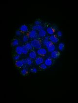
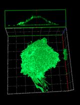
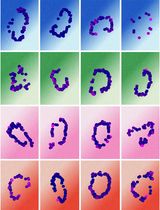
Here, we present a protocol for implementing the fluorogen-activating protein FAST (fluorescence-activating and absorption-shifting tag) in fluorescence lifetime imaging microscopy (FLIM), which allows separating fluorescent species in the same spectral channel based on fluorescence lifetime properties. Previous studies have demonstrated FLIM multiplexing using various combinations of synthetic probes, fluorescent proteins, or self-labeling tags. In this protocol, we utilize engineered FAST point mutation variants that bind fluorogen HBR-2,5-DM. The designed probes possess nearly identical, compact protein sizes (14 kDa), and the resulting protein–fluorogen complexes demonstrate comparable steady-state optical properties and exhibit distinct fluorescence lifetimes, displaying monoexponential fluorescence decay kinetics. When FAST variants are expressed with localization signals, these properties facilitate robust signal separation in regions with co-localized or spatially overlapping labels (nucleus and cytoskeleton in this protocol) in live mammalian cells. This method can be applied to separate other overlapping cellular compartments, such as the nucleus and Golgi apparatus, or mitochondria and cytoskeleton.
Key features
• The protocol employs FAST protein technology for fluorescent labeling.
• Separation of cellular compartments in the green channel (emission wavelength ~500–550 nm) using fluorescence lifetime data.
• Requires no coding skills.
• The protocol is optimized for SPCImage software.
Keywords: FLIM (FLIM)Background
A high density of acquired information is often desired from a single sample, which requires the use of advanced multiplexed imaging systems. One way to achieve this is through the implementation of fluorescence lifetime measurements (FLIM). FLIM measures the fluorescence decay time of molecules. This technique allows for distinguishing spectrally similar probes as long as their excited states exist for significantly different durations before photon emission [1]. In time-correlated single photon counting (TCSPC) FLIM implementation, each pixel of an acquired image consists of a histogram of single photon counts per time bin. These lifetime histograms may be fitted with exponential decay functions. Parameters of the function (an, τn) can then be used for the separation of different fluorophores. Another approach for lifetime separation is the fit-free phasor approach, which uses a 2D representation to cluster pixels with similar lifetimes in phasor space.
Fluorogen-activating proteins are nonfluorescent proteins that can bind specific dyes called fluorogens [2]. Fluorogens in their free form are also nearly nonfluorescent molecules, but binding within protein pockets stabilizes their structure and results in increased fluorescence quantum yield. Fluorescence lifetimes may depend on the fluorophore's surrounding conditions [3,4], and due to this property, fluorogen-activating proteins have proven to be excellent tools in FLIM [5–7]. In this case, the microenvironment of fluorogens is essentially determined by the fluorogen-activating protein binding pocket. The FAST is one of the most extensively studied fluorogen-activating proteins [8,9]. This protein offers several advantages: it is significantly smaller than conventional fluorescent proteins (14 kDa) and requires neither time nor oxygen for maturation. By studying the 3D structure of FAST, we developed a set of point mutants with significant alterations in the characteristics of the resulting protein–fluorogen complexes [10]. Further investigation allowed us to select several FAST variants (R52K, P68T, P68K, and F62L) with similar brightness and dissociation constants of complexes with HBR-2,5-DM fluorogen and to apply them for fluorescence lifetime multiplexing in cellulo (in living cells). Here, we describe a detailed protocol for the separation of two cellular compartments (nucleus and cytoskeleton) using FLIM data.
This protocol is applicable for separating other overlapping compartments across different cell lines. Such applications may require optimization of transfection conditions to obtain compartments with comparable fluorescence signal intensities, which significantly facilitates successful data analysis. When selecting compartment pairs for study, the importance of achieving similar fluorescence intensities should be considered.
The Phasor approach described in data analysis enables separation of triplets of FAST variants; however, in this case, separation would rely on visual control of the expected compartment morphologies.
While other fluorogens can be used in combination with FAST in FLIM, they do not allow for the most successful data analysis approach (the fitting-based approach with fixed τ).
Materials and reagents
Biological materials
1. HeLa cell line (ATCC CCL-2)
Reagents
1. DMSO (Thermo Scientific Chemicals, catalog number: 327182500); store at 4 °C
2. DMEM (PanEco, catalog number: С410п); store at 4 °C
Note: Commercially available international equivalent: DMEM, high glucose (Gibco, catalog number: 11965092)
3. FAST variants constructs with localization signals [5,6]; please contact the corresponding author for constructs
Note: R52K, P68T, P68K, and F62L FAST variants can be used in the protocol. The separation of the vimentin-P68T and H2B-F62L pair is demonstrated as an example in the Data analysis section.
4. Fetal bovine serum (FBS) (Biosera, catalog number: FB-1001/500); store at -20 °C
5. Fluorogen HBR-2,5-DM [(Z)-5-(4-Hydroxy-2,5-dimethylbenzylidene)-2-thioxo-1,3-thiazolidin-4-one] [11]; please contact the corresponding author for fluorogen; store at -20 °C
6. Hanks’ balanced salt solution (HBSS) (PanEco, catalog number: Р020п); store at 4 °C
Note: Commercially available international equivalent: HBSS, calcium, magnesium, no phenol red (Gibco, catalog number: 14025092)
7. HEPES (Gibco, catalog number: 15630056); store at 4 °C
8. Opti-MEM powder (Gibco, catalog number: 22600134); store at 4 °C
9. Penicillin-Streptomycin, 100× solution (PanEco, catalog number: А063п'); store at -20 °C
Note: Commercially available international equivalent: Sigma-Aldrich, catalog number: P4333.
10. Polyethylenimine (PEI) (Polysciences, catalog number: 23966-1); store at -20 °C
11. Sodium bicarbonate (Thermo Scientific Chemicals, catalog number: 424270010); store at room temperature
12. Trypsin, powder (Gibco, catalog number: 27250018); store at 4 °C
13. Versene solution (PanEco, catalog number: Р080п); store at 4 °C
Note: Commercially available international equivalent: Gibco, catalog number: 15040066.
Solutions
1. DMEM complete (see Recipes)
2. HBR-2,5-DM stock solution (see Recipes)
3. HBR-2,5-DM-containing imaging solution (see Recipes)
4. Imaging media (see Recipes)
5. Opti-MEM (see Recipes)
6. Trypsin-Versene solution (see Recipes)
Recipes
1. DMEM complete
| Reagent | Final concentration | Quantity or volume |
|---|---|---|
| DMEM | n/a | 44.5 mL |
| FBS | 10% | 5 mL |
| Penicillin-streptomycin, 100× solution | 1× | 0.5 mL |
Note: Store at 4 °C for up to a month.
2. HBR-2,5-DM stock solution
| Reagent | Final concentration | Quantity or volume |
|---|---|---|
| HBR-2,5-DM | 10 mM | 5.34 mg |
| DMSO | n/a | 2 mL |
Note: Prepare 20 μL aliquots. Store at -20 °C for up to 3 months. Avoid repeated freeze-thaw cycles.
3. HBR-2,5-DM-containing imaging solution
| Reagent | Final concentration | Quantity or volume |
|---|---|---|
| HBR-2,5-DM stock solution | 5 μM | 10 μL |
| DMSO | n/a | 20 mL |
Note: Add the HBR-2,5-DM stock solution to the imaging solution and mix thoroughly by vigorous vortexing. Prepare the fluorogen-containing imaging solution fresh for each experiment, as storage beyond 24 h is not recommended due to potential degradation of the fluorogen.
4. Imaging solution
| Reagent | Final concentration | Quantity or volume |
|---|---|---|
| HBSS | n/a | 49.5 mL |
| HEPES 1 M | 10 mM | 0.5 mL |
Note: Store at 4 °C for up to 3 months.
5. Opti-MEM
| Reagent | Final concentration | Quantity or volume |
|---|---|---|
| Opti-MEM powder | 1.4% | 6.795 g |
| Sodium bicarbonate powder | 2.4 g/L | 1.2 g |
| Distilled water | n/a | 500 mL |
Note: Store at 4 °C for up to 3 months.
6. Trypsin-Versene solution
| Reagent | Final concentration | Quantity or volume |
|---|---|---|
| Versene solution | n/a | 50 mL |
| Trypsin powder | 0.25% | 125 mg |
Note: Store at 4 °C for up to a month.
Laboratory supplies
1. 10 μL filtered pipette tips (Vertex, catalog number: 4117NSF)
2. 10 mL serological pipette (Greiner CELLSTAR, catalog number: 607160)
3. 1,000 μL filtered pipette tips (Vertex, catalog number: 4337NSF)
4. 1.5 mL microtube (SSI Bio, catalog number: 1260-00)
5. 20 μL filtered pipette tips (Vertex, catalog number: 4237NAF)
6. 200 μL filtered pipette tips (Vertex, catalog number: 4237NSF)
7. 35 mm glass-bottomed culture dish (SPL Life Sciences, catalog number: 100350)
8. T25 culture flask (SPL Life Sciences, catalog number: 70025)
Equipment
1. 0.5–10 μL micropipette (Eppendorf, catalog number: 3123000020)
2. 100–1,000 μL micropipette (Eppendorf, catalog number: 3123000063)
3. 2–20 μL micropipette (Eppendorf, catalog number: 3123000039)
4. 20–200 μL micropipette (Eppendorf, catalog number: 3123000055)
5. Cell culture incubator (Sanyo, model: MCO-175)
6. FLIM equipment: Manual TE2000-U microscope (Nikon), confocal scanning system DCS-120 (Becker and Hickl), GVD - 120 galvo scanner control + GDA amplifier (Becker and Hickl) equipped with the 30/70 mirror as beamsplitter, 100× S Fluor 0.5–1.3 oil iris objective (Nikon); Laser WhiteLase SC480-6 (Fianium), AOTF module (Fianium), 480/10× filter (Chroma) (to cut residual “white” spectrum); TCSPC modules SPC-150 (Becker and Hickl), PMC-100-1 detectors (Becker and Hickl), HQ495LP, HQ525/50 (Chroma); Type F immersion oil (Leica, catalog number: 11513859)
Note: Alternative FLIM equipment configurations can be used for this protocol. If necessary, consult with a FLIM specialist.
7. Laminar flow cabinet (Labconco, model: MP-Logic Class II, Type A2 Biological Safety Cabinets)
8. Large capacity pipette (Hangzhou Miu Instruments, model: LEP-100)
9. Vortex Microspin FV-2400 (Biosan, catalog number: BS-010201-AAA)
10. Water bath thermostat (ELMI, model: TW-2.03)
Software and datasets
1. FIJI 1.53t (August 2022); free, open source
2. SPCImage software 8.9 (December 2023); commercial license required
3. SPCM Data Acquisition Software v.9.81 (2023); commercial license required
Procedure
文章信息
稿件历史记录
提交日期: Jun 26, 2025
接收日期: Aug 21, 2025
在线发布日期: Sep 15, 2025
出版日期: Oct 5, 2025
版权信息
© 2025 The Author(s); This is an open access article under the CC BY-NC license (https://creativecommons.org/licenses/by-nc/4.0/).
如何引用
Gilvanov, A. R., Solovyev, I. D., Savitsky, A. P., Baranov, M. S. and Bogdanova, Y. A. (2025). Fluorescence Lifetime-Based Separation of FAST-Labeled Cellular Compartment. Bio-protocol 15(19): e5460. DOI: 10.21769/BioProtoc.5460.
分类
细胞生物学 > 细胞成像 > 荧光
生物物理学 >
您对这篇实验方法有问题吗?
在此处发布您的问题,我们将邀请本文作者来回答。同时,我们会将您的问题发布到Bio-protocol Exchange,以便寻求社区成员的帮助。