

High-Throughput Screening Identification of Chemical Compounds That Affect Cold-Regulated Gene Expression in Arabidopsis thaliana Using an Excised Single Leaf
利用单片叶片高通量筛选影响拟南芥低温调控基因表达的化合物
发布: 2025年05月20日第15卷第10期 DOI: 10.21769/BioProtoc.5319 浏览次数: 1808
评审: Anonymous reviewer(s)
参见作者原研究论文
The authors used this protocol in:

May 2023
Advertisement


Abstract
The identification of chemical compounds that affect intracellular processes has greatly contributed to the understanding of developmental regulation in plants. In this protocol, we describe a method for identifying chemical compounds that affect cold-regulated gene expression in Arabidopsis thaliana. Specifically, we generated Arabidopsis plants harboring a COLD-REGULATED 15A (COR15A) promoter::luciferase (COR15Apro::LUC) construct and grew them in a submerged liquid culture. Using a single true leaf excised from COR15Apro::LUC plants and 96-well culture plates, we performed high-throughput screening of chemical compounds that inhibit cold-induction of COR15Apro::LUC. Luciferase activity was detected using a microplate reader and a chemiluminescence imaging device. This protocol can be easily used for the identification of chemical compounds that regulate other processes, being versatile with respect to equipment.
Key features
• High-throughput screening of chemical compounds that affect cold-regulated gene expression is possible using a single leaf excised from Arabidopsis grown in a submerged culture.
• Screening is based on luciferase activity derived from an excised single leaf.
• Direct measurement of luciferase activity is possible using a microplate reader and a chemiluminescence imaging device.
• This protocol can be easily used for the identification of chemical compounds that regulate other processes.
Keywords: Arabidopsis thaliana (拟南芥)Graphical overview
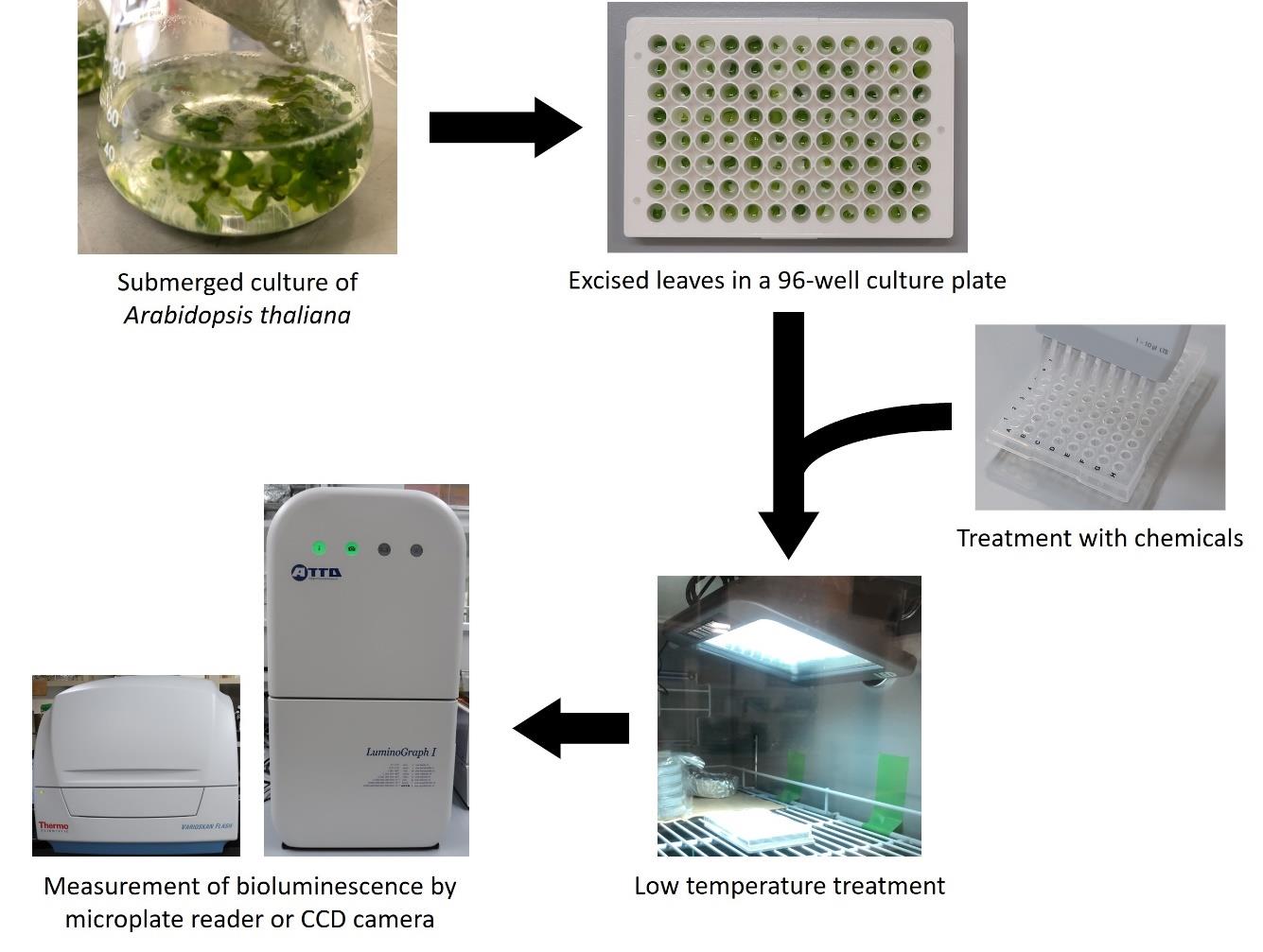
Graphical summary of a screening method for identifying compounds that inhibit low-temperature induction of COR15Apro::LUC
Background
The identification of chemical compounds that affect intracellular processes has greatly contributed to the understanding of developmental regulation in plants. However, high-throughput chemical screening of plant tissues faces some challenges due to the nature of plants. First, most chemical screening uses germinating seedlings (∼1-week-old seedlings at best) or plant cell cultures [1–6] and not mature tissues. Chemical screening using mature plant tissues of Arabidopsis, with a lifetime of more than two months, would be of great benefit to further understand plant growth and development. Second, leaf tissues are surrounded by cuticle layers, and this makes it difficult for compounds to enter the plant cell. Therefore, the establishment of a screening system using mature plant tissues would allow a better understanding of signaling pathways that regulate plant growth and development during the mature stage.
In this study, we describe a high-throughput screening method using single leaves of mature plants to identify small molecules that affect cold-regulated gene expression [7]. In this protocol, we used transgenic Arabidopsis thaliana harboring the COLD-REGULATED 15A (COR15A) promoter::luciferase (COR15Apro::LUC) construct as a model. COR15A was originally identified as a cold-induced gene, but its expression was induced more than 100 times by abscisic acid (ABA) treatment [8]. Moreover, cycloheximide (CHX) has been shown to inhibit cold acclimation in multiple plant species [9,10]. The characteristics of these two compounds made it easy to set up this assay system.
Although we only investigated low-temperature response in this experiment, the protocol can be applied to other plant responses to endogenous (e.g., phytohormones) and environmental cues. The protocol is compatible with imaging devices and microplate readers. The protocol enables the identification of new compounds that control plant growth and development and provides new insights into the molecular mechanisms underlying plant growth and development.
Materials and reagents
Biological materials
1. Arabidopsis thaliana harboring COR15Apro::LUC construct
See General note 1.
Reagents
1. Murashige and Skoog plant salt mixture (MS salt) (Wako, catalog number: 392-00591)
2. Sucrose (Wako, catalog number: 196-00015)
3. 2-Morpholinoethanesulfonic acid monohydrate (MES) (Dojindo, catalog number: 343-01621)
4. D-Luciferin, potassium salt (GOLDBIO, catalog number: LUCK-1G)
5. Cycloheximide (Wako, catalog number: 037-20991)
6. TritonTM X-100 (ICN Biomedicals Inc., catalog number: 807426)
7. Dimethyl sulfoxide (DMSO) (Wako, catalog number: 043-07216)
8. Ethanol (Wako, catalog number: 057-00456)
9. Chlorine-based bleach (e.g., KAO Kitchen Haiter)
10. Autoclaved dH2O
Solutions
1. 0.5× MS liquid medium (see Recipes)
2. 50 mM D-Luciferin (see Recipes)
3. 2 mg/mL cycloheximide (see Recipes)
4. 20% (w/v) Triton X-100 (see Recipes)
5. MS-Luciferin solution (see Recipes)
6. 70% (v/v) ethanol (see Recipes)
7. Seed sterilization solution (see Recipes)
8. 0.1% agar solution (see Recipes)
Recipes
1. 0.5× MS liquid medium
| Reagent | Final concentration | Quantity or Volume |
|---|---|---|
| MS salt | 0.5× | 0.23 g |
| MES | 0.5 g/L | 0.05 g |
| Sucrose | 1% | 1 g |
| dH2O | 100 mL |
This recipe is for two flasks. Adjust the volume to the amount you need. In general, divide the 100 mL of 0.5× MS liquid medium into two flasks (50 mL each) and then autoclave the flasks wrapped with aluminum foil. Autoclaved medium can be stored for at least two weeks at room temperature and under sterile conditions.
2. 50 mM luciferin
| Reagent | Final concentration | Quantity or Volume |
|---|---|---|
| D-Luciferin, potassium salt | 50 mM | 0.2 g |
| dH2O | 12.6 mL |
Smaller amounts can be prepared depending on the objective. Luciferin (50 mM) can be aliquoted into 1.5 mL tubes and stored at -80 °C.
3. 2 mg/mL cycloheximide
| Reagent | Final concentration | Quantity or Volume |
|---|---|---|
| Cycloheximide | 2 mg/mL | 2.4 mg |
| DMSO | 1.2 mL |
In this study, 2 mg/mL of cycloheximide was used because the concentration of the chemical compounds in our library was 2 mg/mL [7]. This concentration can be modified depending on the assay.
4. 20% (w/v) Triton X-100
| Reagent | Final concentration | Quantity or Volume |
|---|---|---|
| TritonTM X-100 | 20% (w/v) | 20 g |
| dH2O | 100 mL |
This recipe is for non-sterile conditions. You may use a commercially available sterile-filtered Triton X-100 if necessary.
5. MS-Luciferin solution (for 100 wells)
| Reagent | Final concentration | Quantity or Volume |
|---|---|---|
| 0.5× MS liquid medium | 0.5× | 20 mL |
| 50 mM luciferin | 0.5 mM | 200 μL |
| 0.1% (w/v) Triton X-100 | 0.001% | 200 μL |
Dilute 20% (w/v) Triton X-100 in dH2O.
6. 70% (v/v) ethanol
| Reagent | Final concentration | Quantity or Volume |
|---|---|---|
| Ethanol | 70% (v/v) | 7 mL |
| dH2O | Up to 10 mL |
7. Seed sterilization solution
| Reagent | Final concentration | Quantity or Volume |
|---|---|---|
| Chlorine-based bleach | 30% | 3 mL |
| 20% (w/v) Triton X-100 | 0.02% | 10 μL |
| dH2O | Up to 10 mL |
8. 0.1% agar solution
Add 0.1 g of agar for plant culture in 100 mL of dH2O and autoclave.
Laboratory supplies
1. 96-well PCR plate (e.g., BIO-BIK, catalog number: 3425-00)
2. 96-well culture plate (e.g., Nunc 96-well flat bottom, catalog number: 136101)
3. Stainless steel scissors
4. Stainless steel tweezers
5. Kimwipe and Kimtowel (Nippon Paper Cresia); these should be autoclaved if the experiments require sterile conditions
6. 100 mL flask (e.g., Iwaki, catalog number: 4980FK100)
7. Microtube 1.5 mL (e.g., SSIbio, catalog number: 1210-00)
8. 15 mL and 50 mL conical tubes (e.g., Falcon)
Equipment
1. Varioskan Flash microplate reader (Thermo Fisher Scientific)
2. LuminoGraph I imaging device (ATTO Co. Ltd., Japan) (Optional)
3. Growth chamber (e.g., Nippon Medical & Chemical Instruments Co., Ltd.)
4. Multichannel pipette
Software and datasets
1. SkanIt Software 2.4.3 (operating software for Varioskan Flash microplate reader)
2. ImageSaver 6 (operating software for LuminoGraph I chemiluminescence imaging device) (Optional)
3. CS Analyzer image analysis software (attached to LuminoGraph I) (Optional)
Procedure
文章信息
稿件历史记录
提交日期: Feb 27, 2025
接收日期: Apr 24, 2025
在线发布日期: May 11, 2025
出版日期: May 20, 2025
版权信息
© 2025 The Author(s); This is an open access article under the CC BY-NC license (https://creativecommons.org/licenses/by-nc/4.0/).
如何引用
Readers should cite both the Bio-protocol article and the original research article where this protocol was used:
- Kitawaki, K., Mihara, R., Ito-Inaba, Y. and Inaba, T. (2025). High-Throughput Screening Identification of Chemical Compounds That Affect Cold-Regulated Gene Expression in Arabidopsis thaliana Using an Excised Single Leaf. Bio-protocol 15(10): e5319. DOI: 10.21769/BioProtoc.5319.
- Kitawaki, K., Mihara, R., Kamimura, S., Sato, A., Ushiyama, M., Ito-Inaba, Y. and Inaba, T. (2023). Chemical screening approach using single leaves identifies compounds that affect cold signaling in Arabidopsis. Plant Physiol. 193(1): 234–245. https://doi.org/10.1093/plphys/kiad280
分类
植物科学 > 植物生理学
植物科学 > 植物分子生物学 > 遗传分析
您对这篇实验方法有问题吗?
在此处发布您的问题,我们将邀请本文作者来回答。同时,我们会将您的问题发布到Bio-protocol Exchange,以便寻求社区成员的帮助。