- EN - English
- CN - 中文
Differentiation, Maintenance, and Contraction Profiling of Human Induced Pluripotent Stem Cell–Derived Cardiomyocytes
人诱导多能干细胞来源心肌细胞的分化、维持及收缩特性分析
发布: 2025年03月05日第15卷第5期 DOI: 10.21769/BioProtoc.5222 浏览次数: 3864
评审: Philipp WörsdörferWilliam C. W. ChenSamantha Haller

相关实验方案

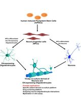
人 iPSC 衍生神经元与少突胶质细胞共培养用于髓鞘形成的小分子筛选分析
Stefanie Elke Chie [...] Maria Consolata Miletta
2025年05月05日 3362 阅读
Abstract
The development of patient-derived cardiac disease models has advanced rapidly due to the progress of human-induced pluripotent stem cell (hiPSC) technologies. Many protocols detail individual parts of the entire workflow, from handling hiPSCs and differentiating them into cardiomyocytes to live contraction imaging via widefield/phase-contrast and fluorescence microscopy. Here, we propose a streamlined protocol that guides users through hiPSC culture, differentiation, expansion, and functional imaging of hiPSC cardiomyocytes. First, hiPSC maintenance and handling procedures are outlined. Differentiation occurs over a two-week period, followed by selective expansion to increase the yield of hiPSC cardiomyocytes. Comprehensive characterization and quantification enable detailed contraction profiling of these cells. Designed to be low-cost, this protocol is suited for applications in drug discovery, screening, and clinical testing of patient-specific phenotypes. The addition of cardiomyocyte expansion and automated analysis distinguishes our protocol from current approaches.
Key features
• Protocol for the maintenance, differentiation, and expansion of hiPSC cardiomyocytes.
• Detailed guidance for characterization and functional imaging of hiPSC cardiomyocytes.
• Streamlined workflow integrating state-of-the-art protocols streamlined into a cost-effective approach.
• A complete timeline from hiPSC culture to contraction imaging achievable in as little as three weeks.
Keywords: hiPSC (hiPSC)Background
In recent years, human-induced pluripotent stem cells (hiPSCs) have become a widely accepted alternative for studying disease and toxicity, offering patient-specific models that mirror the original cells, thereby allowing for drug testing or examination of genetic mutations without direct patient involvement. In cardiovascular research, the use of hiPSC-derived cardiomyocytes has enabled patient-specific treatment advancements. Though still maturing, hiPSC cardiomyocytes are able to spontaneously contract and respond to pacing with electrodes or optogenetic guidance using channelrhodopsins. By utilizing state-of-the-art genetic engineering using CRISPR-Cas9 systems, it is possible to create isogenic iPSC lines that have the patient’s genetic background, further improving our understanding of cardiac disease and progression. 3D cardiac engineering is another field that is quickly progressing, with the possibilities of generating regenerative cardiac patches and 3D-engineered heart tissue. Advantages of this method include upscaling of cardiomyocyte differentiation and automated analysis. Disadvantages are the lack of maturation steps (e.g., electrical pacing) or guided differentiation to atrial cardiomyocytes via the addition of retinoic acid.
This protocol utilizes state-of-the-art techniques regarding hiPSC maintenance and hiPSC cardiomyocyte differentiation and maintenance [1–3]. This protocol describes the generation of assay-ready differentiated hiPSC cardiomyocytes in 15 days, including the expansion of cardiomyocytes afterward. As previously reported by Maas et al. [2], cardiomyocytes can be manipulated into the cell cycle by subjecting them to WNT pathway inhibition and a loss of cell-cell contact early in differentiation. This process allows the upscaling of otherwise non-dividing cells. We also provide a workflow for our in-house automated analysis software that can be used to quantify contractions using brightfield or calcium transient fluorescence recordings.
We optimized the protocol using two in-house hiPSC cell lines: a healthy control cell line and a hypertrophic cardiomyopathy cell line. The performance of differentiated hiPSC cardiomyocytes was compared to that of an industrial standard (Ncyte cardiomyocytes, Ncardia).
Materials and reagents
Biological materials
1. hiPSC (recommended cell line listed on hPSCreg: LUMCi028-A (RRID:CVCL_ZA25)
Reagents
1. Matrigel® hESC-qualified matrix (Corning, catalog number: 354277)
2. mTeSRTM plus (StemCell Technologies, catalog number: 100-0276)
3. ReLeSRTM dissociation reagent (StemCell Technologies, catalog number: 100-0483)
4. DMEM/F12 (Gibco, catalog number: 11554546)
5. Y-27672 (referred to as ROCK inhibitor, 10 mM) (Tocris, catalog number: 1254)
6. PBS (Sigma-Aldrich, catalog number: D8537)
7. DMSO (Sigma-Aldrich, catalog number: 41640)
8. HyClone fetal bovine serum (Cytiva, catalog number: SH30071.02E)
9. Accutase (StemCell Technologies, catalog number: 07920)
10. Trypan blue (Thermo Fisher, catalog number: 15250061)
11. B27 (50×) (Gibco, catalog number: A3582801)
12. B27 minus insulin (50×) (Gibco, catalog number: A1895601)
13. CHIR-99021 (5 mM) (Sigma-Aldrich, catalog number: SML1046)
14. BMP-4 (20 mg/mL) (Sigma-Aldrich, catalog number: SRP3016)
15. Activin-A (20 mg/mL) (Sigma-Aldrich, catalog number: SRP3003)
16. Wnt-C59 (2 mM) (SelleckChem, catalog number: S7037)
17. DL-lactate solution (60% w/w) (Sigma-Aldrich, catalog number: L4263)
18. StableCellTM RPMI-1640 (Sigma-Aldrich, catalog number: R2405)
19. RPMI-1640 minus glucose (Gibco, catalog number: 11879020)
20. Fibronectin (1 mg/mL) (Corning, catalog number: 354008)
21. TrypLETM Select enzyme (10×) (Gibco, catalog number: A1217701)
22. KnockOutTM (KO) serum replacement (Gibco, catalog number: 10828028)
23. RevitaCell supplement (100×) (Gibco, catalog number: A2644501)
24. Pluronic F-127 (10% w/v) (Sigma-Aldrich, catalog number: P2443)
25. Cal-520 AM (2.5 mM) (AAT Bioquest, catalog number: 21130)
Solutions
1. hiPS freezing medium (see Recipes)
2. Differentiation medium A (see Recipes)
3. Differentiation medium B (see Recipes)
4. Maintenance medium (see Recipes)
5. Selection medium (see Recipes)
6. Replating medium (see Recipes)
7. CM freezing medium (see Recipes)
8. CM thawing medium (see Recipes)
9. Imaging medium (see Recipes)
Recipes
1. hiPSC freezing medium
*Note: Volumes indicated are for one cryovial.
| Reagent | Final concentration | Quantity or Volume |
|---|---|---|
| mTeSR Plus | 50% | 550 μL |
| Hyclone fetal bovine serum | 40% | 440 μL |
| DMSO | 10% | 110 μL |
| ROCK inhibitor (10 mM) | 10 μM | 1.1 μL |
| Total* | ~1,100 μL |
2. Differentiation medium A
*Note: Volumes indicated are for one well of a standard six-well plate.
| Reagent | Final concentration | Quantity or Volume |
|---|---|---|
| RPMI-1640 | n/a | 1,954 μL |
| B27 minus insulin (50×) | 1× | 40 μL |
| CHIR-99021 (5 mM) | 5 μM | 2 μL |
| BMP4 (20 μg/mL) | 20 ng/mL | 2 μL |
| Activin-A (20 μg/mL) | 20 ng/mL | 2 μL |
| Total* | 2 mL |
3. Differentiation medium B
*Note: Volumes indicated are for one well of a standard six-well plate.
| Reagent | Final concentration | Quantity or Volume |
|---|---|---|
| RPMI-1640 | n/a | 1958 μL |
| B27 minus insulin (50×) | 1× | 40 μL |
| Wnt-C59 (2 mM) | 2 μM | 2 μL |
| Total* | 2 mL |
4. Maintenance medium
*Note: Volumes indicated are for one well of a standard six-well plate.
| Reagent | Final concentration | Quantity or Volume |
|---|---|---|
| RPMI-1640 | n/a | 1,960 μL |
| B27 (50×) | 1× | 40 μL |
| Total* | 2 mL |
5. Selection medium
*Note: Volumes indicated are for one well of a standard six-well plate.
#Note: 60% (w/w) of DL-lactate equates to a concentration of approximately 7.014 M.
| Reagent | Final concentration | Quantity or Volume |
|---|---|---|
| RPMI-1640 minus glucose | n/a | 1,958 μL |
| B27 (50×) | 1× | 40 μL |
| DL-lactate (60% w/w)# | 4 mM | 1.1 μL |
| Total* | ~2 mL |
6. Replating medium
*Note: Volumes indicated are for one standard well of a six-well plate.
| Reagent | Final concentration | Quantity or Volume |
|---|---|---|
| RPMI-1640 | n/a | 1,758 μL |
| B27 (50×) | 1× | 40 μL |
| KO serum replacement | 10% | 200 μL |
| ROCK inhibitor (10 mM) | 10 μM | 2 μL |
| Total* | 2 mL |
7. CM freezing medium
*Note: Volumes indicated are for one cryovial.
| Reagent | Final concentration | Quantity or Volume |
|---|---|---|
| RPMI-1640 | n/a | 779 μL |
| B27 (50×) | 1× | 20 μL |
| KO serum replacement | 10% | 100 μL |
| ROCK inhibitor (10 mM) | 10 μM | 1 μL |
| DMSO | 10% | 100 μL |
| Total* | 1 mL |
8. CM thawing medium
*Note: Volumes indicated are for one standard well of a six-well plate.
| Reagent | Final concentration | Quantity or Volume |
|---|---|---|
| RPMI-1640 | n/a | 1,758 μL |
| B27 (50×) | 1× | 40 μL |
| RevitaCell supplement (100×) | 1× | 20 μL |
| ROCK inhibitor (10 mM) | 10 μM | 2 μL |
| Total* | 2 mL |
9. Imaging medium
*Note: Volumes indicated are for one standard well of a six-well plate.
| Reagent | Final concentration | Quantity or Volume |
|---|---|---|
| RPMI-1640 | n/a | 1,936 μL |
| B27 (50×) | 1× | 40 μL |
| F-127 (10% w/v) | 0.1% | 20 μL |
| Cal-520 (2.5 mM) | 5 μM | 4 μL |
| Total* | 2 mL |
Laboratory supplies
1. 6-well plates (Greiner Bio-One, catalog number: 10536952)
2. 15 mL tubes (Corning, catalog number: 430791)
3. 50 mL tubes (Greiner Bio-One, catalog number: 227261)
4. 1.5 mL safe-lock tubes (Eppendorf, catalog number: 0030120086)
5. Cryovial tubes (Thermo Fisher, catalog number: 368632)
6. Pipettes (2, 20, 200, 1,000 μL) (Gilson, catalog number: F167360)
7. Filtered pipette tips (10, 20, 200, 1,000 μL) (Greiner Bio-One, catalog numbers: 771363, 773363, 775363, 778363)
8. Pipette controller (Fisher Scientific, catalog number: 03-840-312)
9. 5, 10 mL filtered serological pipettes (Greiner Bio-One, catalog numbers: 606180, 607180)
Equipment
1. Countess 3 automated cell counter (Invitrogen, catalog number: AMQAX2000)
2. Inverted microscope with fluorescence and widefield/phase-contrast (e.g., Ti-Eclipse inverted microscope, Nikon, catalog number: SM100606-MG1)
3. Heated stage top incubator with CO2 + air (e.g., Tokai hit stage top incubator, Tokai Hit, catalog number: ZILCSD-HL)
4. Microscope camera (Photometrics, model: Coolsnap HQ2)
5. Centrifuge for 15–50 mL tubes (e.g., Eppendorf 5810R, Eppendorf, catalog number: EP022628168)
Software and datasets
1. Microscope imaging software, e.g., MetaMorph (version 7.8.0.0)
2. ImageJ (version 1.53t)
3. MATLAB (version R2022A, license required)
4. All data and code have been deposited to GitHub: https://github.com/msnelders1/CardiomyocyteContractionAnalysis (Access date, October 24, 2024)
Procedure
文章信息
稿件历史记录
提交日期: Nov 6, 2024
接收日期: Jan 6, 2025
在线发布日期: Feb 13, 2025
出版日期: Mar 5, 2025
版权信息
© 2025 The Author(s); This is an open access article under the CC BY-NC license (https://creativecommons.org/licenses/by-nc/4.0/).
如何引用
Snelders, M., van der Pluijm, I. and Essers, J. (2025). Differentiation, Maintenance, and Contraction Profiling of Human Induced Pluripotent Stem Cell–Derived Cardiomyocytes. Bio-protocol 15(5): e5222. DOI: 10.21769/BioProtoc.5222.
分类
干细胞 > 多能干细胞 > 细胞分化
细胞生物学 > 细胞分离和培养 > 细胞分化
您对这篇实验方法有问题吗?
在此处发布您的问题,我们将邀请本文作者来回答。同时,我们会将您的问题发布到Bio-protocol Exchange,以便寻求社区成员的帮助。
Share
Bluesky
X
Copy link


