

Confocal Live Imaging of Reproductive Organs Development in Arabidopsis
利用共聚焦活体成像技术观察拟南芥生殖器官的发育
发布: 2025年02月05日第15卷第3期 DOI: 10.21769/BioProtoc.5177 浏览次数: 2742
评审: Pooja VermaHeng ChenTasleem JavaidAnonymous reviewer(s)
参见作者原研究论文
The authors used this protocol in:
Aug 2021
Advertisement


Abstract
Understanding how multicellular organisms are shaped requires high-resolution, quantitative data to unravel how biological structures grow and develop over time. In recent years, confocal live imaging has become an essential tool providing insights into developmental dynamics at cellular resolution in plant organs such as leaves or meristems. In the context of flowers, growth tracking has primarily been limited to sepals, the outermost floral organs, or the post-fertilization gynoecium, which are easily accessible for microscopy. Here, we describe a detailed pipeline for the preparation, dissection, and confocal imaging of the development of internal reproductive floral organs of Arabidopsis thaliana including both the stamen and gynoecium. We also discuss how to acquire high-quality images suitable for efficient 2D and 3D segmentation that allow the quantification of cellular dynamics underlying their development.
Key features
• Fine dissection of tiny and tightly enclosed floral organs.
• Confocal live imaging method allowing long-term observation of plant reproductive morphogenesis.
• Assessing the quality of acquired images for efficient segmentation at cellular resolution in 2D and 3D.
Keywords: Confocal microscopy (共聚焦显微镜)Background
Flowers are essential for plant reproduction and vital for crop yield. The mature flower of Arabidopsis thaliana consists of concentric whorls of floral organs: non-reproductive sepals and petals that surround stamens and carpels (fused together to form gynoecium). The stamen, the male reproductive organ of the flower, consists of anther-producing pollen that is supported by a long filament. The gynoecium, the female reproductive structure, is composed of two fused carpels (forming the ovary) topped by the style and pollen-receiving stigma. After fertilization, the gynoecium develops into fruit, which is crucial for plant propagation.
The development of floral reproductive organs is difficult to observe, as early emerging sepals quickly enclose the initiating stamens and gynoecium making them inaccessible for imaging without dissection. Widely used methods relying on tissue fixing, sectioning, or clonal analysis have provided valuable insights into the development of internal floral organs [e.g., 1–5]. However, these methods lack the spatial and temporal resolution to understand and uncover dynamic developmental processes underlying organogenesis at cellular resolution. While confocal live imaging has been extensively used to follow growth in easily accessible sepals [e.g., 6–10], such an approach has only recently been applied to uncover developmental dynamics in reproductive organs [e.g., 11–13].
In this paper, we describe the confocal live imaging method to observe the growth of Arabidopsis thaliana stamen and gynoecium. We detail the procedures for dissecting and exposing internal floral organs and acquiring confocal images of the same sample over several consecutive days. Furthermore, we provide some hints on how to increase the survival rate of the sample and improve the quality of confocal images suitable for 2D or 3D segmentation. Our protocol enables observations with minimal perturbation of the normal in planta morphogenesis allowing long-term quantitative observation of these hidden floral organs at cellular resolution.
Materials and reagents
Biological materials
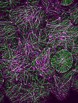
1. Reproductive organs of 4-week-old Arabidopsis thaliana Col-0 carrying a plasma membrane–localized fluorescent marker. Here, we used plants carrying pUBQ10::myr-YFP construct [14]; however, different plasma membrane marker lines can be used
Reagents
1. Murashige and Skoog basal salt mixture (Sigma, catalog number: M5524-50L)
2. Sucrose (Fisher, catalog number: S5-3)
3. Murashige and Skoog vitamin solution (Sigma, catalog number: M3900-50ML)
4. Agar (Fisher, catalog number: BP1423-2)
5. Plant preservative mixture (PPM) (Plant Cell Technology, catalog number: 71806-1)
6. 95% denatured alcohol (Fisher, catalog number: HC-1100-1GL)
7. Propidium iodide (PI) (Sigma, catalog number: P4170) (potential carcinogen)
Solutions
1. 1/2 MS medium (for 1 L) (see Recipes)
2. 0.1% PPM solution (see Recipes)
3. 70% ethanol solution (see Recipes)
4. 0.1% PI staining solution (see Recipes)
Recipes
1. 1/2 MS medium (for 1 L)
2.15 g of Murashige and Skoog basal salt mixture
10 g of sucrose
1 mL of Murashige and Skoog vitamin solution
Add deionized H2O to 1 L
Adjust pH to 5.8
15 g of agar
2. 0.1% PPM solution
1 L of sterile deionized H2O
1 mL of plant preservative mixture (PPM)
3. 70% ethanol solution
737 mL of 95% denatured alcohol
263 mL of sterile deionized H2O
4. 0.1% PI staining solution (potential carcinogen)
1 mg of propidium iodide
1 mL of deionized H2O
Laboratory supplies
1. 35 × 10 mm Petri dishes (SARSTEDT, catalog number: 82.1135.500)
2. Laboratory film (ParafilmTM)
3. 200 μL and 1 mL pipette and corresponding tips
4. Precision tweezers with fine point (Dumont No. 5)
5. Syringe needles, 25 G × 1" (BD®, catalog number: 305125)
6. Tungsten 1 µm probe tips (Lambda, catalog number: T20-10)
7. Low lint tissue wipe (KimwipesTM, Kimberly-Clark)
8. Deionized H2O
9. Scalpel blades (FEATHER, # 15)
10. Surgical tape (MicroporeTM tape 3M)
11. Plastic pots, trays, and lids for plant potting (thermoformed square pots with drainage 2.63" × 2.63" × 2.25", no-hole trays 21" × 11" × 2.5", and compatible plastic dome 21.50" × 11" × 2.10")
12. All-purpose growing soil mixture (ASB Greenworld Grower Mix)
Equipment
1. Autoclave
2. Laminar flow cabinet
3. Dissecting stereomicroscope with a minimum of 4× zoom magnification (Zeiss, model: Stemi 35)
4. Upright confocal microscope (Zeiss, model: LSM800) equipped with long working distance, water-dipping lenses with a good numerical aperture (W Plan-Apochromat 40×/1.0 DIC M27 FWD = 2.5 mm)
5. Growth chamber (Conviron GEN1000)
Software and datasets
1. Zeiss Zen 2.6 blue edition (Carl Zeiss Microscopy GmbH)
Procedure
文章信息
稿件历史记录
提交日期: Aug 29, 2024
接收日期: Nov 27, 2024
在线发布日期: Dec 17, 2024
出版日期: Feb 5, 2025
版权信息
© 2025 The Author(s); This is an open access article under the CC BY license (https://creativecommons.org/licenses/by/4.0/).
如何引用
Readers should cite both the Bio-protocol article and the original research article where this protocol was used:
- Wang, B., Bauer, A., Gómez-Felipe, A., Silveira, S. R. and Kierzkowski, D. (2025). Confocal Live Imaging of Reproductive Organs Development in Arabidopsis. Bio-protocol 15(3): e5177. DOI: 10.21769/BioProtoc.5177.
- Silveira, S. R., Le Gloanec, C., Gómez-Felipe, A., Routier-Kierzkowska, A. L. and Kierzkowski, D. (2022). Live-imaging provides an atlas of cellular growth dynamics in the stamen. Plant Physiol. 188(2): 769–781.
分类
植物科学 > 植物发育生物学 > 形态建成
植物科学 > 植物细胞生物学 > 细胞成像
您对这篇实验方法有问题吗?
在此处发布您的问题,我们将邀请本文作者来回答。同时,我们会将您的问题发布到Bio-protocol Exchange,以便寻求社区成员的帮助。