

Microfluidic Cultures of Basal Forebrain Cholinergic Neurons for Assessing Retrograde Cell Death by Live Imaging
用于活细胞成像评估逆行性细胞死亡的基底前脑胆碱能神经元微流体培养
发布: 2025年01月05日第15卷第1期 DOI: 10.21769/BioProtoc.5149 浏览次数: 1893
评审: Domenico Azarnia TehranAlessandro DidonnaMiaomiao Tian
参见作者原研究论文
The authors used this protocol in:

Aug 2023
Advertisement


Abstract
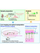
Neurons are highly polarized cells, with axons that may innervate distant target regions. In the brain, basal forebrain cholinergic neurons (BFCNs) possess extensive axons that project to several target regions such as the cortex, hippocampus, and amygdala, and may be exposed to a specific microenvironment in their axon targets that may have retrograde effects on neuronal health. Interestingly, BFCNs express the pan-neurotrophin receptor p75NTR throughout life while also concomitantly co-expressing all Trk receptors, making them capable of responding to both mature and precursor neurotrophins to promote survival or apoptosis, respectively. Levels of these trophic factors may be modulated in the BFCN axon or soma microenvironment under neurodegenerative conditions such as seizure and brain injury. In this protocol, BFCNs are established in microfluidic devices for compartmental culture, with the aim of studying the effects of axon- or soma-specific stimulation of BFCNs for an in vitro representation of distal axon vs. soma environments as seen in vivo. This study further establishes a novel method of tracing and imaging live BFCNs exposed to stimuli in their distal axons with the aim of assessing retrograde cell death. The in vitro compartmental culture system of BFCNs that allows live imaging may be applied to investigate various effects of axon- or soma-specific stimuli that affect BFCN health, maintenance, and death, to model events that occur in the context of brain injury and neurodegenerative disorders.
Key features
• Separation of axons and soma of basal forebrain primary neurons in vitro using microfluidic chambers.
• Compartmental/localized treatment of axons or somas of BFCNs.
• Live imaging of retrogradely labeled BFCNs to assess cell death.
Keywords: Microfluidics (微流体)Background
Neurons may possess long axons that project to distal areas and respond retrogradely to stimuli limited to their axonal microenvironment. Basal forebrain cholinergic neurons (BFCNs) send extensive and long axonal projections to several distal brain areas, such as the cortex, to regulate cognitive functions such as attention, emotion, and memory [1]. Interestingly, throughout their life, BFCNs express the pan-neurotrophin receptor p75NTR as well as all three members of the Trk family of receptor tyrosine kinases. Neurotrophins are essential for BFCNs’ survival, maintenance, differentiation, and function [2]. Neurotrophins in their precursor and mature forms activate different signaling cascades, which lead to contrasting cellular responses. Proneurotrophins are cleaved to form mature neurotrophins, which promote a pro-survival response through Trk signaling [3–6]. In contrast, under conditions of brain insult such as seizures or traumatic brain injuries (TBI), proneurotrophins (proNTs) bind p75NTR and sortilin to promote apoptosis in BFCNs [7]. Interestingly, as BFCNs express all the neurotrophin receptors, these neurons are capable of responding to a changing balance of pro vs. mature neurotrophins. To add to the complexity, the dynamic shift in pro vs. mature neurotrophin balance may occur at the BFCN neuronal target regions such as after cortical injury [8]. Although it has been established that p75NTR promotes BFCN degeneration in mass cultures and after seizure conditions in vivo [7], given the loss of BFCNs after cortical injury it is important to answer whether p75NTR can promote BFCN degeneration retrogradely as well, especially when the BFCN soma is unexposed to induced proneurotrophins [8].
This protocol has been applied by Dasgupta et al. [8] to establish whether the changes in the neurotrophic environment of BFCN axon terminals after TBI have a retrograde effect on survival of these afferent projections by live imaging and cell death analysis of in vitro compartmentally cultured BFCNs. Silicone microfluidic devices allow for the segregation of neuronal axons and soma as well as for compartmental manipulation with localized treatments [9], having been used extensively on several neuronal cell types to investigate a myriad of cell biology questions [10]. Moreover, the transparent material allows for labeling and imaging of live tracers that indicate dying cells after treatments. Major studies have established retrograde degeneration through p75NTR in peripheral neurons [11,12], but this protocol helped establish the role of p75NTR and proneurotrophins on BFCN retrograde degeneration, adding to the evidence that suggests that brain injury induced elevation in cortical proneurotrophin leads to p75NTR signaling in BFCNs initiated at the axon terminals to promote retrograde degeneration [8]. Overall, this protocol can be applied to study cell death and other cellular responses to compartmental stimulation of BFCNs by live imaging and can also be adapted to other neuronal populations.
Materials and reagents
Biological materials
1. C57BL/6, strain# 000664 mice were purchased from The Jackson Laboratory and mated in-house. Timed pregnant mice at a pregnancy age of E15 were used for the basal forebrain dissection.
Reagents
1. Poly-d-lysine (Sigma-Aldrich, catalog number: A-003-M)
2. Glucose (Sigma-Aldrich, catalog number: G7021)
3. Transferrin (Sigma-Aldrich, catalog number: T4382)
4. Insulin (Sigma-Aldrich, catalog number: I5500)
5. Putrescine (Sigma-Aldrich, catalog number: P7505)
6. Selenium (Sigma-Aldrich, catalog number: S-5261)
7. Progesterone (Sigma-Aldrich, catalog number: P0130)
8. Penicillin and Streptomycin (10,000 IU) (Sigma-Aldrich, catalog number: P4333)
9. Minimum essential medium (MEM) (Gibco, catalog number: 11095080)
10. Ham’s F-12 media (Gibco, catalog number: 11765054)
11. B-27TM Plus supplement (50×) (Gibco, catalog number: A3582801)
12. Sterile PBS (1×) (Gibco, catalog number: 10010023)
13. Cholera toxin subunit B (recombinant), Alexa FluorTM 488 conjugate (CTB) (Invitrogen, catalog number: C34775)
14. Propidium iodide (PI) (Molecular Probes, catalog number: P1304MP)
15. Sylgard 184 Silicone Elastomer kit (Dow, catalog number: 4019862)
16. Isopropanol (Sigma-Aldrich, catalog number: I9030)
17. Paraformaldehyde (Sigma-Aldrich, catalog number: P6148)
Solutions
1. Serum-free media (SFM) (see Recipes)
Recipes
1. Serum-free media (SFM)
1:1 mixture of Eagle's MEM and Ham's F-12 supplemented with glucose (6 mg/mL), progesterone (20 nm), putrescine (60 μm), transferrin (100 μg/mL), selenium (30 nm), penicillin (0.5 U/mL), and streptomycin (0.5 μg/mL) [4]. Alternatively, you may use commercially available Neurobasal media.
Laboratory supplies
1. Sterile Petri plates (60 mm) (NuncTM EasYDishTM Dishes, catalog number: 150462)
2. Sterile surgical tweezer set (DUMONT Fine Science Tools, catalog number: 11251-10)
3. Surgical scissors (Millipore Sigma, catalog number: S3146)
4. Surgical scalpels (WPI Scalpel Handle, model: #4, catalog number: 500237-G)
5. Sterile scalpel blades (Cincinnati Surgical, catalog number: 00SME11)
6. Spatula (Sigma-Aldrich, catalog number: Z513342)
7. Rapid core puncher, ID 6.0 mm, OD 6.5 mm, white (Well Tech, Ted Pella Inc, catalog number: 15115-12)
8. Hemocytometer (Fisher Scientific, catalog number: 02-671-51B)
9. Sterile serological pipettes (VWR, catalog number: 89130-898, 10 mL)
10. Sterile glass coverslips (25 mm) (Deckglaser, Carolina coverglass, catalog number: 633037)
11. Falcon tube (15 mL) (Fisher Scientific, catalog number: 14-959-53A)
Equipment
1. Laminar flow culture hood (Edgegard) (GMI, catalog number: 8038-30-1041)
2. Nikon SMZ1000 dissecting microscope (Nikon, catalog number: SMZ1000-P)
3. CO2 incubator (Sanyo, model: MCO-17AC)
4. Isotemp oven (Fisher Scientific, catalog number: 13-247-750G)
5. Vertical rotating mixer (Atlantis Bioscience, catalog number: VM-80)
6. Scienceware® vacuum desiccator (Sigma-Aldrich, catalog number: Z119016)
7. Confocal microscope for live imaging (Zeiss, model: LSM 510 microscope)
Software and datasets
1. Fiji: ImageJ (Open Source; https://github.com/imagej) (https://imagej.net/software/fiji/)
Procedure
文章信息
稿件历史记录
提交日期: Aug 12, 2024
接收日期: Oct 30, 2024
在线发布日期: Nov 17, 2024
出版日期: Jan 5, 2025
版权信息
© 2025 The Author(s); This is an open access article under the CC BY license (https://creativecommons.org/licenses/by/4.0/).
如何引用
Readers should cite both the Bio-protocol article and the original research article where this protocol was used:
- Dasgupta, S., Pandya, M. A. and Friedman, W. J. (2025). Microfluidic Cultures of Basal Forebrain Cholinergic Neurons for Assessing Retrograde Cell Death by Live Imaging. Bio-protocol 15(1): e5149. DOI: 10.21769/BioProtoc.5149.
- Dasgupta, S., Montroull, L. E., Pandya, M. A., Zanin, J. P., Wang, W., Wu, Z. and Friedman, W. J. (2023). Cortical Brain Injury Causes Retrograde Degeneration of Afferent Basal Forebrain Cholinergic Neurons via the p75NTR. eNeuro. 10(8): ENEURO.0067–23.2023.
分类
神经科学 > 细胞机理 > 细胞分离和培养
细胞生物学 > 细胞分离和培养 > 微流体细胞培养
细胞生物学 > 细胞活力 > 细胞死亡
您对这篇实验方法有问题吗?
在此处发布您的问题,我们将邀请本文作者来回答。同时,我们会将您的问题发布到Bio-protocol Exchange,以便寻求社区成员的帮助。